Abstract
Background
Chimeric antigen receptor (CAR) T cell therapy has proven its clinical utility in hematological malignancies. Optimization is still required for its application in solid tumors. Here, the lack of cancer-specific structures along with tumor heterogeneity represent a critical barrier to safety and efficacy. Modular CAR T cells indirectly binding the tumor antigen through CAR-adaptor molecules have the potential to reduce adverse events and to overcome antigen heterogeneity. We hypothesized that a platform utilizing unique traits of clinical grade antibodies for selective CAR targeting would come with significant advantages. Thus, we developed a P329G-directed CAR targeting the P329G mutation in the Fc part of tumor-targeting human antibodies containing P329G L234A/L235A (LALA) mutations for Fc silencing.
Methods
A single chain variable fragment-based second generation P329G-targeting CAR was retrovirally transduced into primary human T cells. These CAR T cells were combined with IgG1 antibodies carrying P329G LALA mutations in their Fc part targeting epidermal growth factor receptor (EGFR), mesothelin (MSLN) or HER2/neu. Mesothelioma, pancreatic and breast cancer cell lines expressing the respective antigens were used as target cell lines. Efficacy was evaluated in vitro and in vivo in xenograft mouse models.
Results
Unlike CD16-CAR T cells, which bind human IgG in a non-selective manner, P329G-targeting CAR T cells revealed specific effector functions only when combined with antibodies carrying P329G LALA mutations in their Fc part. P329G-targeting CAR T cells cannot be activated by an excess of human IgG. P329G-directed CAR T cells combined with a MSLN-targeting P329G-mutated antibody mediated pronounced in vitro and in vivo antitumor efficacy in mesothelioma and pancreatic cancer models. Combined with a HER2-targeting antibody, P329G-targeting CAR T cells showed substantial in vitro activation, proliferation, cytokine production and cytotoxicity against HER2-expressing breast cancer cell lines and induced complete tumor eradication in a breast cancer xenograft mouse model. The ability of the platform to target multiple antigens sequentially was shown in vitro and in vivo.
Conclusions
P329G-targeting CAR T cells combined with antigen-binding human IgG1 antibodies containing the P329G Fc mutation mediate pronounced in vitro and in vivo effector functions in different solid tumor models, warranting further clinical translation of this concept.
Keywords: Immunotherapy; Immunotherapy, Adoptive; Receptors, Chimeric Antigen
WHAT IS ALREADY KNOWN ON THIS TOPIC.
Treatment-associated toxicities and antigen-heterogeneity limit the therapeutic success of chimeric antigen receptor (CAR) T cell therapy in solid tumors.
Modular CAR T cell platforms are required to further improve this promising treatment approach, but their progress is limited by the need for dual development of both individual CAR-adaptor molecules and cellular products.
The P329G mutation is a validated Fc mutation that is broadly applied clinically and can easily be introduced into Fc-inert therapeutic antibody adaptors without introduction of additional tags or posttranslational (chemical) modification.
WHAT THIS STUDY ADDS
We developed a CAR targeting this specific mutation to enable a simple and straightforward tumor antigen targeting by using already developed P329G-mutated antibodies.
HOW THIS STUDY MIGHT AFFECT RESEARCH, PRACTICE OR POLICY
We herein demonstrated specific and efficient effector functions against mesothelioma, pancreatic, and breast cancer models as well as modularity and reversibility of this novel CAR T cell platform in vitro and in vivo, setting the rationale for further translation of this modular CAR T cell platform for the treatment of patients with cancer.
Background
Chimeric antigen receptor (CAR) T cell therapy has achieved remarkable response rates in patients suffering from B and plasma cell malignancies, but their application to solid tumors is underwhelming.1 Unlike leukemia or lymphoma, where lineage antigens such as CD19 or B cell maturation antigen (BCMA) allow selective targeting, no such structures exist in most non-hematological cancers.2 Typically, solid tumors express tumor associated antigens (TAA) on their cell surface, which are amplified or upregulated compared with healthy tissues, but are not unique to cancer cells.2 This is further complicated by the high heterogeneity of many tumors, where uniform antigen expression is rare. Even when such expression seems homogeneous, therapeutic pressure then selects for negative escape variants.1–3 In other words, cell therapy in solid oncology faces two critical hurdles: on-target off-tumor toxicity and primary or acquired antigenic escape.1–3 Strategies have been developed to overcome these limitations by using bispecific (tandem) CARs or by expressing multiple CARs targeting different antigens in one T cell, thus potentially allowing better control of T cell activity.3 Besides that, modular cell therapeutic platforms have been developed which comprise both a T cell product and an intracellular or extracellular adaptor molecule.4–6 A direct consequence of such progress is the need to develop a cellular product and suitable adaptor molecules, both of which come with their own regulatory hurdles and issues. An adaptor cell therapy platform using an already approved or extensively tested molecule for retargeting, would come with clear advantages for translation into the clinic.
A recent development in the antibody field are effector-silenced antibodies generated by introducing P329G and L234A/L235A (LALA) mutations into the Fc part. These mutations eliminate unwanted immune effector functions of the antibody by disrupting its interaction with Fcγ receptors (FcγR).7 8 Multiple such Fc-silenced therapeutic IgG1 antibodies have entered clinical trials.9–14 Most advanced are the T cell bispecific antibodies (TCB) glofitamab targeting CD2012 and cibisatamab targeting carcinoembryonic antigen (CEA).11 The Ang-2/VEGF bispecific antibody (BiAb) faricimab was recently approved.14 For the clinically established anti-CD20 antibodies rituximab and obinutuzumab (GA101), effector-silenced versions carrying P329G LALA mutations have been developed.15 Notably, even the effector-silent version of obinutuzumab retains antitumor efficacy.15 P329G LALA mutations are unique to the administered antibody and will not be found on any endogenous antibodies or structures,7 creating an opportunity of selective, yet reversible targeting of specific cell types.
We reasoned that such P329G LALA-modified antibodies would allow selective redirection of CAR T cells that target the P329G mutation. At the same time, any activity of said CAR T cells should vanish with antibody decay, and targeting could be easily switched by administering other already developed P329G LALA-modified tumor antigen-targeting antibodies. We developed P329G-targeting CAR T cells and could show selective activation to kill tumor cells only in the presence of such P329G LALA Fc-mutated antibodies. We here demonstrate antitumor efficacy in different solid tumor models. Importantly, we showcase the modularity and the reversibility of this novel CAR T cell platform, underscoring the translational potential of this approach.
Methods
Cell lines
Cell lines used in this work are shown in online supplemental table 1. SUIT-2-mesothelin (SUIT-2-MSLN), MIA PaCa-2-mesothelin (MIA PaCa-2-MSLN) and MSTO-211H-mesothelin-luciferase (MSTO-MSLN-LUC) have been previously described16 as well as HCC1569.17 A HER2-overexpressing MCF-7 (MCF-7 HER2 OE) cell line was generated by transducing MCF-7 cells18 with pMP71 containing full-length human HER2. Cell lines were regularly checked for mycoplasma contamination. Authentication of cell lines by short tandem repeat (STR) profiling analysis was conducted in-house.
jitc-2022-005054supp001.pdf (2.2MB, pdf)
Antibody development and production
All human antibodies including Fc-mutated P329G L234A/L235A (numbering according to EU nomenclature) and glycoengineered (GE) variants were produced by transient expression in HEK293/CHO cells using the corresponding expression vectors for heavy and light chains, as described previously.10 Antibodies were subsequently purified via protein A, followed by size exclusion chromatography and characterized for absence of aggregates and endotoxins analytically. Antibodies against epidermal growth factor receptor (EGFR) based on the humanized antibody GA201,19 mesothelin (MSLN) based on the humanized mesothelin antibody SS120 21 or human epidermal growth factor receptor 2 (HER2/neu/ErbB2) based on trastuzumab were used. EGFR-targeting antibodies were used with a P329G LALA-mutated Fc part (EGFR-PG) or GE Fc part (EGFR-GE) for enhanced CD16 binding as previously described.22 Both the MSLN binder and the HER2 binder were endowed with P329G LALA Fc mutations. Binders were used at a concentration of 1 µg/mL in the assays if not otherwise indicated at which binding to antigen-positive cells is saturated. The fluorescence resonance energy transfer (FRET) competition assay is described in online supplemental methods section.
Generation of CAR constructs
All CAR constructs were second generation CAR vector systems containing CD28 and CD3ζ signaling domains as previously described.23 24 Constructs were either generated using conventional cloning techniques or codon-optimized and cloned into pMP71 retroviral vectors using commercial cloning services (Twist Bioscience, USA). Development of the single chain variable fragment (scFv) extracellular domain (ECD) of the P329G-targeting CAR (referred to as P329G-CAR) was previously described.8 The CAR composed of the CD16 ECD was used with the high-affinity CD16 variant 158 V and 48 H (referred to as CD16-ECD-CAR or CD16-CAR) due to improved recognition of human IgG1 Fc as previously described.22 Single-cell clones were generated and screened for the highest level of virus production by determining transduction efficiency in primary T cells. This method was used to generate the producer cell lines 293Vec-RD114 for the P329G-CAR, MSLN-targeting CAR (referred to as MSLN-CAR), HER2-targeting CAR (referred to as HER2-CAR), CD33-targeting CAR (referred to as CD33-CAR) and CD16-ECD-CAR constructs as previously described.24 For detection of CAR expression, all CAR constructs (except CD16-ECD-CAR) contained c-Myc tags.
CAR T cell generation and expansion
For virus production, retroviral pMP71 (kindly provided by C. Baum, Hannover) vectors carrying the sequence of the relevant CAR were stably introduced in the packaging cell lines 293Vec-Galv and 293Vec-RD114, as previously described.25 Human T cell transduction has been previously described.24 Detailed information is provided in online supplemental methods section.
Cytotoxicity assay
Killing was assessed by impedance-based real-time killing assays using a xCELLigence system (Agilent, USA) as previously described16 or luciferase-based killing assays using Bio-Glo Luciferase Assay System according to manufacturer’s protocol (Promega Corporation, USA). Detailed information is provided in online supplemental methods section.
T cell stimulation assay using plate-bound recombinant protein
Flat bottom 96-well plates were coated overnight at 4°C with Fc-tagged recombinant human MSLN or recombinant HER2/ErbB2 (SinoBiological, China) in a concentration of 5 µg/mL diluted in 50 or 100 µL phosphate-buffered saline (PBS). On the next day, plates were blocked with 2% bovine serum albumin (BSA) dissolved in PBS for 30 min and washed with PBS. After another washing step, 2×10⁵ T cells resuspended in hTCM without cytokines were added to the plate. Cell suspension was supplemented with different antibodies or recombinant MSLN. A second and third plate were coated accordingly after 24 hours and 48 hours for the 48 hours and 72 hours readouts, respectively. Following an incubation time, supernatants were obtained. Additional information for the modularity experiment is provided in online supplemental methods section.
Cytokine protein level quantification
IFN-gamma (IFN-γ) and Interleukin (IL)-2 release in co-culture supernatants was quantified by ELISA according to manufacturer’s protocol (BD Biosciences, USA).
Flow cytometry
Cells were stained with antibodies against immunophenotypic markers and analyzed on a BD FACSCanto or BD LSRFortessa II flow cytometer. Quantification of absolute cell counts was carried out by using Count Bright Absolute Counting Beads (Thermo Fisher Scientific, USA). Antigen density was assessed using a Quantitative Analysis Kit (QIFIKIT) and calculated according to the manufacturer’s protocol (Agilent Dako, USA). Additional information is provided in online supplemental methods section.
Animal experiments
All experimental studies were approved and performed in accordance with guidelines and regulations implemented by the Regierung von Oberbayern (ROB). NOD.Cg-Prkdcscid Il2rgtm1Wjl/SzJ (NSG) mice were originally bought from Charles River Laboratories or Janvier, bred at the local facilities, housed in specific pathogen-free facilities and in groups of 2–5 animals per cage. We used female mice aged 2–5 months (in total n=83) as recipients of matching appropriate tumor cell lines as described for each experiment. The 106 tumor cells in 100 µL of PBS were subcutaneously injected into the flanks. In accordance with the animal experiment application, tumor growth calculated as tumor volume [(length × width²)/2] and health status of mice were checked every 2–3 days. The 107 CAR T cells or PBS only were injected intravenously in 100 µL PBS once average group tumor size had reached 5 mm in length and 5 mm in width. Binders were administered intraperitoneally (i.p.) twice a week. All experiments were carried out randomized with adequate controls. Tumor measurements and endpoints were registered by an observer blinded to the treatment groups. No time points or mice were excluded from the experiments presented in the study. The detailed experimental procedure can be found in the results section as a scheme before each experiment. For ethical reasons, endpoints of survival studies were defined as tumor ulceration, tumor sizes exceeding 15 mm in any dimension, or clinical signs of distress including weight loss. For survival analyses, the above-defined criteria were taken as surrogates for survival and recorded in Kaplan-Meier plots.
Preparation of single-cell suspensions
Harvested spleens, tumors and blood of mice were processed to single-cell suspensions as previously described.24 Further details are provided in online supplemental methods section.
Statistical analysis
At least three biological replicates for each experiment and triplicates for each group were performed unless otherwise indicated in the figure legends. Two-tailed Student’s t-test was used for comparisons between two groups, while two-way analysis of variance (ANOVA) with Bonferroni posttest (multiple time points) or one-way ANOVA with Tukey’s post-test (single time points) was used for comparisons across multiple groups. A log-rank (Mantel-Cox) test was used to compare survival curves. Sample size was determined by t-test (two-tails) using the software G*Power 3.1 with given alpha, power, and effect size. All statistical tests were performed with GraphPad Prism 9 (GraphPad Software, USA). P values<0.05 were considered statistically significant and represented as *p<0.05, **p<0.01, ***p<0.001 and ****p<0.0001. Graphs were designed with GraphPad Prism V.9 (GraphPad Software, USA) and Adobe Illustrator V.26.0.2 (Adobe, USA). Where not otherwise indicated, results are presented as mean±standard error of the mean (SEM).
Results
Binding and functionality of the P329G LALA-mutated binder
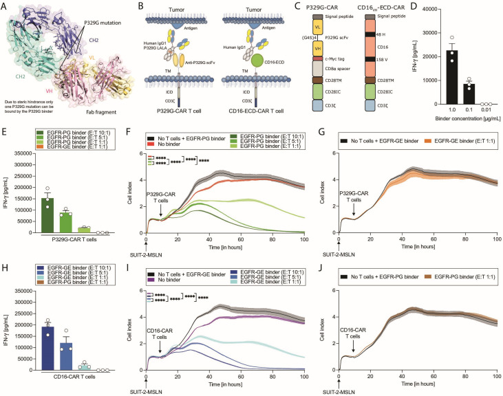
We initially evaluated the binding properties of an anti-P329G antibody. The crystal structure of the purified protein complex of Fc P329G with anti-P329G Fab8 implies that the interaction of the anti-P329G Fab fragment with the Fc region can only occur with 1:1 stoichiometry. This prevents the simultaneous binding of two different P329G-targeting CARs to one Fc fragment due to steric hindrance and excludes the possibility of unspecific CAR T cell dimerization in the absence of tumor antigen binding (figure 1A). Surface plasmon resonance analysis determined the equilibrium binding affinity of the P329G antibody to the P329G Fc portion to be 15 nM.8 The effector-silenced tumor antigen-targeting antibody carrying the P329G LALA Fc mutations thus acts as an adaptor, mediating a crosslinking between P329G-recognizing CAR T cells and antigen-expressing tumor cells (figure 1B).
Figure 1.
P329G-targeting CAR T cells perform similarly to CD16-ECD-CAR T cells. (A) Structure of P329G (PG) Leu234Ala/Leu235Ala (LALA) Fc/anti-P329G Fab complex. The heavy chain (VH) of the Fab fragment is shown in light pink and the light chain (VL) in yellow orange, the two chains of the Fc portion are shown in green/blue with the P329G mutation indicated. Steric hindrance on binding allows exclusively 1:1 binding of the anti-P329G Fab to the Fc region.8 (B) Schematic representation of P329G-targeting CAR (P329G-CAR) and CD16-ECD-CAR (CD16-CAR) T cell-mediated tumor binding. A CAR consists of an extracellular domain (ECD) for antigen recognition, conventionally a single chain variable fragment (scFv), which is linked via a hinge region to a transmembrane domain (TM), followed by an intracellular costimulatory domain (ICD) and a CD3zeta (CD3ζ) signaling domain. P329G LALA-mutated and unmutated glycoengineered human IgG1 serve as adaptor molecules. (C) Schematic representation of P329G-CAR and CD16VH-ECD-CAR. (D) IFN-γ ELISA after 72 hours of co-culture for three independent donors (n=3). P329G-CAR T cells were cultivated with different mesothelin (MSLN) binder concentrations and SUIT-2-MSLN cells. (E) IFN-γ ELISA after 48 hours of co-culture for three independent donors (n=3). P329G-CAR T cells were cultivated with P329G LALA-mutated (EGFR-PG) or glycoengineered EGFR (EGFR-GE) binders and SUIT-2-MSLN cells. (F+G) SUIT-2-MSLN tumor cell killing by different P329G-CAR T cell conditions measured over time through xCELLigence technology. (H) IFN-γ ELISA after 48 hours of co-culture for three independent donors (n=3). CD16-CAR T cells were cultivated with EGFR-PG or EGFR-GE binders and SUIT-2-MSLN cells. (I, J) SUIT-2-MSLN tumor cell killing by different CD16-CAR T cell conditions measured over time through xCELLigence technology. Each experiment (D–J) was performed in triplicates. Subfigures (F–G, I–J) show data of one representative donor out of three independent experiments. Values in all graphs represent means±SEM (*p<0.05, **p<0.01, ***p<0.001, ****p<0.0001). P values for xCELLigence data in (F–G, I–J) are shown for last time point. Only selected p values are shown.
Specificity of this new modular CAR T cell platform
Unlike Fc-binding CD16-ECD-CAR (CD16-CAR) interacting with human IgG in a non-selective manner, P329G-targeting CAR (referred to as P329G-CAR) T cells can only bind to P329G-mutated Fc parts. To demonstrate this specificity, the activity of P329G-CAR and CD16-CAR T cells was evaluated in the presence of EGFR-targeting P329G LALA-mutated (EGFR-PG) and GE (EGFR-GE) binders (figure 1B). Both constructs consist of the same CAR backbone with different extracellular targeting portions (figure 1C). We found that P329G Fc-mutated antibodies trigger specific activation of P329G-CAR T cells (figure 1D). As SUIT-2-MSLN cells had a high EGFR expression, they were employed for subsequent evaluation of EGFR binders (online supplemental figures S1A–C). P329G-CAR T cells only produced IFN-γ combined with EGFR-PG but not with EGFR-GE binder (figure 1E). P329G-CAR T cells efficiently killed SUIT-2-MSLN cells in an effector to target cell ratio dependent manner (figure 1F). Importantly, killing was not seen without binder (figure 1F) or with EGFR-GE binder (figure 1G). In direct comparison, CD16-CAR T cells showed IFN-γ production (figure 1H) and cytotoxicity (figure 1I–J) only with EGFR-GE but not with EGFR-PG binder. Effector functions of P329G-CAR T cells were comparable to CD16-CAR T cells. In the absence of a binder or of tumor cells, no relevant cytokine production was seen (online supplemental figures S1D–F).
Influence of human immunoglobulin G on P329G-CAR T cells and CD16-CAR T cells
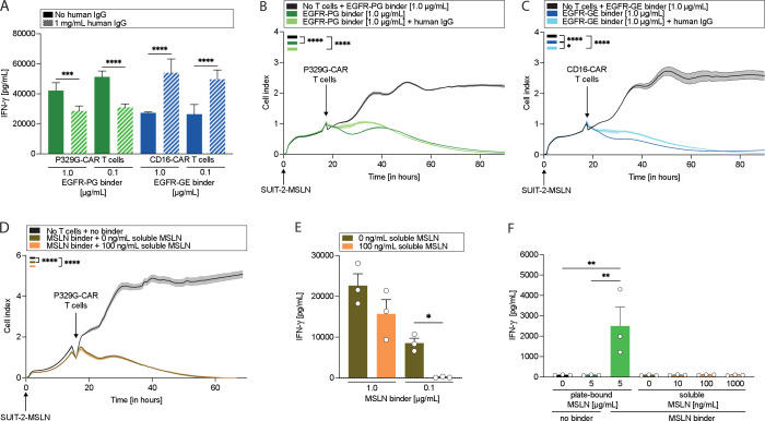
As we are addressing a subtle change in amino acid sequence compared with wild type IgG, we next sought to investigate factors that could detrimentally affect the platform. Numerous aspects can influence the safety, activation, and efficacy of P329G-CAR or CD16-CAR T cells in patients. The most relevant components influencing CAR T cells are immunoglobulins and other immune cells. To evaluate the influence of unmodified, polyclonal immunoglobulins on the interaction of P329G-CAR or CD16-CAR T cells with their corresponding binders, we investigated the impact of human polyclonal immunoglobulin fraction as they occur in the serum/plasma on CAR T cell activity in the presence or absence of different EGFR binders (figure 2A–C). We used a concentration of 1 mg/mL IgG for comparative studies, as on-tumor activation of CD16-ECD-CAR T cells with the high-affinity CD16 variant 158 V in the presence of higher serum concentrations of polyclonal human antibodies (10 mg/mL) was previously described.22 Competition with polyclonal IgG decreased IFN-γ production for P329G-CAR T cells in the presence of normal (p=0.0008) and low (p<0.0001) binder concentrations likely due to a low affinity intermediate interaction of the P329G-CAR with wild type IgG1 Fc regions (figure 2A). In contrast, and as previously reported, CD16-CAR T cells showed increased IFN-γ production when IgG was added to normal (p<0.0001) and low (p<0.0001) binder concentrations (figure 2A). Interestingly, killing capacity against SUIT-2-MSLN cells was similar for P329G-CAR and CD16-CAR T cells (figure 2B, C) regardless of the presence or absence of human IgG, respectively. This was also seen if lower binder concentrations were used (online supplemental figures S2A, S2B). We additionally performed a FRET competition assay to confirm that a wild type human IgG1 Fc is not recognized by this antibody. It could be shown that only the antibody with the P329G mutation can compete with the binding of the anti-P329G Fab to the labeled antibody with P329G mutation, whereas a human wild type IgG1 antibody did not compete with binding up to concentrations of 10 mg/mL. At very high concentrations a decrease of the fluorescent signal is observed with the wild type antibody, but the same decrease was observed for BSA and is therefore considered due to the protein concentration reducing the fluorescent signal and not to a specific interaction (online supplemental figure S2C).
Figure 2.
IgG and soluble recombinant MSLN do not activate P329G-targeting CAR T cells. (A) IFN-γ ELISA after 96 hours of co-culture for one representative donor out of three independent donors. CAR T cells were cultivated with SUIT-2-MSLN cells and P329G LALA-mutated EGFR (EGFR-PG) or glycoengineered EGFR (EGFR-GE) binder in a concentration of 1.0 or 0.1 µg/mL in the absence or presence of IgG at a concentration of 1 mg/mL. SUIT-2-MSLN tumor cell killing by (B) P329G-targeting CAR (P329G-CAR) T cells and (C) CD16-ECD-CAR (CD16-CAR) T cells in different EGFR binder conditions measured over time through xCELLigence technology. (D) SUIT-2-MSLN tumor cell killing by P329G-CAR T cells and MSLN binder in absence or presence of soluble MSLN (100 ng/mL). (E) IFN-γ ELISA after 96 hours of co-culture for three independent donors (n=3). P329G-CAR T cells and MSLN binder (1.0 or 0.1 µg/mL) were cultivated with SUIT-2-MSLN in presence or absence of soluble MSLN (100 ng/mL). (F) IFN-γ ELISA after 24 hours of co-culture for three independent donors (n=3). P329G-CAR T cells and MSLN binder were cultivated in presence or absence of plate-bound MSLN (5 µg/mL) or soluble MSLN (0, 10, 100, or 1000 ng/mL). Each experiment was performed in triplicates. Subfigures (A–D) show data of one representative donor out of three independent experiments. Values in all graphs represent means±SEM (*p<0.05, **p<0.01, ***p<0.001, ****p<0.0001). P values for xCELLigence data in (B–D) are shown for last time point. Only selected p values are shown.
Influence of soluble MSLN on P329G-CAR T cells
As some antibody targets might exist both in a membrane-bound and soluble form, we aimed to investigate the influence of soluble antigens on the platform’s function. One of the most investigated TAA is MSLN, which exists both in soluble and membranous variants.26 We thus took advantage of MSLN-targeting antibodies bearing P329G LALA Fc mutations to investigate the impact of a soluble antigen on the platform. We used reported physiological concentrations of 10 ng/mL and 100 ng/mL as well as the supraphysiological concentration of 1000 ng/mL MSLN (figure 2D–F and online supplemental figures S3A–D).26 Soluble MSLN did not reduce the killing of SUIT-2-MSLN tumor cells by P329G-CAR T cells at any concentration (figure 2D and online supplemental figures S3A, S3B). IFN-γ production was not significantly changed at a binder concentration of 1 µg/mL (figure 2E and online supplemental figure S3C). However, at lower binder concentrations of 0.1 µg/mL, cytokine production dropped in the presence of soluble MSLN (p=0.04), hinting at a competition for antibody binding between bound and soluble MSLN (figure 2E). P329G-CAR T cells, however, could only be activated in the presence of both plate-bound MSLN and the MSLN binder, as evidenced by IFN-γ (figure 2F) and IL-2 (online supplemental figure S3D) production. These results inform on the high discriminatory capacity of the P329G-CAR T cell platform toward immobilized target-antibody complexes.
In vitro characterization of P329G-CAR T cells with a MSLN binder
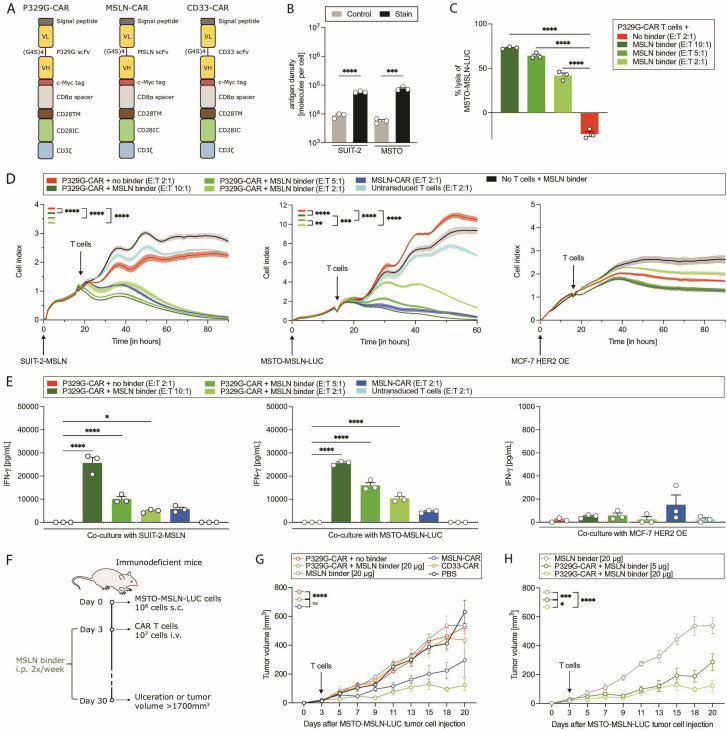
To further evaluate the application of P329G-CAR T cells for solid tumors, we performed in vitro characterization in the presence of both MSLN-targeting antibodies carrying the P329G LALA Fc mutations (MSLN binder) and MSLN-expressing target cell lines. Functionality of P329G-CAR T cells was compared with MSLN-targeting CAR (referred to as MSLN-CAR) T cells as positive control and to CD33-targeting CAR (referred to as CD33-CAR) T cells as negative control, both of which have the same CAR backbone (figure 3A). The pancreatic cancer cell line SUIT-2-MSLN and the mesothelioma cell line MSTO-MSLN-LUC were chosen due to their relevant MSLN expression (figure 3B). P329G-CAR T cells combined with the MSLN binder mediated efficient killing of MSTO-MSLN-LUC (figure 3C, D) and SUIT-2-MSLN tumor cells (figure 3D) at different E:T ratios. No killing was seen in the absence of a binder (figure 3C, D) or with untransduced T cells (figure 3D). P329G-CAR T cells had no relevant cytotoxic capacity against low-MSLN-expressing (data not shown) MCF-7 HER2 OE cells (figure 3D). In contrast to conditions without binder or untransduced T cells, P329G-CAR T cells combined with the MSLN binder produced relevant IFN-γ levels when cultivated with MSLN-expressing cell lines (figure 3E). Cytokine release was dependent on the amount of T cells added to the culture (figure 3E). Effector functions of P329G-CAR T cells were comparable to those of MSLN-CAR T cells, when cultivated at the same E:T ratio (figure 3D, E), indicating that the use of a CAR-adaptor molecule does not lower the therapeutic potential of P329G-CAR T cells.
Figure 3.
P329G-targeting CAR T cells combined with the MSLN binder mediate in vitro and in vivo effector functions. (A) Schematic representation of P329G-targeting CAR (P329G-CAR), MSLN-targeting CAR (MSLN-CAR), and CD33-targeting CAR (CD33-CAR). (B) Density of MSLN surface expression on SUIT-2-MSLN and MSTO-MSLN-LUC cells. (C) MSTO-MSLN-LUC tumor cell killing by different P329G-CAR T cell conditions measured by luciferase-based assay after 64 hours of co-culture for three independent donors (n=3). (D) SUIT-2-MSLN, MSTO-MSLN-LUC and MCF-7 HER2 OE tumor cell killing by different T cell conditions measured over time through xCELLigence. (E) IFN-γ ELISA after 48 hours of co-culture with SUIT-2-MSLN, MSTO-MSLN-LUC and MCF-7 HER2 OE for one representative donor out of three independent donors. (F) Experimental layout for (G, H). MSLN binder was administered intraperitoneally (i.p.) twice a week. (G, H) MSTO-MSLN-LUC tumor growth curves of mice treated with a single intravenous injection of phosphate-buffered saline (PBS) or 107 CAR T cells (n=5 mice per group). (G, H) MSLN binder-treated mice received 20 µg or 5 µg per mouse. Each experiment was performed in triplicates. Subfigures (D, E) show data of one representative donor out of three independent experiments. Values in all graphs represent means±SEM (*p<0.05, **p<0.01, ***p<0.001, ****p<0.0001). P values for xCELLigence data in (D) are shown for last time point. Only selected p values are shown.
In vivo characterization of P329G-CAR T cells with a MSLN binder
To further evaluate the functionality of our modular CAR T cell platform, we next performed a xenograft treatment experiment. MSTO-MSLN-LUC cells were subcutaneously injected into immunodeficient mice 3 days prior to CAR T cell injection (figure 3F). Treatment of mice with P329G-CAR T cells and the MSLN binder (20 µg) significantly reduced tumor growth (p<0.0001) compared with P329G-CAR T cells without a binder or CD33-CAR T cells (figure 3G). Remarkably, the reduction in tumor volume was comparable for mice treated with P329G-CAR T cells and the MSLN binder or with MSLN-CAR T cells (figure 3G). Titration of the binder dose was performed showing also efficient reduction of tumor growth at lower binder doses (figure 3H). As this tumor model is highly ulcerative, only a slight survival benefit (data not shown) and no complete tumor eradication could be seen. This set the rationale to prove the concept with another target antigen in a different tumor model. Together these results indicate equal in vitro and in vivo potency of the P329G-CAR T cell platform compared with conventional CAR T cells.
In vitro characterization of P329G-CAR T cells redirected with a HER2 binder
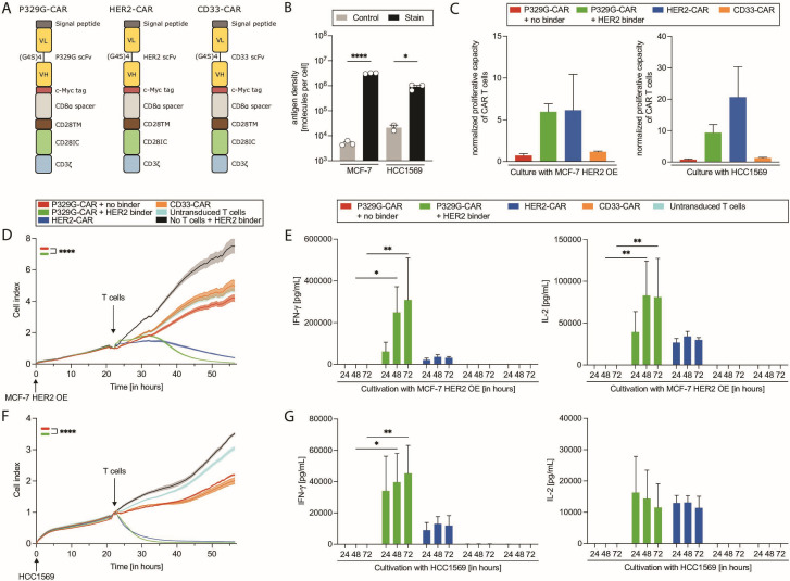
P329G-CAR T cells were further characterized in vitro in the presence of a HER2-targeting antibody carrying P329G LALA Fc mutations (HER2 binder) and HER2-expressing target cell lines. The functionality of P329G-CAR T cells was compared with HER2-targeting CAR (referred to as HER2-CAR) T cells as positive control and to CD33-CAR T cells as negative control with the same CAR backbone (figure 4A). The breast cancer cell lines HCC1569 and MCF-7 HER2 OE were chosen as target cell lines based on their HER2 expression (figure 4B). Only P329G-CAR T cells combined with the HER2 binder as well as HER2-CAR T cells were able to proliferate while being co-cultured with antigen-positive tumor cells (figure 4C). No sufficient proliferation was noted in the absence of tumor cells (online supplemental figure S4A). P329G-CAR T cells together with the HER2 binder demonstrated effective tumor cell killing and cytokine production when co-cultured with MCF-7 HER2 OE (figure 4D, E) and HCC1569 tumor cells (figure 4F, G) in contrast to controls. Notably, the killing capacity of P329G-CAR T cells was similar to HER2-CAR T cells. No relevant cytokine production was noted in the absence of tumor cells (online supplemental figures S4B, S4C).
Figure 4.
P329G-targeting CAR T cells combined with a HER2 binder mediated sufficient in vitro effector functions. (A) Schematic representation of P329G-targeting CAR (P329G-CAR), HER2-targeting CAR (HER2-CAR), and CD33-targeting CAR (CD33-CAR). (B) Density of HER2 surface expression on MCF-7 HER2 OE and HCC1569. (C) Proliferative capacity after 72 hours of co-culture normalized to CAR T cells per beads measured by FACS after 24 hours co-culture in the presence of HCC1569 or MCF-7 HER2 OE of three independent donors (n=3). (D) MCF-7 HER2 OE tumor cell killing by different T cell conditions plated at E:T ratio of 2:1 measured over time through xCELLigence. (E) IFN-γ and IL-2 ELISA after 24 hours, 48 hours, and 72 hours of co-culture with MCF-7 HER2 OE for three independent donors (n=3). (F) HCC1569 tumor cell killing by different T cell conditions plated at a E:T ratio of 2:1 measured over time through xCELLigence. (G) IFN-γ and IL-2 ELISA after 24 hours, 48 hours, and 72 hours co-culture with HCC1569 supernatants for three independent donors (n=3). Each experiment was performed in triplicates. Subfigures (D, F) show data of one representative donor out of three independent experiments. Values in all graphs represent means±SEM (*p<0.05, **p<0.01, ***p<0.001, ****p<0.0001). P values for xCELLigence data in (D, F) are shown for last time point. Only selected p values are shown.
Immunophenotypic changes of P329G-CAR T cells after stimulation
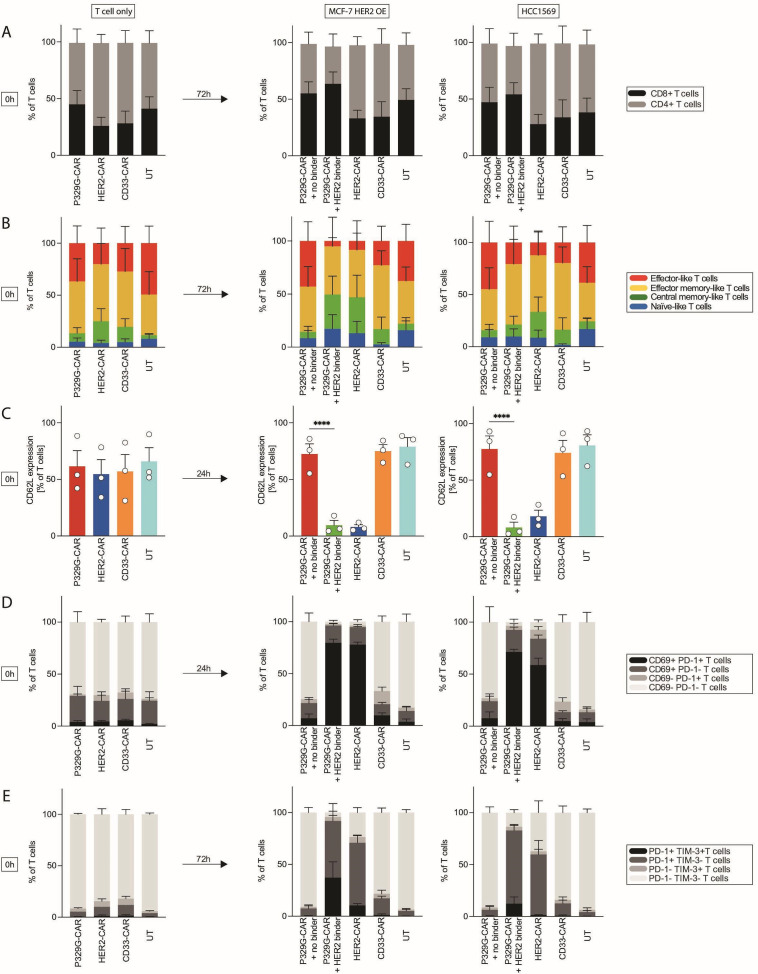
The immunophenotype of activated P329G-CAR T cells was determined after 24 hours and 72 hours of co-culture with HER2-expressing tumor cell lines (figure 5). P329G-CAR T cells showed a more balanced CD4+ T helper (TH) cell to CD8+ cytotoxic T cell ratio (CD4:CD8 ratio) compared with HER2-CAR and CD33-CAR T cells, which were more skewed toward a CD4+ T cell dominance prior to activation (figure 5A). The CD4:CD8 ratio was conserved on stimulation with tumor cells irrespective of the presence of a binder (figure 5A). P329G-CAR T cells alone, CD33-CAR T cells and untransduced T cells did not reveal relevant changes in their phenotype after 72 hours of co-culture with HER2-expressing tumor cells, as expected. In contrast, P329G-CAR T cells with the HER2 binder and HER2-CAR T cells evolved towards a higher content of central memory T cells (TCM) to the detriment of effector T cells (Teff) (figure 5B). These changes in T cell subpopulations were accompanied by a decreased expression of the lymph node homing marker CD62L (figure 5C), particularly on P329G-CAR T cells cultured with the binder (p<0.0001). P329G-CAR T cells cultivated with the HER2-targeting binder and HER2-CAR T cells both upregulated CD69 as well as programed cell death protein 1 (PD-1) and slightly upregulated T cell immunoglobulin mucin-3 (TIM-3) on activation (figure 5D, E). However, HER2-CAR T cells showed no relevant increase of TIM-3 expression when cultivated with HCC1569 (figure 5E). P329G-CAR alone, CD33-CAR and untransduced T cells did not demonstrate relevant CD69, PD-1, and TIM-3 upregulation (figure 5D, E), consistent with their lack of specific activation in co-culture.
Figure 5.
Phenotypic changes of CAR T cells after activation. Phenotype of P329G-targeting CAR (P329G-CAR), HER2-targeting CAR (HER2-CAR), and CD33-targeting CAR (CD33-CAR) T cells or untransduced T cells (UT) are shown at start of co-culture (0 hours) and after 24 hours or 72 hours of co-culture in the presence of MCF-7 HER2 OE or HCC1569 of three donors (n=3). (A) CD8+ and CD4+ T cells are shown. (B) T cell subpopulations are shown. Naïve-like T (TN) cells were defined as CD45RA+ CCR7+, central memory-like T (TCM) cells as CD45RA- CCR7+, effector memory-like T (TEM) cells as CD45RA- CCR7- and effector-like T (Teff) cells as CD45RA+ CCR7-. Changes in T cell subpopulations were not significant (NS). (C) Expression of CD62L is shown. (D) Expression of CD69 and PD-1 is shown. P329G-CAR T cells in the presence or absence of HER2 binder were compared for MCF-7 HER2 OE and for HCC1569: p<0.0001 for CD69+ PD-1+, NS for CD69+ PD-1-, NS for CD69- PD-1+, p<0.0001 for CD69- PD-1-. (E) Expression of PD-1 and TIM-3 is shown. P329G-CAR T cells in the presence or absence of HER2 binder where compared for MCF-7 HER2 OE: p<0.05 for PD-1+ TIM-3+, p<0.01 for PD-1+ TIM-3-, NS for PD-1- TIM-3+, p<0.0001 for PD-1- TIM-3-; and for HCC1569: NS for PD-1+ TIM-3+, p<0.0001 for PD-1+ TIM-3-, NS for PD-1- TIM-3+, p<0.0001 for PD-1- TIM-3-. Each experiment was performed in triplicates. Values in all graphs represent means±SEM (*p<0.05, **p<0.01, ***p<0.001, ****p<0.0001). Selected p values are shown in the figures or described in the legend.
P329G-CAR T cells combined with a HER2 binder in a xenograft mouse model
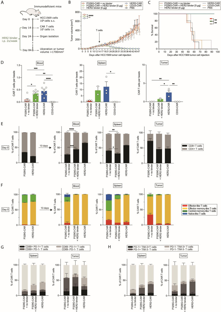
To substantiate the broad applicability of this novel CAR T cell platform, we performed further xenograft mouse experiments. HER2-expressing HCC1569 were subcutaneously injected into immunodeficient mice 14 days prior to CAR T cell injection (figure 6A). Mice treated with P329G-CAR T cells and the HER2 binder or with HER2-CAR T cells experienced complete tumor eradication (figure 6B) for more than 70 days (data shown until day 47 post tumor cell injection), leading to an overall survival of 100% of these mice (figure 6C). Interestingly, in mice treated with HER2-CAR T cells, more CAR T cells were detected in the blood, spleen, and especially in the tumor 10 days after CAR T cell injection (figure 6D). In mice treated with P329G-CAR T cells alone or with CD33-CAR T cells, only a few cells were still detectable at that time (figure 6D). Phenotypic analysis revealed that P329G-CAR T cells and HER2-CAR T cells enriched for CD8+ cytotoxic T cells particularly at the tumor site (figure 6E). Analysis of subpopulations of T cells detected in blood, spleen and tumor, revealed fewer less-differentiated T cells and terminally differentiated effector-like T cells and more effector memory-like T cells, particularly when comparing mice treated with P329G-CAR T cells with or without the HER2 binder (figure 6F). CAR T cells in mice treated with P329G-CAR T cells and the HER2 binder or with HER2-CAR T cells had a similar CD62L expression in both spleen and tumor (online supplemental figures S4D, S4E). Even 10 days after T cell injection, CAR T cells still showed signs of activation, as evidenced by CD69, PD-1, and TIM-3 expression at the tumor site (figure 6G, H). These results confirm the in vivo potency of P329G-CAR T cells.
Figure 6.
P329G-targeting CAR T cells with HER2 binder mediate sufficient in vivo effector functions. (A) Experimental layout for (B–H). P329G-targeting CAR (P329G-CAR), HER2-targeting CAR (HER2-CAR), CD33-targeting CAR (CD33-CAR) T cells or phosphate-buffered saline (PBS) were intravenously injected. HER2 binder (5 µg per mouse) was administered intraperitoneally (i.p.) twice a week. 10 days after CAR T cell injection, bleeding was performed, and 2–3 sentinel mice per group were used for organ isolation. (B) HCC1569 tumor growth curves of mice treated with a single intravenous injection of PBS (n=5 mice) or 107 CAR T cells (n=4 mice per group). (C) Survival curves of (B). (D) CAR T cells per beads for blood, spleen, and tumor of treated mice 10 days after CAR T cell injection. (E) CD8+ and CD4+ T cells and (F) T cell subpopulations are shown for CAR T cells on day of CAR T cell injection and for human CAR T cells detected in blood, spleen, and tumor of treated mice 10 days after CAR T cell injection. Naïve-like T (TN) cells were defined as CD45RA+ CCR7+, central memory-like T (TCM) cells as CD45RA- CCR7+, effector memory-like T (TEM) cells as CD45RA- CCR7- and effector-like T (Teff) cells as CD45RA+ CCR7-. P329G-CAR T cells in the presence or absence of HER2 binder were compared for blood: p<0.05 for TN, p<0.001 for TCM, p<0.0001 for TEM and NS for Teff; for spleen: p<0.0001 for TN, p<0.0001 for TCM, p<0.0001 for TEM and p<0.01 for Teff; and for tumor: NS for TN, NS for TCM, p<0.0001 for TEM and p<0.0001 for Teff. (G) Expression of CD69 and PD-1, and (H) expression of PD-1 and TIM-3 on CAR T cells detected in spleens, and tumors of treated mice 10 days after CAR T cell injection. (G) P329G-CAR T cells in the presence or absence of HER2 binder were compared for spleen: NS for CD69+ PD-1+, NS for CD69+ PD-1-, p<0.01 for CD69- PD-1+, p<0.001 for CD69- PD-1-; and for tumor: NS for all subsets. (H) P329G-CAR T cells in the presence or absence of HER2 binder were compared for spleen: NS for PD-1+ TIM-3+, p<0.0001 for PD-1+ TIM-3-, NS for PD-1- TIM-3+, p<0.001 for PD-1- TIM-3-; and for tumor: NS for all subsets. Values in all graphs represent means±SEM (*p<0.05, **p<0.01, ***p<0.001, ****p<0.0001). Selected p values are shown in the figures or described in the legend.
In vitro modularity and reversibility of the P329G-CAR T cell platform
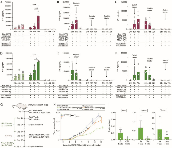
To demonstrate that, as predicted, the P329G-CAR T cell platform is indeed modular and its activity can be reversed, we next conducted a set of in vitro experiments. CAR T cells were seeded onto recombinant HER2-coated plates (figure 7A–C). In the presence of the HER2 binder, P329G-CAR T cells released IFN-γ (figure 7A). Depletion of the HER2 binder led to a decrease of cytokine production in the following culture period (figure 7B). When P329G-CAR T cells were cultivated with the MSLN binder, no cytokine secretion was seen. Switching from the MSLN binder to the HER2 binder restored cytokine production (figure 7C). Along the same lines, the switch from the HER2 binder towards the MSLN binder led to a decrease in cytokine production (figure 7C). Similar effects were also observed when plates were coated with recombinant MSLN (figure 7D–F). These results substantiate the selectivity, in vitro modularity, and reversibility of the platform.
Figure 7.
P329G-targeting CAR T cells mediated effector functions in a modular and reversible fashion. (A–C) IFN-γ ELISA after co-culture of P329G-targeting CAR (P329G-CAR), HER2-targeting CAR (HER2-CAR), MSLN-targeting CAR (MSLN-CAR) T cells or untransduced T cells (UT) with recombinant HER2 and different conditions of binders for three independent donors (n=3). (D–F) IFN-γ ELISA after co-culture of CAR T cells or UT with recombinant MSLN and different conditions of binders for three independent donors (n=3). (G) Experimental layout for (H). HER2 or MSLN binder (5 µg per mouse) were administered intraperitoneally (i.p.) twice a week. Bleeding and organ isolation was performed for mice still alive on day 28 after MSTO-MSLN-LUC tumor cell injection. (H) MSTO-MSLN-LUC tumor growth curves of survivor mice re-challenged with the tumor (n=4 mice per group) and treatment naïve mice (n=4–5 mice per group). (I) Human T cells and CAR T cells per beads ratio for blood, spleen, and tumor of survivor mice treated with P329G-CAR T cells and the binders. Each experiment of subfigures (A–F) was performed in triplicates. Values in all graphs represent means±SEM (*p<0.05, **p<0.01, ***p<0.001, ****p<0.0001). Only selected p values are shown.
In vivo modularity of the P329G-CAR T cell platform
Finally, to confirm modularity in vivo, we re-challenged the surviving HCC1569-bearing immunodeficient mice, which were initially treated with P329G-CAR T cells and the HER2 binder or with HER2-CAR T cells (figure 6A), with another tumor cell line expressing a different tumor antigen (figure 7G). MSTO-MSLN-LUC cells were subcutaneously injected into the unaffected flank of these surviving mice 89 days after initial HCC1569 tumor cell injection. No binder was injected for 5–6 days prior to and after tumor rechallenge. After this short resting period of binder application, mice were treated with the MSLN binder (figure 7G). Tumor-bearing control mice never received T cells. Rechallenged mice treated with P329G-CAR T cells and the MSLN binder demonstrated reduced tumor growth (figure 7H) compared with HER2-CAR T cell-treated mice (p<0.0001), binder only treated mice (p=0.0001) and untreated mice (p<0.0001) (figure 7H). This reduction in tumor growth is comparable to the results seen in the primary treatment of MSTO-MSLN-LUC tumors using the P329G-CAR T cell platform (figure 3F–H). Human T cells were still detectable in the blood, spleen, and tumor of mice treated with P329G-CAR T cells and binder more than 100 days after T cell injection (figure 7I) showing a long-term persistence of P329G-CAR T cells.
Discussion
Here, we could demonstrate the activity and potency of a novel P329G-targeting CAR T cell platform against different solid tumor models. The modularity of this novel platform could be substantiated in vitro and in vivo. Our results provide evidence that this universal and modular platform could further help to improve CAR T cell therapy beyond hematology.
Conventional CAR T cells contain a scFv as an extracellular antigen recognition domain and can only bind a single target antigen. Modular CAR T cell platforms, however, uncouple antigen targeting and signaling, leading to a dichotomous system consisting of a CAR and a CAR-adaptor molecule.4–6 The major advantages of modular CAR T cell platforms are the possibility to stop administration of the adaptor molecule in the case of undesired side effects as well as the ability to target multiple antigens by administering different CAR-adaptor molecules. Importantly, only one universal T cell product would need to be engineered for and could then be combined with various adaptor molecules, leading to a reduction of labor-intensive and cost-intensive CAR development.4–6 Modular CAR T cell platforms can be subdivided into Fc-binding CARs, Tag-specific CARs and BiAb-binding CARs.4–6 The clear advantage of CARs binding the Fc part of IgG antibodies is that they can be combined with clinically approved monoclonal antibodies like rituximab, trastuzumab, or cetuximab.22 27 However, such platforms, which are typically based on the extracellular portion of CD16, cannot possibly discriminate between endogenous antibodies and therapeutic compounds. In fact, we could previously show that endogenous IgG can trigger CD16-based responses in a therapeutically relevant manner, generating safety and efficacy concerns.22 Thus, more selective targeting is required. Tag-specific CARs, in contrast, are designed to target tags coupled chemically, enzymatically, or genetically to a tumor-targeting moiety. This includes commonly available tags like the synthetic dye fluorescein isothiocyanate (FITC), and biotin or peptide tags directly introduced by genetic fusion.4–6 An universal CAR (UniCAR) platform, which combines a CAR targeting an unique peptide epitope (5B9) derived from a nuclear protein and a CAR-adaptor molecule directed against the TAA and the epitope (5B9) has been used in a pre-clinical solid tumor model.28 Another UniCAR T cell using a soluble adaptor consisting of the UniCAR epitope linked to a scFv directed against CD123 (TM123) is currently investigated in a phase 1a trial in relapsed/refractory AML patients with ≥20% CD123+ bone marrow blasts (NCT04230265).29 Such tags come with the need for additional quality control steps of the adaptor molecule and also bear the risk of increasing the immunogenicity of the therapy. Similarly, BiAbs are able to redirect T cells by targeting the synthetic receptor introduced genetically in the therapeutic cell.6 We could previously demonstrate the potency of BiAbs to redirect synthetic agonistic receptor transduced T cells.16 30 However, all these more selective approaches come with the burden of developing new adaptors for each product and antigen as well as the use of non-natural/non-human tags or post-translational modification of antibodies in the case of chemical conjugation. An allogeneic universal CAR T cell strategy is the UCART19 approach using adoptive T cells targeting CD19+ malignancies by performing a gene knockout of TRAC and CD52 to prevent TCR mediated recognition of patient’s HLA antigens and to permit alemtuzumab usage in lymphodepletion.31 However, healthy volunteers must be identified and no long-term data exist for now.
A recent development in antibody technologies are Fc-silenced antibodies generated through the introduction of P329G mutations in their Fc part, and a growing number of such antibodies are in clinical development and trials.9–15 Given the wealth of data on such antibodies, we reasoned that these P329G-mutated antibodies would enable the efficient redirection of P329G-targeting CAR T cells without requiring de novo development of antibodies or adaptors, thus easing translation. Antibodies based on the human IgG1 isotype with P329G (LALA) mutations lack FcγR binding, but have retained FcRn binding, thus they are supposed to typically show IgG-like pharmacokinetics in man with half-lives of 2–3 weeks.7 We argue that our results encourage this path, as we demonstrate P329G-targeting CAR T cells to be efficiently redirected against a range of different cancer entities and antigens.
MSLN is a tumor differentiation antigen, which is expressed on mesothelial cells in the pericardium, pleura, and peritoneum.32 Importantly, MSLN expression in healthy tissues is limited, while it is highly expressed in multiple human malignancies such as pancreatic, ovarian and lung adenocarcinoma as well as malignant mesothelioma.32 Based on these characteristics, MSLN is regarded as a suitable target for CAR T cell therapy.32 MSLN-targeting CAR T cells are currently under evaluation in multiple clinical trials assessing safety and feasibility.32 However, persistence and long-term efficacy of MSLN-targeting CAR T cells have been found to be limited.32 Multiple strategies were brought forward to improve treatment with these CAR T cells including regional delivery32 or genetic engineering for the expression of specific chemokine receptors23 24 or for resistance against hypofunction.24 These novel strategies are either being tested in clinical trials or are still undergoing preclinical testing. We could demonstrate that P329G-targeting CAR T cells combined with a MSLN binder have similar in vitro and in vivo effector functions to conventional MSLN-targeting CAR T cells. Due to a highly ulcerative tumor model, no complete tumor eradication could be seen. Higher T cell numbers or preloading of P329G-CAR T cells with the binder might improve antitumor efficacy. This needs to be contrasted with the antibody being able to immediately bind with 15 nM affinity to the P329G-CAR T cells in circulation and occupy the CAR to a certain percentage, depending on the dose. However, as the affinity for the tumor antigen is higher than to the P329G-CAR, it will bind bivalently. It will preferentially accumulate in the tumor and bind there so that free P329G-CAR T cells can bind too in the tumor microenvironment (TME). An open question remains whether P329G-targeting CAR T cells would come with the same limitations as MSLN-directed or other targeted CAR T cells.
To further investigate the safety of the proposed therapy, we assessed the impact of soluble MSLN on P329G-targeting CAR T cells. It was reported that healthy donors typically have serum MSLN concentrations under 10 ng/mL, while mesothelioma and ovarian cancer patients can have concentrations higher than 50 ng/mL.26 We did not observe unspecific activation or decrease of cytotoxicity of P329G-CAR T cells in the presence of soluble MSLN, even at supraphysiological concentrations. At low binder concentrations, however, soluble MSLN competed with membrane bound MSLN for antibody binding and subsequently decreased T cell activation, suggesting that patients with high MSLN serum concentrations might require higher therapeutic antibody doses.33
Another important target antigen for immunotherapy in solid tumors is HER2/neu, a member of the EGFR family. HER2 expression plays an important role for the targeted therapy of breast cancer as well as gastric and gastroesophageal cancers.34 HER2 is also expressed on other cancers, and HER2-targeted therapy mediated responses in colorectal, biliary, pancreatic, lung and bladder cancer.34 Although HER2-targeted therapy is well established in the treatment of breast cancer, therapeutic failure or relapse is still frequently seen.35 A major advantage of HER2-targeting CAR T cells compared with antibody-based therapy is that CAR T cells might not be subject to the same resistance mechanisms as antibody therapy, and importantly possess their own cytolytic activity.36 37 We could demonstrate that P329G-targeting CAR T cells combined with a trastuzumab-derived HER2 binder carrying P329G LALA mutations in the Fc part mediated efficient in vitro proliferation and effector functions. Phenotypic analysis after CAR T cell activation revealed that P329G-CAR T cells had a more balanced CD4:CD8 ratio compared with HER2-targeting CAR T cells independent of activation. CD4+ TH cells are important for persistence of CAR T cells, as well as for the secondary expansion and memory formation of CD8+ cytotoxic T cells.38 Thus, a balanced CD4:CD8 ratio is deemed highly beneficial for cancer eradication.39 To this end, CAR T cell products with a defined CD4:CD8 ratio have been developed.40 Additionally, T cell differentiation status also plays an important role in the success of CAR T therapy.41 While profound in vitro antitumor efficacy could be detected by terminally differentiated T cells, CAR T cell activation, proliferation and persistence of such cells can be insufficient in vivo.42 Naïve-like T cells (TN cells) and stem cell memory-like T cells (TSCM cells) became more important for CAR T cell therapy due to their ability for self-renewal and differentiation into all T cell subsets leading to long-lasting antitumor efficacy.41 Co-culture of P329G-CAR T cells with HER2-expressing cells mediated a decrease of Teff cells and reduced CD62L expression, leading to a modified composition of T cell subpopulations. The inhibitory TME might also limit the response rates of CAR T cell therapy in certain malignancies.3 The observed upregulation of the inhibitory receptors PD-1 and TIM-3 on P329G-targeting CAR T cells, even when compared with HER2-targeting CAR T cells, was accompanied by complete tumor eradication in vivo, indicating a different impact of these molecules on the functionality of P329G-targeting CAR T cells. It should be noted, however, that while HER2 served as a target to show proof-of-concept in vitro and in vivo, trastuzumab is not mouse cross-reactive so that on-target off-tumor toxicity of the P329G-CAR T cell approach on essential normal tissues could not be investigated.
In general, for severe treatment-associated adverse events, safety switches for CAR T cell therapy are required, which should not impair CAR T cell survival. Possible strategies to overcome CAR-associated toxicities are to reduce antigen-binding domain affinity and decrease cytokine secretion or CAR immunogenicity by modulating the hinge, transmembrane (TM) or costimulatory domain(s) of the CAR.43 As a CAR ‘off-switch’ in case of severe adverse events, CD20-expressing CAR T cells can be targeted by the CD20-directed antibody rituximab,44 or the tyrosine-kinase inhibitor dasatinib can temporarily be applied to decrease the functionality of administered CAR T cells.45 As the presence of an adaptor molecule is mandatory for modular CAR T cells, the pharmacokinetic properties, biodistribution and binding affinity of these adaptor molecules directly influence CAR T cell activity.4–6 In this context, administration of IgG-based adaptors may raise concerns regarding safety management.6 However, like for antibody-based therapies if patients suffer from severe adverse events, administration of the antibody can be paused to diminish CAR T cell activity, particularly during phase 1 dose escalation clinical trials. Typically, for first in human trials, dose escalation of the P329G adaptor antibody starting with subtoxic doses would be implemented and then step-up-dosing regimens would be performed to limit CRS as it is being applied for TCB. In line with this it has been shown that stopping the application of the adaptor molecule is an effective way to turn off CAR T cell activity.6 Theoretically, administration of complex-forming counter-adaptors such as an inert Fc portion with the PG LALA mutations could be administered as an potential antidote.4–6 These approaches can help to manage the safety and feasibility of P329G-CAR T cells and address some of the most important limitations currently observed clinically. As transient rest of CAR T cells can improve persistence and durability, it would be interesting to explore if resting P329G-targeting CAR T cells by modifying their dose and administration schedule would optimize their safety, potency and persistance.46
Another important limitation of CAR T cell therapy is that tumor cells might become resistant to single targeting CAR T cells by selecting antigen-negative variants. Between 30%–70% of relapsed ALL patients had a CD19-negative relapse after receiving well-established CD19-CAR T cell treatment.43 In addition, patients with multiple myeloma treated with anti-BCMA CAR T cells demonstrated antigen loss or downregulation, eventually leading to tumor relapse.43 Targeting multiple antigens might be a possible strategy to overcome this issue. Bispecific CAR T cells targeting, for example, CD19 and CD2047 48 or CD19 and CD2249 50 were developed. Here, we showed that P329G-targeting CAR T cells could be successfully re-directed to different target antigens, leading to reduced tumor growth in a xenograft mouse model. In the case of target antigen downregulation or complete antigen escape, administration of a novel antibody could be feasible, and patients could still benefit from circulating CAR T cells as suggested by our in vitro and in vivo experiments.
In summary, we described a novel universal and modular CAR T cell platform for application in solid tumors. Modular CAR T cells provide promising solutions for current problems of conventional CAR T cells. P329G-targeting CAR T cells combine the advantages of CAR T cell therapy and antibody-based therapy, leading to improvement of therapy-associated toxicities and efficacy limitations. Importantly, multiple effector-silenced antibodies with P329G Fc mutations have been developed preclinically and clinically14 15 and a first BiAb containing P329G LALA mutations has been approved validating the use of the P329G mutation (faricimab).14 Currently, such IgG-based modular Claudin 18.2-targeting P329G-directed CAR T cells are already being investigated in a clinical trial (NCT05199519). Further preclinical evaluation is required to show the translational potential of this promising approach.
Acknowledgments
SS was supported by the Else Kröner-Fresenius Clinician Scientist Program Cancer Immunotherapy, the Munich Clinician Scientist Program (MCSP) and the DKTK School of Oncology. A-KK was supported by a grant from the Förderprogramm für Forschung und Lehre (FöFoLe) of the Ludwig Maximilian University (LMU) of Munich. The authors wish to thank Manuel Spaeni and Erwin van Puijenbroek (Roche) for the antibody production. Additionally, the authors wish to thank Regula Buser (Roche) for performing the FRET competition assay. We acknowledge the iFlow Core Facility of the University Hospital of the Ludwig Maximilian University (LMU) Munich for assistance with the generation of flow cytometry data. We acknowledge the Core Facility Flow Cytometry of the University Hospital of the Ludwig Maximilian University (LMU) of Munich, funded by the Deutsche Forschungsgemeinschaft (DFG, German Research Foundation)-499549016.
Footnotes
Twitter: @SophiaStock_, @MRbenmebarek
SS and M-RB contributed equally.
Contributors: SS, M-RB and A-KK designed the study, performed the experiments, and analyzed the data; SS designed the figures and wrote the primary manuscript; DD, CJ, K-GS, JB, AF-G. EM, PU and CK initially conceptualized and designed the P329G-CAR. DD, CJ, K-GS, AF-G. CK developed and provided the antibodies; JB analyzed structural data; AF-G performed surface plasmon resonance; M-RB, A-KK, DD, CK and SK edited the manuscript; CK discussed experimental design; MS and SE reviewed the manuscript; SK supervised the project and was responsible for the overall content; all authors have read and agreed to the published version of the manuscript.
Funding: SS, M-RB and A-KK: Declare no funding. DD, CJ, K-GS, JB, AF-G, EM, PU and CK: Declare no funding except by Roche. MS received research funding from a German Research Foundation (DFG) grant (451580403), the SFB-TRR 388/1 2021-452881907, the Bavarian Elite Graduate Training Network, the Wilhelm Sander-Stiftung (20180871), the Else Kröner-Fresenius-Stiftung and the Bavarian Center for Cancer Research (BZKF). This study was supported by the Marie Sklodowska-Curie Program Training Network for Optimizing Adoptive T Cell Therapy of Cancer funded by the H2020 Program of the European Union (Grant 955575, to SK); by the Hector Foundation (to SK); by the International Doctoral Program i-Target: Immunotargeting of Cancer funded by the Elite Network of Bavaria (to SK and SE); by Melanoma Research Alliance Grants 409510 (to SK); by the Else Kröner-Fresenius-Stiftung (2021_EKFK_01, to SK); by the German Cancer Aid (to SK); by the Ernst-Jung-Stiftung (to SK); by the LMU Munich’s Institutional Strategy LMUexcellent within the framework of the German Excellence Initiative (to SE and SK); by the Bundesministerium für Bildung und Forschung (to SE and SK); by the Go-Bio Initiative (to SK), by the m4 award of the Bavarian Ministry for Economical Affairs (to SK); by the Wilhelm Sander-Stiftung (2022.051.1, to SK); by the European Research Council Grant 756017, ARMOR-T (to SK); by the German Research Foundation (DFG) (to SK); by the SFB-TRR 338/1 2021–452881907 (to SK); by the Fritz-Bender Foundation (to SK) and by the Deutsche José Carreras Leukämie-Stiftung (to SK).
Competing interests: SS, M-RB and A-KK declare that they have no competing interests. DD and CJ declare previous employment and patents with Roche (Switzerland). DD declares current employment, patents, and stock ownership with Innovent Biologics (China) and CJ declares current employment, patents and ownership interest in Athebio AG (Switzerland). K-GS, JB, AF-G, EM, PU and CK declare employment, patents, and stock ownership with Roche (Switzerland). MS received honoraria from AMGEN, BMS, Janssen, Kite/Gilead, Roche, Novartis, Pfizer, Celgene and Takeda. MS received research support from AMGEN, BMS, Janssen, Kite/Gilead, Miltenyi, MorphoSys, Novartis, Roche and Seattle Genetics for work unrelated to the manuscript. MS declare consultancy for AMGEN, Celgene, Janssen, Kite/Gilead, Novartis, and Takeda. SK has received honoraria from TCR2 Inc., Novartis, BMS and GSK. SK and SE are inventors of several patents in the field of immuno-oncology. SK and SE received license fees from TCR2 Inc. and Carina Biotech. SK and SE received research support from TCR2 Inc., Tabby Therapeutics and Arcus Bioscience for work unrelated to the manuscript. P329G-CAR© is a trademark by Roche (Switzerland) and being developed by Roche (Switzerland) and by Innovent Biologics (China).
Provenance and peer review: Not commissioned; externally peer reviewed.
Supplemental material: This content has been supplied by the author(s). It has not been vetted by BMJ Publishing Group Limited (BMJ) and may not have been peer-reviewed. Any opinions or recommendations discussed are solely those of the author(s) and are not endorsed by BMJ. BMJ disclaims all liability and responsibility arising from any reliance placed on the content. Where the content includes any translated material, BMJ does not warrant the accuracy and reliability of the translations (including but not limited to local regulations, clinical guidelines, terminology, drug names and drug dosages), and is not responsible for any error and/or omissions arising from translation and adaptation or otherwise.
Data availability statement
Data are available on reasonable request. All data relevant to the study are included in the article or uploaded as online supplemental information.
Ethics statements
Patient consent for publication
Not applicable.
References
- 1.Lesch S, Benmebarek M-R, Cadilha BL, et al. Determinants of response and resistance to CAR T cell therapy. Semin Cancer Biol 2020;65:80–90. 10.1016/j.semcancer.2019.11.004 [DOI] [PubMed] [Google Scholar]
- 2.Martinez M, Moon EK. Car T cells for solid tumors: new strategies for finding, infiltrating, and surviving in the tumor microenvironment. Front Immunol 2019;10:128. 10.3389/fimmu.2019.00128 [DOI] [PMC free article] [PubMed] [Google Scholar]
- 3.Stoiber S, Cadilha BL, Benmebarek M-R, et al. Limitations in the design of chimeric antigen receptors for cancer therapy. Cells 2019;8. 10.3390/cells8050472. [Epub ahead of print: 17 05 2019]. [DOI] [PMC free article] [PubMed] [Google Scholar]
- 4.Darowski D, Kobold S, Jost C, et al. Combining the best of two worlds: highly flexible chimeric antigen receptor adaptor molecules (CAR-adaptors) for the recruitment of chimeric antigen receptor T cells. MAbs 2019;11:621–31. 10.1080/19420862.2019.1596511 [DOI] [PMC free article] [PubMed] [Google Scholar]
- 5.Sutherland AR, Owens MN, Geyer CR. Modular chimeric antigen receptor systems for universal CAR T cell retargeting. Int J Mol Sci 2020;21. 10.3390/ijms21197222. [Epub ahead of print: 30 Sep 2020]. [DOI] [PMC free article] [PubMed] [Google Scholar]
- 6.Arndt C, Fasslrinner F, Loureiro LR, et al. Adaptor CAR Platforms-Next generation of T cell-based cancer immunotherapy. Cancers 2020;12. 10.3390/cancers12051302. [Epub ahead of print: 21 05 2020]. [DOI] [PMC free article] [PubMed] [Google Scholar]
- 7.Schlothauer T, Herter S, Koller CF, et al. Novel human IgG1 and IgG4 Fc-engineered antibodies with completely abolished immune effector functions. Protein Eng Des Sel 2016;29:457–66. 10.1093/protein/gzw040 [DOI] [PubMed] [Google Scholar]
- 8.Darowski D, Jost C, Stubenrauch K, et al. P329G-CAR-J: a novel Jurkat-NFAT-based CAR-T reporter system recognizing the P329G Fc mutation. Protein Eng Des Sel 2019;32:207–18. 10.1093/protein/gzz027 [DOI] [PubMed] [Google Scholar]
- 9.Regula JT, Lundh von Leithner P, Foxton R, et al. Targeting key angiogenic pathways with a bispecific CrossMAb optimized for neovascular eye diseases. EMBO Mol Med 2016;8:1265–88. 10.15252/emmm.201505889 [DOI] [PMC free article] [PubMed] [Google Scholar]
- 10.Lehmann S, Perera R, Grimm H-P, et al. In vivo fluorescence imaging of the activity of CEA TCB, a novel T-cell bispecific antibody, reveals highly specific tumor targeting and fast induction of T-cell-mediated tumor killing. Clin Cancer Res 2016;22:4417–27. 10.1158/1078-0432.CCR-15-2622 [DOI] [PubMed] [Google Scholar]
- 11.Bacac M, Fauti T, Sam J, et al. A novel carcinoembryonic antigen T-cell bispecific antibody (CEA TCB) for the treatment of solid tumors. Clin Cancer Res 2016;22:3286–97. 10.1158/1078-0432.CCR-15-1696 [DOI] [PubMed] [Google Scholar]
- 12.Hutchings M, Morschhauser F, Iacoboni G, et al. Glofitamab, a novel, bivalent CD20-Targeting T-cell-engaging bispecific antibody, induces durable complete remissions in relapsed or refractory B-cell lymphoma: a phase I trial. J Clin Oncol 2021;39:1959–70. 10.1200/JCO.20.03175 [DOI] [PMC free article] [PubMed] [Google Scholar]
- 13.Surowka M, Schaefer W, Klein C. Ten years in the making: application of CrossMab technology for the development of therapeutic bispecific antibodies and antibody fusion proteins. MAbs 2021;13:1967714. 10.1080/19420862.2021.1967714 [DOI] [PMC free article] [PubMed] [Google Scholar]
- 14.Sharma A, Kumar N, Parachuri N, et al. Faricimab phase 3 DME trial significance of personalized treatment intervals (PTI) regime for future DME trials. Eye 2022;36:679–80. 10.1038/s41433-021-01831-4 [DOI] [PMC free article] [PubMed] [Google Scholar]
- 15.Herter S, Herting F, Muth G, et al. GA101 P329GLALA, a variant of obinutuzumab with abolished ADCC, ADCP and CDC function but retained cell death induction, is as efficient as rituximab in B-cell depletion and antitumor activity. Haematologica 2018;103:e78–81. 10.3324/haematol.2017.178996 [DOI] [PMC free article] [PubMed] [Google Scholar]
- 16.Karches CH, Benmebarek M-R, Schmidbauer ML, et al. Bispecific antibodies enable synthetic agonistic receptor-transduced T cells for tumor immunotherapy. Clin Cancer Res 2019;25:5890–900. 10.1158/1078-0432.CCR-18-3927 [DOI] [PMC free article] [PubMed] [Google Scholar]
- 17.Darwich A, Silvestri A, Benmebarek M-R, et al. Paralysis of the cytotoxic granule machinery is a new cancer immune evasion mechanism mediated by chitinase 3-like-1. J Immunother Cancer 2021;9:e003224. 10.1136/jitc-2021-003224 [DOI] [PMC free article] [PubMed] [Google Scholar]
- 18.Voigt C, May P, Gottschlich A, et al. Cancer cells induce interleukin-22 production from memory CD4+ T cells via interleukin-1 to promote tumor growth. Proc Natl Acad Sci U S A 2017;114:12994–9. 10.1073/pnas.1705165114 [DOI] [PMC free article] [PubMed] [Google Scholar]
- 19.Gerdes CA, Nicolini VG, Herter S, et al. GA201 (RG7160): a novel, humanized, glycoengineered anti-EGFR antibody with enhanced ADCC and superior in vivo efficacy compared with cetuximab. Clin Cancer Res 2013;19:1126–38. 10.1158/1078-0432.CCR-12-0989 [DOI] [PubMed] [Google Scholar]
- 20.Bauss F, Lechmann M, Krippendorff B-F, et al. Characterization of a re-engineered, mesothelin-targeted Pseudomonas exotoxin fusion protein for lung cancer therapy. Mol Oncol 2016;10:1317–29. 10.1016/j.molonc.2016.07.003 [DOI] [PMC free article] [PubMed] [Google Scholar]
- 21.Alewine C, Xiang L, Yamori T, et al. Efficacy of RG7787, a next-generation mesothelin-targeted immunotoxin, against triple-negative breast and gastric cancers. Mol Cancer Ther 2014;13:2653–61. 10.1158/1535-7163.MCT-14-0132 [DOI] [PMC free article] [PubMed] [Google Scholar]
- 22.Rataj F, Jacobi SJ, Stoiber S, et al. High-Affinity CD16-polymorphism and Fc-engineered antibodies enable activity of CD16-chimeric antigen receptor-modified T cells for cancer therapy. Br J Cancer 2019;120:79–87. 10.1038/s41416-018-0341-1 [DOI] [PMC free article] [PubMed] [Google Scholar]
- 23.Lesch S, Blumenberg V, Stoiber S, et al. T cells armed with C-X-C chemokine receptor type 6 enhance adoptive cell therapy for pancreatic tumours. Nat Biomed Eng 2021;5:1246–60. 10.1038/s41551-021-00737-6 [DOI] [PMC free article] [PubMed] [Google Scholar]
- 24.Cadilha BL, Benmebarek M-R, Dorman K, et al. Combined tumor-directed recruitment and protection from immune suppression enable CAR T cell efficacy in solid tumors. Sci Adv 2021;7. 10.1126/sciadv.abi5781. [Epub ahead of print: 09 06 2021]. [DOI] [PMC free article] [PubMed] [Google Scholar]
- 25.Ghani K, Wang X, de Campos-Lima PO, et al. Efficient human hematopoietic cell transduction using RD114- and GALV-pseudotyped retroviral vectors produced in suspension and serum-free media. Hum Gene Ther 2009;20:966–74. 10.1089/hum.2009.001 [DOI] [PMC free article] [PubMed] [Google Scholar]
- 26.Hassan R, Remaley AT, Sampson ML, et al. Detection and quantitation of serum mesothelin, a tumor marker for patients with mesothelioma and ovarian cancer. Clin Cancer Res 2006;12:447–53. 10.1158/1078-0432.CCR-05-1477 [DOI] [PubMed] [Google Scholar]
- 27.Kudo K, Imai C, Lorenzini P, et al. T lymphocytes expressing a CD16 signaling receptor exert antibody-dependent cancer cell killing. Cancer Res 2014;74:93–103. 10.1158/0008-5472.CAN-13-1365 [DOI] [PubMed] [Google Scholar]
- 28.Pishali Bejestani E, Cartellieri M, Bergmann R, et al. Characterization of a switchable chimeric antigen receptor platform in a pre-clinical solid tumor model. Oncoimmunology 2017;6:e1342909. 10.1080/2162402X.2017.1342909 [DOI] [PMC free article] [PubMed] [Google Scholar]
- 29.Wermke M, Kraus S, Ehninger A, et al. Proof of concept for a rapidly switchable universal CAR-T platform with UniCAR-T-CD123 in relapsed/refractory AML. Blood 2021;137:3145–8. 10.1182/blood.2020009759 [DOI] [PMC free article] [PubMed] [Google Scholar]
- 30.Benmebarek M-R, Cadilha BL, Herrmann M, et al. A modular and controllable T cell therapy platform for acute myeloid leukemia. Leukemia 2021;35:2243–57. 10.1038/s41375-020-01109-w [DOI] [PMC free article] [PubMed] [Google Scholar]
- 31.Benjamin R, Graham C, Yallop D, et al. Genome-edited, donor-derived allogeneic anti-CD19 chimeric antigen receptor T cells in paediatric and adult B-cell acute lymphoblastic leukaemia: results of two phase 1 studies. Lancet 2020;396:1885–94. 10.1016/S0140-6736(20)32334-5 [DOI] [PMC free article] [PubMed] [Google Scholar]
- 32.Klampatsa A, Dimou V, Albelda SM. Mesothelin-targeted CAR-T cell therapy for solid tumors. Expert Opin Biol Ther 2021;21:473–86. 10.1080/14712598.2021.1843628 [DOI] [PubMed] [Google Scholar]
- 33.Sun L, Gao F, Gao Z, et al. Shed antigen-induced blocking effect on CAR-T cells targeting glypican-3 in hepatocellular carcinoma. J Immunother Cancer 2021;9:e001875. 10.1136/jitc-2020-001875 [DOI] [PMC free article] [PubMed] [Google Scholar]
- 34.Meric-Bernstam F, Johnson AM, Dumbrava EEI, et al. Advances in HER2-targeted therapy: novel agents and opportunities beyond breast and gastric cancer. Clin Cancer Res 2019;25:2033–41. 10.1158/1078-0432.CCR-18-2275 [DOI] [PMC free article] [PubMed] [Google Scholar]
- 35.Pondé N, Aftimos P, Piccart M. Antibody-drug conjugates in breast cancer: a comprehensive review. Curr Treat Options Oncol 2019;20:37. 10.1007/s11864-019-0633-6 [DOI] [PubMed] [Google Scholar]
- 36.Szöőr Árpád, Tóth G, Zsebik B, et al. Trastuzumab derived HER2-specific cars for the treatment of trastuzumab-resistant breast cancer: CAR T cells penetrate and eradicate tumors that are not accessible to antibodies. Cancer Lett 2020;484:1–8. 10.1016/j.canlet.2020.04.008 [DOI] [PubMed] [Google Scholar]
- 37.Li H, Yuan W, Bin S, et al. Overcome trastuzumab resistance of breast cancer using anti-HER2 chimeric antigen receptor T cells and PD1 blockade. Am J Cancer Res 2020;10:688–703. [PMC free article] [PubMed] [Google Scholar]
- 38.Janssen EM, Lemmens EE, Wolfe T, et al. Cd4+ T cells are required for secondary expansion and memory in CD8+ T lymphocytes. Nature 2003;421:852–6. 10.1038/nature01441 [DOI] [PubMed] [Google Scholar]
- 39.Sommermeyer D, Hudecek M, Kosasih PL, et al. Chimeric antigen receptor-modified T cells derived from defined CD8+ and CD4+ subsets confer superior antitumor reactivity in vivo. Leukemia 2016;30:492–500. 10.1038/leu.2015.247 [DOI] [PMC free article] [PubMed] [Google Scholar]
- 40.Turtle CJ, Hanafi L-A, Berger C, et al. CD19 CAR-T cells of defined CD4+:CD8+ composition in adult B cell all patients. J Clin Invest 2016;126:2123–38. 10.1172/JCI85309 [DOI] [PMC free article] [PubMed] [Google Scholar]
- 41.Stock S, Schmitt M, Sellner L. Optimizing manufacturing protocols of chimeric antigen receptor T cells for improved anticancer immunotherapy. Int J Mol Sci 2019;20. 10.3390/ijms20246223. [Epub ahead of print: 10 Dec 2019]. [DOI] [PMC free article] [PubMed] [Google Scholar]
- 42.Gattinoni L, Klebanoff CA, Palmer DC, et al. Acquisition of full effector function in vitro paradoxically impairs the in vivo antitumor efficacy of adoptively transferred CD8+ T cells. J Clin Invest 2005;115:1616–26. 10.1172/JCI24480 [DOI] [PMC free article] [PubMed] [Google Scholar]
- 43.Sterner RC, Sterner RM. Car-T cell therapy: current limitations and potential strategies. Blood Cancer J 2021;11:69. 10.1038/s41408-021-00459-7 [DOI] [PMC free article] [PubMed] [Google Scholar]
- 44.Philip B, Kokalaki E, Mekkaoui L, et al. A highly compact epitope-based marker/suicide gene for easier and safer T-cell therapy. Blood 2014;124:1277–87. 10.1182/blood-2014-01-545020 [DOI] [PubMed] [Google Scholar]
- 45.Mestermann K, Giavridis T, Weber J, et al. The tyrosine kinase inhibitor dasatinib acts as a pharmacologic on/off switch for CAR T cells. Sci Transl Med 2019;11. 10.1126/scitranslmed.aau5907. [Epub ahead of print: 03 07 2019]. [DOI] [PMC free article] [PubMed] [Google Scholar]
- 46.Weber EW, Parker KR, Sotillo E, et al. Transient rest restores functionality in exhausted CAR-T cells through epigenetic remodeling. Science 2021;372. 10.1126/science.aba1786. [Epub ahead of print: 02 04 2021]. [DOI] [PMC free article] [PubMed] [Google Scholar]
- 47.Zah E, Lin M-Y, Silva-Benedict A, et al. T cells expressing CD19/CD20 bispecific chimeric antigen receptors prevent antigen escape by malignant B cells. Cancer Immunol Res 2016;4:498–508. 10.1158/2326-6066.CIR-15-0231 [DOI] [PMC free article] [PubMed] [Google Scholar]
- 48.Martyniszyn A, Krahl A-C, André MC, et al. CD20-CD19 bispecific CAR T cells for the treatment of B-cell malignancies. Hum Gene Ther 2017;28:1147–57. 10.1089/hum.2017.126 [DOI] [PubMed] [Google Scholar]
- 49.Fry TJ, Shah NN, Orentas RJ, et al. CD22-targeted CAR T cells induce remission in B-ALL that is naive or resistant to CD19-targeted CAR immunotherapy. Nat Med 2018;24:20–8. 10.1038/nm.4441 [DOI] [PMC free article] [PubMed] [Google Scholar]
- 50.Dai H, Wu Z, Jia H, et al. Bispecific CAR-T cells targeting both CD19 and CD22 for therapy of adults with relapsed or refractory B cell acute lymphoblastic leukemia. J Hematol Oncol 2020;13:30. 10.1186/s13045-020-00856-8 [DOI] [PMC free article] [PubMed] [Google Scholar]
Associated Data
This section collects any data citations, data availability statements, or supplementary materials included in this article.
Supplementary Materials
jitc-2022-005054supp001.pdf (2.2MB, pdf)
Data Availability Statement
Data are available on reasonable request. All data relevant to the study are included in the article or uploaded as online supplemental information.


