Upon fertilization, the animal genome is often inactive and development is driven by proteins and transcripts that are stored in the egg during oogenesis. In a recent Cell Research paper, Shi et al. show that the RNA helicase Ddx3xb facilitates the translation of stored transcripts in zebrafish embryos by a phase separation-dependent process.
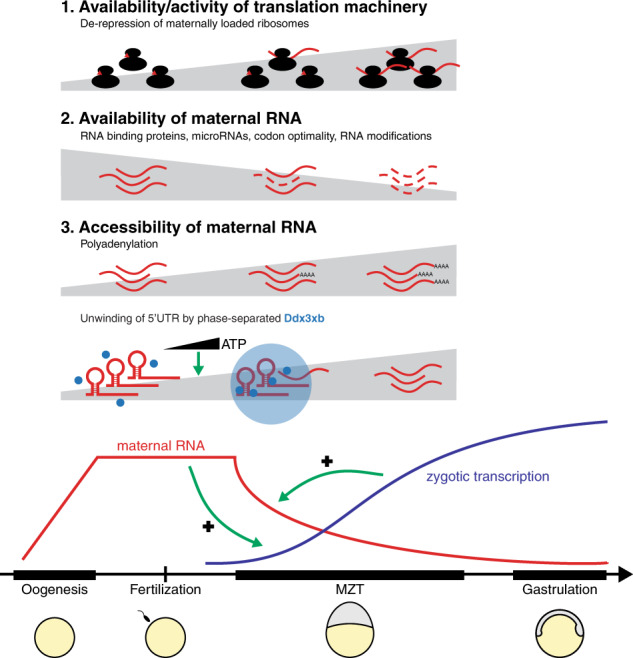
The development of animal embryos is initially directed by maternally provided proteins and RNAs. Developmental control is gradually handed over to the embryo during the maternal-to-zygotic transition, when the zygotic genome is activated.1 To ensure a successful change from the maternal to the zygotic gene expression program, maternal transcripts are degraded during this transition. The changes in gene expression are precisely controlled and highly interconnected (Fig. 1). In zebrafish, for example, the translation of maternally loaded RNAs is required to activate zygotic transcription, while the zygotic transcription of a microRNA is required to initiate the degradation of many maternal transcripts. This raises an interesting conundrum: maternally loaded RNAs need to be translated before they are degraded.
Fig. 1. Processes that determine whether maternally loaded RNAs are translated.

During the maternal-to-zygotic transition (MZT), maternally loaded RNAs are degraded, and zygotic transcription begins. Whether or not an RNA is translated depends on the availability and activity of the translation machinery (1) as well as on the availability (2) and accessibility (3) of the transcript itself. Shown are mechanisms known to be involved in these processes.
Whether or not an RNA is translated depends on the availability of the translation machinery, the availability of transcripts, and the accessibility of these transcripts (Fig. 1). With respect to the translation machinery, recent work has shown that maternally provided ribosomes are maintained in a dormant state until they are gradually activated by the release of an inhibitor.2 The degradation, and thus availability, of transcripts is impacted by RNA-binding proteins,3 microRNAs,4 RNA modifications,5 and codon optimality.6 Accessibility of transcripts for translation has long been known to be increased by cytoplasmic polyadenylation.7 Transcripts, however, are often stored in cytoplasmic granules with their 5′UTR folded, and it has been unclear how they are unwound to be translated. In a recent paper in Cell Research, Shi et al. used an elegant combination of genetics, genomics, and biochemistry approaches to show that the RNA helicase Ddx3xb facilitates the translation of stored transcripts by a phase separation-dependent process.8
Shi et al. identified the RNA helicase Ddx3xb as a protein of interest based on its abundant expression during early zebrafish development and its well-known role in unwinding RNA structures.9 The analysis of embryos in which Ddx3xb was lacking showed that the protein indeed plays a role in the maternal-to-zygotic transition. To determine the mechanism by which Ddx3xb acts, Shi et al. used RIP-seq to identify transcripts that are bound by Ddx3xb, and ribosome profiling to show that the translation efficiency of specifically these transcripts is strongly reduced in mutant embryos. Target RNAs were enriched for a specific motif in their 5′UTR, suggesting that they are targeted for unwinding by the helicase Ddx3xb. Indeed, using icSHAPE data that they previously generated,10 Shi et al. were able to show that Ddx3xb targets were more unwound than non-targets. Thus, unwinding of maternal RNAs by Ddx3xb facilitates their translation.
Perhaps the most exciting part of the work is related to the localization of Ddx3xb in cytoplasmic granules. Granules were visualized in zebrafish embryos by injecting mRNA coding for fluorescently labeled Ddx3xb, and a series of experiments confirmed that they were phase separated, as had been shown previously.9 Both RNA and ATP promote Ddx3xb condensation in vitro, and the appearance of Ddx3xb condensates coincides with an increase in ATP levels in vivo, suggesting a role for ATP in the temporal regulation of condensate formation and the unwinding of RNAs.
Ddx3xb is composed of a central helicase domain and N- and C-terminal intrinsically disordered regions (IDRs). IDRs are known to facilitate phase separation. Removing the N-terminal IDR of Ddx3xb showed that this domain is essential for the formation of cytoplasmic Ddx3xb condensates in vivo. Importantly, however, the N-terminal IDR is not involved in the protein’s helicase activity per se, which was shown by replacing it with IDRs from other phase separating proteins. This allowed Shi et al. to perturb the phase separation behavior of Ddx3xb independently of its helicase activity and thereby address the relevance of Ddx3xb phase separation for helicase function. This was important because while there are lots of studies investigating phase separation, its relevance for biological function is not always clear. Using this very powerful approach, Shi et al. were able to show that phase separation of Ddx3xb is required for all the phenotypes that they observed in the Ddx3xb mutants, such as the reduced unwinding of maternally loaded RNAs, the reduced translation efficiency, and the developmental delay. Thus, the RNA helicase Ddx3xb facilitates the translation of stored transcripts by a phase separation-dependent process.
This study emphasizes the power of using embryonic systems to address fundamental questions of cell biology. It represents a major contribution to our understanding of the role of phase separation in biological processes, as well as the dramatic changes in gene expression as embryos transition from maternal to zygotic control. In the context of the embryo, it would be interesting to investigate whether specific RNAs are preferentially targeted by Ddx3xb. There are many maternally loaded transcripts and only some of these are important for developmental progression, such as those coding for pluripotency factors required to activate zygotic transcription. Are these preferentially unwound? A related question concerns the relationship between translation and degradation of specific transcripts. We expect there to be an interplay between translation-facilitating and degradation-inducing mechanisms, allowing the embryo to make sure that important RNAs are translated before they are degraded.
References
- 1.Vastenhouw NL, Cao WX, Lipshitz HD. Development. 2019;146:dev161471. doi: 10.1242/dev.161471. [DOI] [PubMed] [Google Scholar]
- 2.Leesch, F. et al. bioRxiv10.1101/2021.11.03.467131 (2021).
- 3.Despic V, et al. Genome Res. 2017;27:1184–1194. doi: 10.1101/gr.215954.116. [DOI] [PMC free article] [PubMed] [Google Scholar]
- 4.Giraldez AJ, et al. Science. 2006;312:75–79. doi: 10.1126/science.1122689. [DOI] [PubMed] [Google Scholar]
- 5.Zhao BS, et al. Nature. 2017;542:475–478. doi: 10.1038/nature21355. [DOI] [PMC free article] [PubMed] [Google Scholar]
- 6.Bazzini AA, et al. EMBO J. 2016;35:2087–2103. doi: 10.15252/embj.201694699. [DOI] [PMC free article] [PubMed] [Google Scholar]
- 7.Winata CL, Korzh V. FEBS Lett. 2018;592:3007–3023. doi: 10.1002/1873-3468.13183. [DOI] [PMC free article] [PubMed] [Google Scholar]
- 8.Shi, B. et al. Cell Res. 10.1038/s41422-022-00655-5 (2022).
- 9.Hondele M, et al. Nature. 2019;573:144–148. doi: 10.1038/s41586-019-1502-y. [DOI] [PMC free article] [PubMed] [Google Scholar]
- 10.Shi B, et al. Genome Biol. 2020;21:120. doi: 10.1186/s13059-020-02022-2. [DOI] [PMC free article] [PubMed] [Google Scholar]


