Basophils, though representing a minor population of leukocytes, have diverse roles in various pathophysiologies. A recent report provides an additional evidence on the pathogenic role of basophils in promoting kidney fibrosis by secreting IL-6 and promoting the Th17 cell differentiation.
Basophils are rare granulocytes that participate in T helper (Th) type 2 immunity, principally through the secretion of interleukins IL-4 and IL-13. Besides their role in the defense against helminth infections, basophils play a major role in the pathogenesis of various allergic inflammatory diseases, autoimmune diseases, and cancer.1 Basophils express a diverse array of receptors, and hence could sense signals from various sources including cytokines, toll-like receptor agonists, and allergens, and subsequently undergo rapid activation and release inflammatory mediators. In addition to IL-4 and IL-13, basophils produce histamine, prostaglandins, leukotrienes, IL-6 and chemokines in response to the stimuli. All these inflammatory mediators are potential contributors of basophil-mediated pathogenesis.
Fibrosis is characterized by the development of fibrous connective tissue to scar injury or tissue damage. Renal fibrosis (RF) is described as the altered kidney’s ability to regenerate after injury leading to a gradual loss of renal function, potentially evolving to a life-threatening renal failure and a requirement for dialysis or kidney transplantation. Our comprehension of immune cell interactions that trigger RF is still unfolding. Previous studies in mice have suggested a possible role for basophils in mediating cardiac allograft fibrosis by triggering fibroblast activation, expanding myofibroblasts, promoting the production of extracellular matrix proteins and remodeling of the fibrotic organ.2 Now, by applying single-cell sequencing in a murine model of kidney fibrosis (KF), Katalin Susztak and colleagues report a mechanistic evidence on the pathogenic role of basophils in promoting fibrosis.3
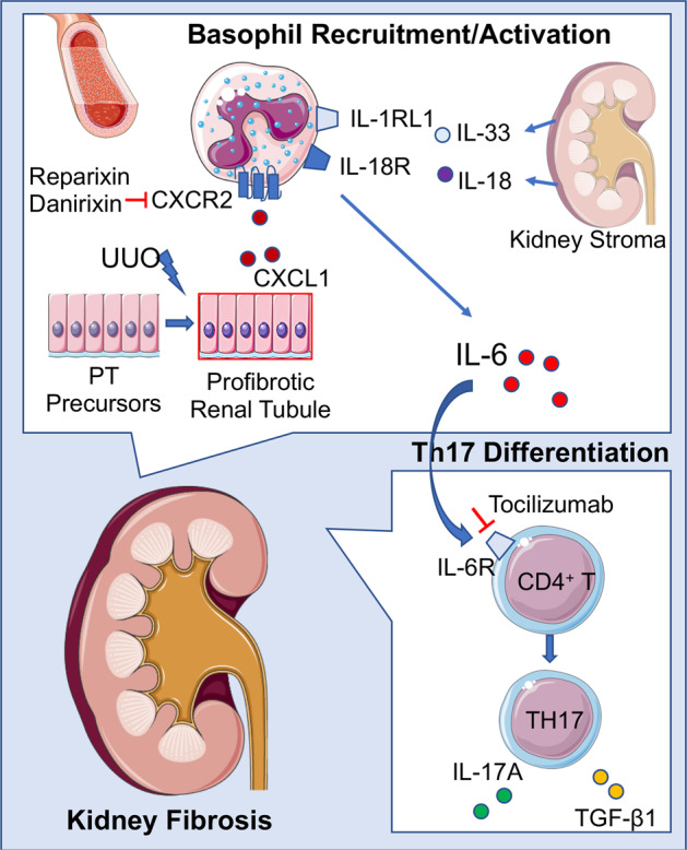
To understand the pathogenesis of KF, Doke et al. compared the transcriptome profile of kidney of wild-type mice (C57BL/6) subjected to sham or unilateral ureter obstruction (UUO) surgery leading to fibrosis. They identified the genes enriched in basophil cluster in the fibrotic kidneys of UUO mice and further studied the unappreciated role of these granulocytes in RF (Fig. 1).3
Fig. 1. Pathogenic role of basophils in KF and possible therapeutic interventions.

During KF, basophils are recruited by profibrotic renal tubules via CXCR2/CXCL1 axis and undergo activation by kidney stroma-produced IL-18 and IL-33. These activated basophils produce IL-6 that induces the differentiation of Th17 cells. IL-6 and Th17 cells act as downstream mediators of KF. The pathogenic roles of IL-6 could be blocked by monoclonal antibodies that target IL-6 receptor (e.g., Tocilizumab). CXCR2/CXCL1 axis that recruits basophils could be pharmacologically targeted though redundancy in the chemokine system is the major drawback. PT, proximal tubules. Parts of the figure were drawn by using pictures from Servier Medical Art. Servier Medical Art by Servier is licensed under a Creative Commons Attribution 3.0 Unported License (https://creativecommons.org/licenses/by/3.0/).
Following subclustering analysis of the proximal tubules (PT) in the UUO kidneys, the authors described a subset of profibrotic PT cells that evolve from PT precursors and display an increased expression of genes encoding fibroinflammatory cytokines and chemokines. Doke et al. used in silico analysis and in situ hybridization to further analyze the cell interaction between the profibrotic PT cells and immune cells, and noted that fibrotic PT cells could contribute to the recruitment of CXCR2+ basophils in UUO kidneys by secreting CXCL1.3
To prove the role of basophils in the pathogenesis of KF, Doke et al. resorted to two complementary approaches of depletion of these cells in vivo by using either conditional Mcpt8Cre/DTR mice or by injecting MAR-1 antibody that depletes FcεRIα+ cells (all basophils intensely express this molecule). In line with their hypothesis, basophil depletion led to the reduced expression of fibroblast markers Col1a1, Col3a1, Timp1 and Acta2 in UUO kidneys, and lessened RF and severity of tubular interstitial damage.
How do basophils contribute to the pathogenesis of KF? Previous study in the chronic cardiac allograft rejection model has identified basophil-derived IL-4 as a major profibrotic cytokine.2 However, subsequent study by the same group identified IL-3 as a key profibrotic cytokine that exerted profibrotic functions and organ remodeling, even if IL-4 was completely suppressed.4 Mechanistically, IL-3 exerted profibrotic effects by activating and inducing IL-6 in the infiltrating basophils. In line with this report, single-cell analysis and in situ hybridization identified an enhanced expression of Il6 in the basophils of UUO kidneys. In addition, basophils of UUO kidneys displayed higher expression of the receptors (Il18r1 and Il1rl1) for the cytokines IL-18 and IL-33 that are known to activate basophils. Bulk RNA-seq data from UUO kidneys also confirmed these results but no difference in the expression of Il4 was observed between UUO and sham kidneys.3 Additional investigations documented that stroma cells in UUO kidneys contribute IL-18 and IL-33. In vitro basophil stimulation with IL-18 or IL-33 led to the induction of IL-6.3 Further investigations on the signals that activate stroma cells are required.
It is well known that IL-6 supports the differentiation of Th17 subset of CD4+ T cells.5 Therefore, the authors were aimed at pinpointing whether Th17 cells and their products are the further downstream mediators of KF. The expression of IL-6 receptor transcripts was higher in CD4+ T cells and Th17 cells from UUO kidneys, and RNA velocity analysis highlighted an enhanced differentiation of Th17 cells. Genetic depletion of basophils reduced Th17 cells and Th17 signatures in UUO kidneys.3
All these data together indicated that CXCR2+ basophils migrate to the kidneys in response to CXCL1 secreted by profibrotic PT cells in UUO mice. These basophils undergo activation by stroma-secreted IL-18 and IL-33 and produce IL-6 that in turn supports Th17 response. Based on these findings and as a proof of concept, the authors pre-treated mice with IL-6 receptor antagonists before inducing UUO. In line with the findings, IL-6 receptor antagonism reduced the expression levels of various fibrosis markers and the accumulation of collagen in the kidneys.3
Are these observations relevant for the humans? To address this, the authors used kidney samples from the patients with chronic kidney disease. It was noteworthy that basophil number, IL6, CXCL1, IL18, IL33 and IL17d transcripts were strongly correlated with the degree of RF,3 thus validating the translational value of the experimental data.
Though basophils are implicated in the pathogenesis of several diseases, the signals that drive basophil activation vary. While basophil activation was induced by T cell-derived IL-3 in cancer progression6 or in chronically rejecting allografts,4 Doke et al. described basophil activation via IL-18 and IL-33 in KF. On the other hand, thymic stromal lymphopoietin is implicated in eliciting basophil responses in eosinophilic esophagitis.7 Though human basophils can be activated by IL-33, in contrast to mouse basophils, they have been shown not to respond to IL-18 even though they constitutively express IL-18 receptors.8 Moreover, IL-3 and IL-33 activate distinct signaling pathways in blood basophils suggesting different activation states or functions of cytokine-activated basophils in these pathologies.
While there are currently no drugs for fibrosis or chronic kidney disease that would specifically target the kidney, the study of Doke et al. provides certain clues on how to improve KF by targeting either basophils, the CXCR2/CXCL1 axis that recruits basophils or IL-6, the downstream mediator of KF. Blocking CXCR2 by an inhibitor (Reparixin) reduced neutrophil accumulation in the kidneys and maintained kidney function in a reperfusion injury model.9 Another CXCR2 inhibitor Danirixin is currently being investigated in the clinical trials.10 The same strategy could be used to reduce the recruitment of basophils during KF (Fig. 1). But redundancy in the chemokine axis is the main drawback. Though monoclonal antibody-based basophil depletion has been achieved in experimental models, it is too far from the human application. However, tocilizumab, a humanized monoclonal antibody against IL-6 receptor is already in the clinic for many years, and this drug can be examined immediately in the chronic kidney disease. Though tocilizumab treatment may not alter the basophils or their activation, it can reduce the number of Th17 cells as shown in rheumatoid arthritis.11
Competing interests
The authors declare no competing interests. The authors have received financial support from the Agence Nationale de la Recherche, France; ANR-19-CE17-0021 (BASIN) for their work on the basophils.
References
- 1.Karasuyama H, Shibata S, Yoshikawa S, Miyake K. Int. Immunol. 2021;33:809–813. doi: 10.1093/intimm/dxab021. [DOI] [PubMed] [Google Scholar]
- 2.Schiechl G, et al. Am. J. Transplant. 2016;16:2574–2588. doi: 10.1111/ajt.13764. [DOI] [PubMed] [Google Scholar]
- 3.Doke, T. et al. Nat. Immunol. 23, 947–959 (2022). [DOI] [PubMed]
- 4.Balam S, et al. J. Immunol. 2019;202:3514–3523. doi: 10.4049/jimmunol.1801269. [DOI] [PubMed] [Google Scholar]
- 5.Maddur MS, Miossec P, Kaveri SV, Bayry J. Am. J. Pathol. 2012;181:8–18. doi: 10.1016/j.ajpath.2012.03.044. [DOI] [PubMed] [Google Scholar]
- 6.De Monte L, et al. Cancer Res. 2016;76:1792–1803. doi: 10.1158/0008-5472.CAN-15-1801-T. [DOI] [PubMed] [Google Scholar]
- 7.Noti M, et al. Nat. Med. 2013;19:1005–1013. doi: 10.1038/nm.3281. [DOI] [PMC free article] [PubMed] [Google Scholar]
- 8.Pecaric-Petkovic T, Didichenko SA, Kaempfer S, Spiegl N, Dahinden CA. Blood. 2009;113:1526–1534. doi: 10.1182/blood-2008-05-157818. [DOI] [PMC free article] [PubMed] [Google Scholar]
- 9.Cugini D, et al. Kidney Int. 2005;67:1753–1761. doi: 10.1111/j.1523-1755.2005.00272.x. [DOI] [PubMed] [Google Scholar]
- 10.Madan A, et al. Open Forum Infect. Dis. 2019;6:ofz163. doi: 10.1093/ofid/ofz163. [DOI] [PMC free article] [PubMed] [Google Scholar]
- 11.Samson M, et al. Arthritis Rheum. 2012;64:2499–2503. doi: 10.1002/art.34477. [DOI] [PubMed] [Google Scholar]


