Abstract
Purpose of Review
The unique pathophysiological changes of constrictive pericarditis (CP) can now be identified with better imaging modalities, thereby helping in its early diagnosis. Through this review, we outline the pathophysiology of CP and its translation into symptomology and various imaging findings which then are used for both diagnosis and guiding treatment options for CP.
Recent Findings
Multimodality imaging has provided us with the capability to recognize early stages of the disease and identify patients with a potential for reversibility and can be treated with medical management. Additionally, peri-procedural planning and prediction of post-operative complications has been made possible with the use of advanced imaging techniques.
Summary
Advanced imaging has the potential to play a greater role in identification of patients with reversible disease process and provide peri-procedural risk stratification, thereby improving outcomes for patients with CP.
Keywords: Constrictive pericarditis, Multimodality imaging, Pathophysiology, Cardiac magnetic resonance imaging, Pre-operative imaging
Introduction
Constrictive pericarditis (CP) is caused by fibrosis and calcification of the pericardium, which indirectly worsen lusitropy and impedes normal diastolic filling, masquerading as diastolic heart failure [1–3]. Complex pathophysiology and unique hemodynamic derangements of CP translate into characteristic findings on non-invasive and invasive diagnostic modalities and help to differentiate it from other causes of diastolic heart failure especially restrictive cardiomyopathy (RCM) [1, 4–6]. The accurate diagnosis of CP is vital to ensure amenability for the existent potential of medical and surgical treatment options [7]. This article will review the concepts of pathophysiological and hemodynamic basis of CP and their effect on the characteristic findings on various forms of multimodality imaging.
Etiology
Various insults have been identified as causative factors for CP; however, the leading causes differ according to geographic location [8, 9]. Where tuberculosis is said to be the most frequent cause of constrictive pericarditis in Africa and Asia, it is rare in Western Countries, with reports ranging from less than 1 to 5.6% of cases [1, 10–12]. In the Western world, indeterminate/idiopathic causes remain the most common etiology of CP, followed by post-cardiac surgery and radiation-induced [1, 10]. Other uncommon etiologies may include rheumatological diseases, trauma, malignancy, or infections [8, 9]. Only 1.8% of acute pericarditis cases progress to constrictive pericarditis with low risk (< 1%) for viral and idiopathic pericarditis, moderate risk (2–5%) for neoplastic pericardial diseases and immune-mediated pericarditis, and high risk (20–30%) for bacterial pericarditis, especially if purulent [13].
Pathology
The pericardium is comprised of two avascular layers, an outer parietal layer and an inner visceral layer, adherent to epicardium [14]. Pathological changes in CP most commonly affect the parietal pericardium but may also involve visceral pericardium or even epicardium and may lead to the development of adhesions between the parietal layer and epicardium [1, 15]. Due to proximity to the myocardium, these alterations in the pericardial elasticity lead to compromised cardiac filling [14].
There are three subtypes of CP: 1. transient constrictive pericarditis (TCP), 2. chronic constrictive pericarditis (CCP), 3. effusive-constrictive pericarditis (ECP) [13, 14]. TCP is characterized by a temporary form of constriction due to underlying inflammation, which may either resolve spontaneously or after medical therapy. CCP, on the other hand, is a result of chronic inflammation and scarring, which leads to fibrosis and calcification [14, 16]. There is an increasing belief that TCP may be an early and reversible manifestation in the natural history of CCP [14]. ECP is an uncommon clinical syndrome characterized by the coexistence of tense pericardial effusion and constriction of the heart by the visceral pericardium. The hallmark of ECP is persistent elevated right atrial pressure after pericardiocentesis [17–19].
The common link between all these types of CP is the development of inelasticity of the pericardium, therefore making it non-compliant, thus preventing ventricular filling. In ECP, tense pericardial fluid plays an additional role in preventing ventricular filling. The degree and type of histopathological changes in the pericardium depend upon the sub-type of CP. TCP demonstrates fluctuating pericardial edema, inflammation, and fibrin deposition as seen in acute pericarditis, whereas in CCP, there is more fibrosis and calcification [14]. This calcification may extend deep into the myocardium and sub endocardium [20]. Pericardial thickening is presumed to be the consequence of all pathologies. It can, however, be absent in up to 12–18% of patients. Patients with a thin pericardium and CP have less fibrosis and inflammation, and may represent cases where CP is not only due to fibro-calcification but also due to shrinkage in pericardial volume causing constriction of the heart [21, 22].
Pathophysiology
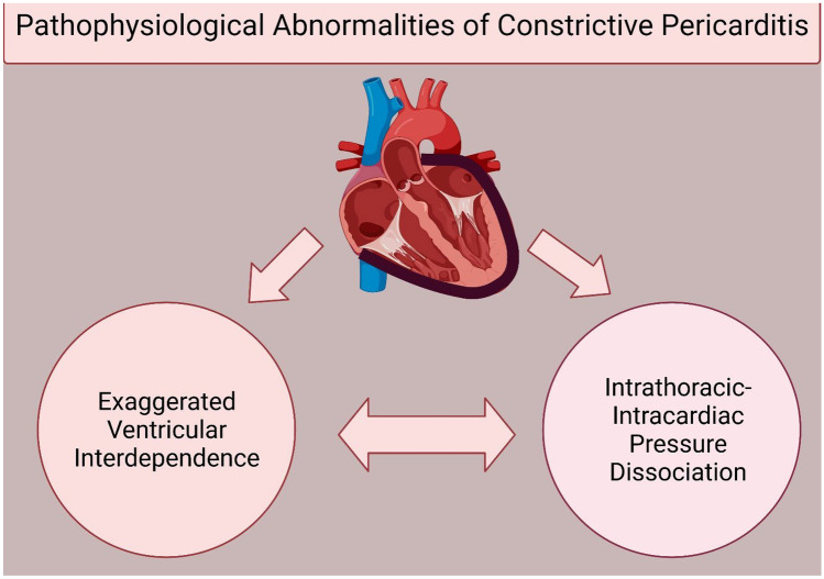
The pathological decrease in compliance of the pericardium is responsible for the exaggerated physiological changes in the heart, which give rise to symptoms of CP. The stiff ventricular-pericardial unit impedes ventricular relaxation causing a rapid rise in filling pressures for given venous return translating to elevated and equalized diastolic pressures. The fibrotic, non-compliant, often thickened and calcified pericardium in CP which encases the heart not only prevents ventricular filling, but also prevents the transmission of pressures from the thoracic cavity to the inside of the heart, enabling two pathophysiological phenomena: 1. exaggerated ventricular interdependence and, 2. intrathoracic-intracardiac pressure dissociation (Fig. 1). These phenomena result in dynamic respirophasic changes, which are responsible for characteristics findings on invasive and non-invasive diagnostic modalities and partake in symptomatology [1, 3].
Fig. 1.
Schematic representation of pathophysiology of constrictive pericarditis. The phenomena of pericardial thickening (in black), exaggerated ventricular interdependence, and intracardiac-intrathoracic pressure dissociation play role in the clinical features and diagnosis of constrictive pericarditis. (Created with BioRender.com)
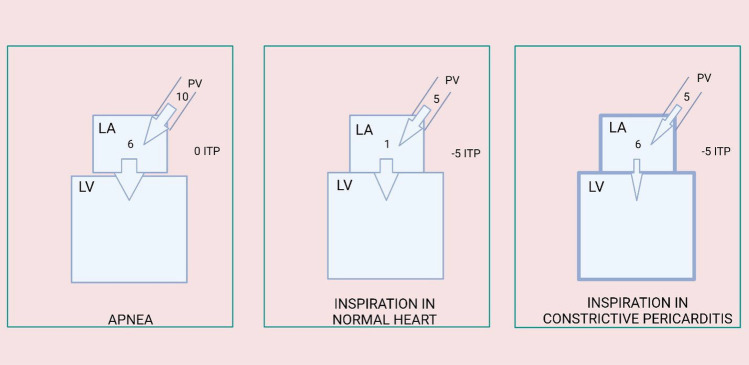
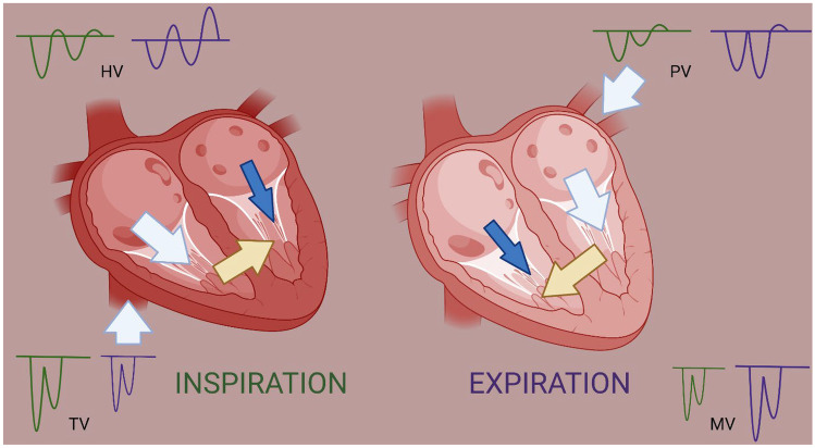
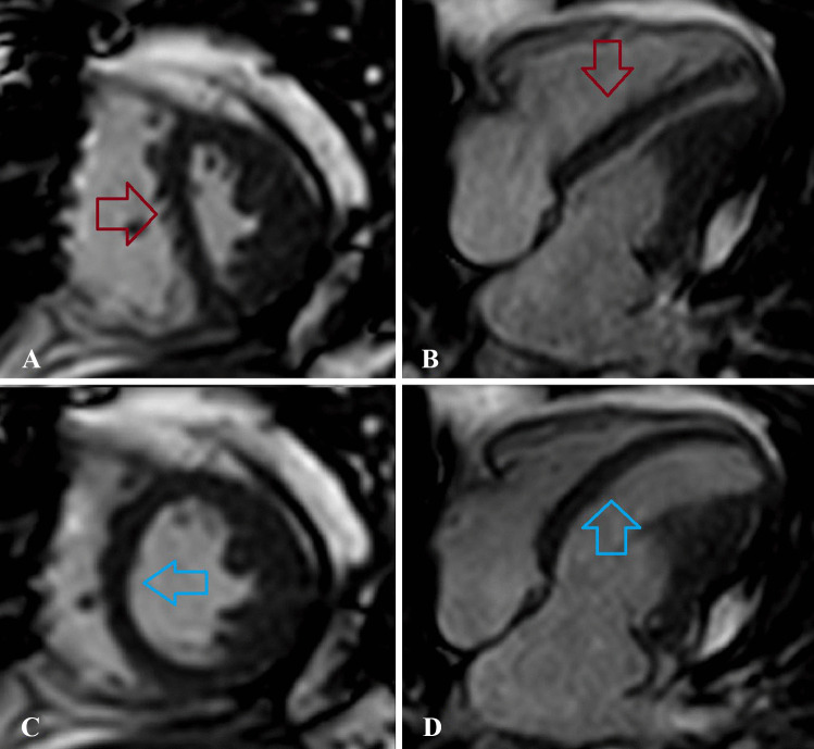
During inspiration, the intrathoracic pressure (ITP) decreases, which in a normal heart affects and reduces both the left ventricular (LV) filling pressure and LV diastolic pressure; therefore, the pressure gradient remains constant during respiration [23]. However, in constrictive pericarditis, the drop in ITP is transmitted to the pulmonary veins (not encapsulated by pericardium), but not to the heart due to fibrous pericardium. This intrathoracic-intracardiac pressure dissociation leads to reduction in the filling pressure but not in the left ventricular diastolic pressure resulting in a decreased pressure gradient causing decreased LV filling. The interventricular septum, due to decreased LV filling shifts to the left side during inspiration, favoring right heart filling and thus showing exaggerated ventricular interdependence (Fig. 2) [1–3, 5]. Simultaneously during inspiration, increased venous return from the inferior vena cava increases filling of the right ventricle (RV). The pericardial reserve volume in a normal heart accommodates this increased right heart filling [24]. But in CP, the expansion of the right ventricle is limited due to fibrous pericardium, shifting the septum towards the left side, additionally contributing to the ventricular interdependence (Fig. 3) [4, 5, 23]. The opposite occurs during expiration, wherein positive ITP increases the gradient and therefore LV filling, causing a rightward movement of the interventricular septum. This further leads to underfilling of the RV and backflow of blood into hepatic veins [1, 4]. This impairment of ventricular filling is further exaggerated in ECP patients where the tense pericardial fluid compounds the effects of CP.
Fig. 2.
Schematic representation of intracardiac-intrathoracic pressure dissociation. In apnea, the pressure gradient between PV and LA/LV leads to ventricular filling. During inspiration in a normal heart, reduction in intrathoracic pressure is equally distributed to extra-pericardial structures (PV) and intrapericardial structures (LA/LV) but the gradient remains. In constrictive pericarditis, the reduction in ITP is not transferred to intrapericardial structures due to thick pericardium leading to reduction of gradient and thus decreased ventricular filling. (Abbreviations: LA, left atrium; LV, left ventricle; PV, pulmonary vein; ITP, intrathoracic pressure) (Created with BioRender.com)
Fig. 3.
Schematic representation of ventricular interdependence. During inspiration (green color), the blood return to the right side (white arrow) increases leading to higher tricuspid valve inflow velocities (green TV Doppler) but thickened pericardium prevents outward expansion of right ventricle leading to shifting of septum (yellow arrow) towards left side, thereby contributing to decreased blood flow on left side (blue arrow) and lower mitral inflow velocities (green MV Doppler). During expiration (purple color), the inflow to left ventricle increases (white arrow) depicted by increased mitral inflow velocity (purple MV Doppler), causing shifting of septum (yellow arrow) towards right side contributing to decreased blood flow on right side (blue arrow) and lower tricuspid inflow velocities (purple TV Doppler) (Created with BioRender.com)
The impaired filling of ventricles during the diastole leads to symptoms of congestion and signs of low cardiac index. These pathophysiological phenomena may (1) dissipate either spontaneously or by medical treatment in patients with TCP, (2) improve but remain persistent after pericardiocentesis as in ECP, or (3) become permanent as in CCP potentially necessitating surgical interventions.
Role of Multimodality Imaging in Diagnosis of CP
Patients with CP usually present with heart failure symptoms, but right-sided heart failure features predominate over left-sided features [25]. Dyspnea on exertion and edema are the most common symptoms, but patients may present with other signs and symptoms of systemic vascular congestion such as ascites or hepatomegaly, atrial arrhythmias, or pleural effusion [1, 3, 10]. Most of the patients with CP have elevated jugular venous pressure (JVP) on examination [10], but preserved, rapid, and deepy descent differentiates it from findings of elevated JVP in cardiac tamponade [3, 24]. Moreover, persistent inspiratory increase in JVP, defined as Kussmaul’s sign, is more commonly seen in CP than cardiac tamponade [3, 24]. These symptoms are not specific for CP and can be found in other pathological conditions as well; therefore, a high degree of suspicion is needed for the diagnosis of CP.
The use of non-invasive diagnostic modalities plays a vital role in the accurate diagnosis of CP in addition to clinical features and invasive hemodynamic assessment. These diagnostic measures also play a significant role in differentiating CP from other diagnoses such as RCM, cardiac tamponade, and other causes of diastolic heart failure. The pathophysiological principles of exaggerated ventricular interdependence and intrathoracic-intracardiac pressure dissociation during respiratory cycle, formation of adhesions or even extension of calcification to the myocardium, and histopathological changes in the pericardium form the principles that help to make the diagnosis and categorize the subtypes of CP.
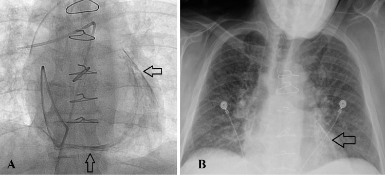
A chest radiograph or fluoroscopic images with evidence of calcium around the heart is strongly suggestive of CCP; however, this finding is only present in a small number of patients (Fig. 4) [1]. Echocardiogram including 2D, Doppler and strain imaging, computed tomography (CT), cardiovascular magnetic resonance (CMR), and positron emission tomographic imaging are the non-invasive diagnostic modalities commonly used for diagnosis and management of CP and will be discussed in detail.
Fig. 4.
Fluoroscopic (A) and chest X-ray (B) images of a patient with constrictive pericarditis showing calcification (arrows)
Echocardiogram
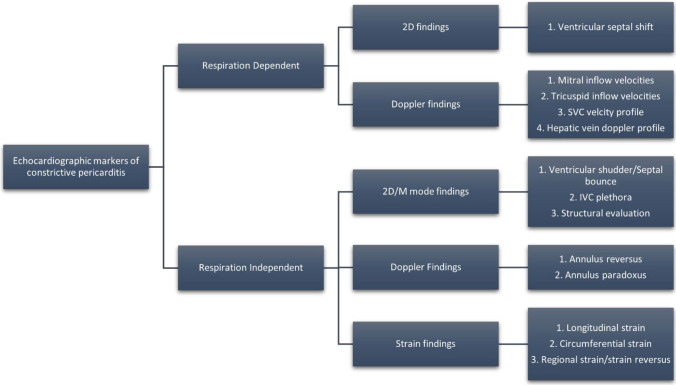
The echocardiographic findings in CP can be broadly classified based on their alteration with respiration. Some of the findings on echocardiography are only seen with respiratory variation, whereas few are independent of respiration (Fig. 5). Among various echocardiographic parameters, Welch et al. demonstrated respiration-related ventricular septal shift, preserved or increased medial mitral annular e′ velocity, and prominent hepatic vein expiratory diastolic flow reversal as important criteria in the diagnosis of CP. They reported that the presence of ventricular septal shift and either medial e′ velocity ≥ 9 cm/s or hepatic vein expiratory diastolic reversal ratio ≥ 0.79 corresponded to a desirable combination of sensitivity (87%) and specificity (91%), while requiring all 3 criteria to be present increased specificity further (97%) at the expense of reduced sensitivity (64%) [26].
Fig. 5.
Respiration dependent and independent echocardiographic markers of constrictive pericarditis
Respiration dependent variables:
-
(i)
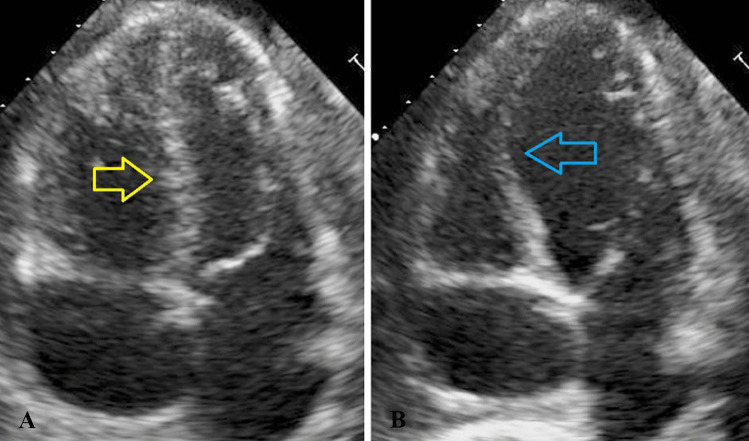
Ventricular septal shift: Inspiratory deviation of the septum towards the left side and expiratory shift towards right side, widely described as ventricular septal shift (VSS) is often one of the first clues and is one of the most sensitive findings, present in almost 93% of patients with CP. This finding is best appreciated using long acquisitions of 2D wall motion from multiple imaging windows and also from M-mode in PLAX view (Fig. 6) [1, 27].
-
(ii)
Pulsed wave Doppler of mitral and tricuspid inflow velocities: As per the pathophysiology discussed above, LV filling decreases in inspiration and increases in expiration and vice versa for RV. This finding can be captured on pulsed wave Doppler of the mitral and tricuspid inflow. The consensus for calculating percentage respiratory variation in CP is (expiration-inspiration)/expiration. The peak mitral inflow drop usually exceeds 25% with respiratory variation, whereas there is an increase of more than 40% on tricuspid inflow E wave [24, 28]. The absence of this finding does not exclude the diagnosis of CP as respiratory variation may not be present in 50% of patient as filling pressures may be altered by marked diuresis [28]. On the other hand, labored respiration may also lead to these changes such as in COPD exacerbation. Finally, the patients with CP are often tachycardic, with fusion of E and A waves, thereby hindering the calculation. However, when present, these features may help to differentiate CP from RCM as this variation is absent in RCM [29, 30].
-
(iii)
Hepatic vein Doppler profile: As discussed earlier, during expiration, the inflow on the left side increases, and the septum shifts to the right side causing decreased filling of the right ventricle. This results in a reduction of hepatic vein diastolic forward flow with prominent reversal of flow at the end of diastole. Diastolic reversal ratio is defined as reversal velocity divided by forward velocity. The finding of a reversal ratio ≥ 0.79 has been described as one of the most specific findings for CP [26].
-
(iv)
Superior vena cava velocity (SVC) profiles: The variation of inflow velocities in SVC has been described in various diseases, including chronic obstructive pulmonary disease (COPD). It is important to differentiate CP from COPD in terms of echocardiographic findings from exaggerated ventricular interdependence. In COPD, there are exaggerated swings in ITP with increased respiratory effort leading to transmission of these to cardiac structures, and therefore exaggerated ventricular interdependence causing respiratory variation in inflow velocities in SVC and across MV and TV [31, 32]. As SVC is an intrathoracic structure, its velocities have been documented to correlate with right atrial pressures, and therefore Doppler recordings from SVC show an increase in forward flow during inspiration [31, 33]. However, in CP, due to restriction to ventricular filling and dissociation of intrathoracic pressure and intracardiac pressures, the right atrial pressures remain elevated throughout the respiratory cycle, and thus, there is minimal forward flow velocity changes in SVC. This manifests as Kussmaul’s sign on clinical examination. Therefore, the absence of variation in SVC forward flow velocity may help differentiate CP from COPD, which has increased variation in flow velocities on inspiration [3, 31].
Fig. 6.
Echocardiographic representation of the phenomenon of ventricular interdependence. Yellow arrow shows leftward shift of septum during inspiration (A) and blue arrow shows rightward shift of septum during expiration (B)
Respiration independent variables:
-
(i)
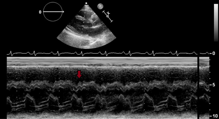
Ventricular shudder/septal bounce: Ventricular shudder, popularly known as the septal bounce is one of the phenomena present in almost 96% of the patients and is best seen on M mode (Fig. 7) [27]. This is an abnormal oscillatory beat-to-beat diastolic septal motion related to ventricular interdependence occurring on a millisecond scale due to subtle differences in the timing of tricuspid and mitral valve opening and right and left atrial contraction. These subtle changes were further described with high fidelity manometric catheters, which showed an abrupt rise in early diastole of the RV pressure curve and overtaking the LV pressure, which on M mode appears as septum shifting briskly from RV to LV. As RV pressure plateaus, LV pressure increases, shifting the septum back. Therefore, differential filling time and pericardial constraint, lead to rapid cessation of filling resulting in characteristic septal bounce. This feature is absent in RCM; however, it can be present in large number (44%) of patients without CP including patients with conduction abnormalities or post-operative abnormal septal motion [27, 34].
-
(ii)
Preserved mitral annular relaxation velocity on tissue Doppler imaging (TDI): The use of TDI relies on the mechano-elastic properties of the myocardium, which are preserved in CP [3] Therefore, a patient with diastolic heart failure with a finding of a normal or increased e′ velocity (≥ 9 cm/s) should suggest a diagnosis of CP [1]. Moreover, increased mitral e′ velocity is also associated with better outcomes in patients with CP [35].
-
(iii)
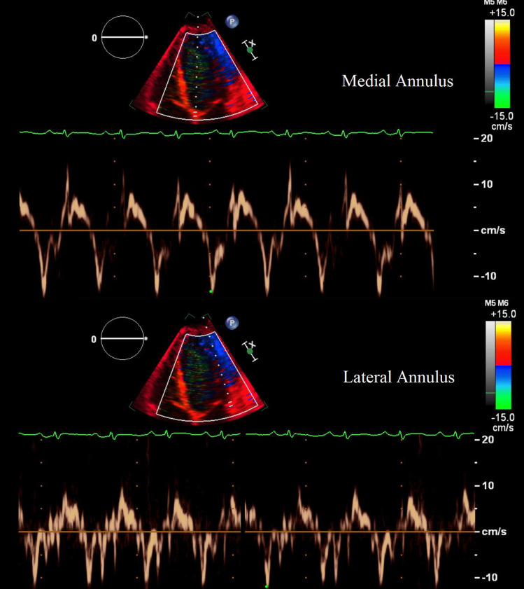
Annulus reversus: Though the mechano-elastic properties of the myocardium are preserved in CP, there might be tethering of the lateral myocardium to the adjacent fibrotic and scarred pericardium influencing the lateral mitral annulus relaxation with compensatory increased relaxation of the septal/medial myocardium [1, 3, 36]. In a normal heart, the lateral e′ velocity often exceeds the medial e′ velocity. However, in CP, due to the phenomenon described above, the opposite pattern is seen with higher medial e′ velocities than the lateral velocities. This phenomenon has been described as annulus reversus (Fig. 8) [36]. In absence of annulus reversus from lateral wall TDI, the anterior, inferior, and inferolateral mitral annular e′ velocities to medial e′ ratios may reveal tethering; however, its diagnostic utility has not been tested [4]. Improvement in lateral/septal (medial) e′ ratio on serial monitoring has been identified as a marker of clinical improvement after anti-inflammatory treatment [37•].
-
(iv)
Annulus paradoxus: The ratio of transmitral flow velocity (E) to e' is a predictor of LV filling pressures and has a strong positive correlation with pulmonary capillary wedge pressure (PCWP). This usually is secondary to decreased “e” in patients with diseased states such as restrictive cardiomyopathy and heart failure with reduced ejection fraction. However, this “e” is well preserved in patients with CP and therefore, despite having higher filling pressures, E/e' ratio is usually on the lower side due to elevated e'. Furthermore, this relation shows a paradoxical relation as medial e' progressively increases with worsening of CP therefore, showing an inverse relationship between PCWP and E/e′, thus known as annulus paradoxus [1, 38, 39].
-
(v)
2D strain echocardiography: Strain measures myocardial deformation in three dimensions. Longitudinal strain measures shortening from base to apex, circumferential strain measures systolic shortening of the short axis of the ventricle, and lastly, radial strain measures myocardial thickening from endocardium to epicardium [40]. The sub-epicardial region is responsible for circumferential shortening, whereas the sub-endocardial region is responsible for longitudinal shortening. Towards the apex, the muscle gets thin, decreasing this distinction [41, 42]. The pericardial layers play an important role in controlling the magnitude of the circumferential and longitudinal expansion. They may allow expansion at lower LV volumes but they resist further expansion at higher volumes [43]. Sengupta et al. [42] noted that the circumferential strain was decreased in patients with CP (due to involvement of subepicardial region), whereas the longitudinal strain was preserved as compared to patients with RCM. Whereas in RCM, the circumferential strain was preserved but the longitudinal strain was decreased compared to CP which might be speculated to be due to the involvement of subendocardial region. Towards the apex, longitudinal strains were equally decreased in both CP and RCM, which might be explained by the loss of distinction between sub-endocardial and sub-epicardial regions due to thinning of the wall [42]. Therefore, global longitudinal strain and global circumferential strain can be used to differentiate between cases of CP and RCM.
The use of regional strain also plays an important role in diagnosis of CP. Due to the tethering of the free walls, especially the lateral wall, longitudinal strain in those areas is decreased as compared to the septal wall, where the strain is high. This phenomenon is analogous to the annulus reversus seen by TDI and hence has been named strain reversus [44, 45]. This has also been described as “hot septum” sign of CP [46]. In addition to tissue Doppler imaging, improvement in lateral/septal (medial) strain ratio on serial monitoring has also been identified as a marker of clinical improvement after anti-inflammatory treatment [37•].
-
(vi)
Additional echocardiography and Doppler findings: Doppler echocardiography usually shows a restrictive LV and RV filling pattern characterized by high E velocity, a shortened deceleration time and a reduced atrial wave [24]. Transthoracic echocardiogram (TTE) is not a reliable measure of pericardial thickness, but transesophageal echocardiogram has a better correlation of pericardial thickness with CT findings [3]. Premature opening of the pulmonary valve [47], plethora of IVC, distortion of ventricular contour by constricted pericardium, and tethering of right ventricular free wall at its interface with the liver are some of the other findings that can be visualized on echocardiogram [1, 4].
Fig. 7.
Phenomenon of septal bounce (red arrow) on M mode echocardiography
Fig. 8.
Tissue Doppler imaging on echocardiography demonstrating the phenomenon of annulus reversus. The Doppler velocity of medial annulus here is higher (13 cm/s) than lateral annulus (10 cm/s)
Computed Tomography
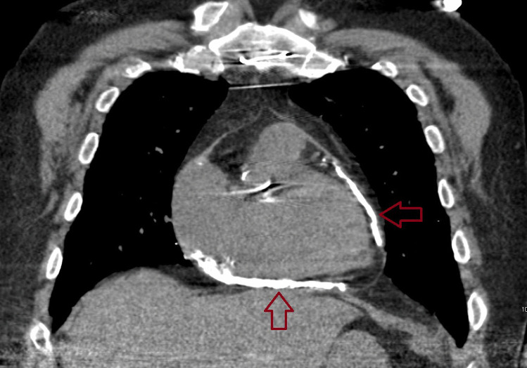
The use of CT imaging in the diagnosis and management of CP is limited. CT scanning can be used for structural evaluation, but pathophysiological phenomena described earlier cannot be adequately assessed with this modality [4]. Cardiac CT can be used to accurately measure pericardial thickness and visualize calcification (Fig. 9) [1]. It may also reveal characteristic morphology of cardiovascular structures such as elongation or tubular configuration of the ventricles [48]. While interpreting results from CT imaging, it should be noted that neither the absence of pericardial thickening and calcification rule out the diagnosis of CP nor does its presence confirm its diagnosis [1, 4, 24].
Fig. 9.
Calcification (arrows) of pericardium on computed tomographic imaging
Cardiovascular Magnetic Resonance (CMR) Imaging
Over the last several years, CMR imaging techniques have improved exponentially and have provided us with the ability to visualize the morphology of cardiac structures, but also help in tissue characterization, assess hemodynamic changes, study ventricular inflow and venous flow patterns, and predict response to treatment [13]. Similar to echocardiogram, CMR evaluation of CP can also be divided into respiration-dependent and -independent parameters. The phenomenon of VSS can be visualized on real-time CMR imaging during free-breathing protocols [4, 49, 50] (Fig. 10). Relative septal excursion which is based on the principle of VSS is derived by dividing the distance between the RV free wall and the septum by the biventricular distance in inspiration and expiration. It can differentiate between CP and RCM when above 11.8% [4, 50, 51]. Velocity encoded phase-contrast imaging can be used to assess ventricular inflow and venous flow patterns, enabling assessment of mitral and tricuspid inflow velocities, which can be used to diagnose CP and differentiate it from RCM [4, 13, 50, 52].
Fig. 10.
Phenomenon of ventricular interdependence on free-breathing protocol of magnetic resonance imaging (MRI). Left panel shows short axis views whereas right panel shows long axis views during MRI red arrow shows leftward shift of septum during inspiration (A, B) and blue arrow shows rightward shift of septum during expiration (C, D)
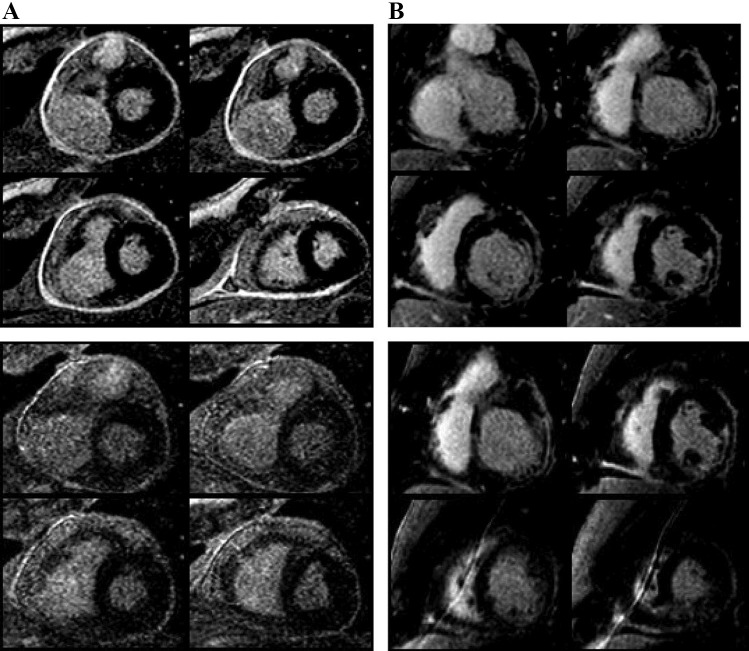
The most important respiration independent variable on CMR imaging is structural evaluation. Pericardial thickness and its characterization can be studied in detail. On CMR, normal pericardium on T1-weighted imaging appears as a thin hypointense (low signal) structure in contrast to the hyperintense (high signal) epicardial and mediastinal fat [13, 50]. A normal pericardium will not retain gadolinium due to its avascular nature, whereas an injured pericardium will have neovascularization, fibroblast proliferation, and expanded extracellular space that accumulates gadolinium and delays its washout resulting in late or delayed hyperenhancement (LGE or DHE) [53••]. The edema weighted T2 short tau inversion recovery (T2-STIR) sequence and late gadolinium enhancement (LGE) sequence show increased pericardial signals on T2-STIR and pericardial LGE, indicating edema and inflammation, respectively (Fig. 11) [4, 50]. Findings of edema or inflammation in a patient with suspected CP suggest an acute or sub-acute process which are potentially reversible with anti-inflammatory medications or reverts spontaneously, precluding pericardiectomy as demonstrated in several studies (Fig. 11) [54–56]. Quantitative assessment of pericardial delayed hyperenhancement when added to clinical and laboratory inflammatory markers can provide information to predict clinical improvement with anti-inflammatory therapy in patients with CP [57]. These findings highlight the importance of detecting inflammation on CMR and modulating lifesaving therapy by adding anti-inflammatory therapy and preventing surgery for these patients with CP.
Fig. 11.
Delayed enhancement of pericardium on cardiac magnetic resonance imaging in 2 patients. A Severe intensity of pericardial delayed enhancement before (left) and moderate intensity after (right) anti-inflammatory medication in a 55-year-old man with constrictive pericarditis who recovered clinically and echocardiographically after medical therapy. B Mild intensity of pericardial delayed enhancement before (left) and persistent mild intensity after (right) anti-inflammatory medication in a 61-year-old man who underwent pericardiectomy because of persistent symptoms and clinical and echocardiographic evidence of constrictive pericarditis after medical therapy. Subendocardial myocardial infarct was also noticed in the inferior and inferolateral walls. (From: DaLi Feng, et al. Circulation. 2011 Oct 25;124(17):1830–7, with permission of Wolters Kluwer Health, Inc.) [85]
Myocardial tagging demonstrating pericardial-myocardial adherence, dilated IVC, abnormal contours of the ventricles, diastolic septal bounce are other variables that can be identified during CMR [58, 59]. American Society of Echocardiography clinical recommendations of multimodality imaging of pericardial diseases suggests the use of CMR, especially in cases where the duration of symptoms is less than 3 months or in the presence of elevated inflammatory markers. This approach can help to identify inflammation, and therefore anti-inflammatory therapy can be considered [24]. Myocardial tissue tracking with CMR has also been utilized and has been found helpful in differentiating cases of CP from RCM, but its use has not been widely adopted due to limited availability [4, 60, 61].
Positron Emission Tomography (PET)
PET imaging relies on 18-fluorodeoxyglucose (18-FDG) uptake as its diagnostic marker instead of anatomical or functional parameters. Active inflammatory process leads to the metabolism of 18-FDG. Therefore, its uptake can be recorded in areas of active inflammation [62]. A small study of 16 patients by Chang et al. indicated the utility of 18 FDG PET imaging to predict responses to steroid therapy in CP; however, its utilization is still limited and more studies are required to validate these findings [63, 64].
Role of Multimodality Imaging in Subtyping of the Constrictive Pericarditis
As discussed earlier, there are various subtypes of CP which can be identified with the use of various interventional and imaging techniques and further prognosticate and guide the treatment modalities. In order to utilize imaging to differentiate between these subtypes, it is important to understand the relation between them. Klein and Cremer [18] introduced the concept of approaching inflammation and hemodynamics separately when evaluating patients with pericardial diseases. The interplay between inflammation and hemodynamic response may lead to categorization of disease pattern. In simple words, patients with effusive pericarditis (inflammation) may or may not develop constrictive physiology (hemodynamics). This constrictive physiology may further be temporary or permanent. Therefore, what one may believe as separate entities, actually represent a spectrum or a continuum of a disease process, thereby making ECP as one end of the spectrum of pericarditis with effusion [18, 65]. Early identification of ECP in this spectrum may change the treatment approach and may, ultimately, also change clinical course for these patients.
The persistent elevated right atrial pressure after pericardiocentesis has been used to diagnose ECP and relies on the interventional hemodynamic assessment during pericardiocentesis which was studied in detail by Sagristà-Sauleda et al. [17] in their evaluation of 190 patients undergoing simultaneous pericardiocentesis and cardiac catheterization. But instead of relying on invasive hemodynamics, the use of pre-pericardiocentesis echo-Doppler parameters may also help identify patients with ECP and was studied by Kim et al. [54] in a cohort of 205 patients undergoing pericardiocentesis. The diagnostic criteria in this study included respiratory variation in the mitral inflow velocity by >25% in combination with at least 1 of the following: expiratory diastolic flow reversal of hepatic vein, respirophasic interventricular shift, or augmented early diastolic mitral septal annular velocity (e′) to a level higher than lateral e′ velocity. Using this criterion in the post-percardiocentesis echocardiographic studies, patients with constrictive physiology were identified, and thus diagnosed with ECP and then compared these markers to the pre-pericardiocentesis echocardiographic data. The study suggested that patients with ECP might have these distinct echo-Doppler features even before pericardiocentesis, thereby helping in early identification of these patients [54]. Therefore, early utilization of echocardiography may identify constrictive physiology in patients with pericardial effusion and further use of aforementioned diagnostic modalities like CMR and PET can demarcate active inflammation, guide anti-inflammatory therapy, and aid in the diagnosis of ECP.
While many of the diagnostic modalities we discussed will help us to diagnose patients with isolated CP, there will be patients in real-world practice who will have mixed disease in setting of underlying myocardial diseases. This is especially true in older patients with multiple comorbidities like cardiomyopathy, prior surgeries, or patients who have CP after radiation therapy. Besides, technical challenges in diagnosis of such mixed pericarditis patients, their outcomes also differ from patients with isolated CP. Yamada et al. [66] in their analysis of 38 patients with mixed physiology showed a statistically significant poor survival rates in patients with mixed physiology as compared to patients with pure constriction. This was further confirmed by Ha et al. [67] in their analysis of 40 post-pericardiectomy cases in which patients with abnormal relaxation, shown by abnormal time constant of isovolumic relaxation (tau) and contractility measured by peak positive rate of increase of LV pressure (+dP/dt), had significantly worse post-operative outcomes, with no death in patients with normal +dP/dt and tau. These parameters were also predictive of poor long-term survival at 2 years with 45% survival rate in patients with abnormal tau and +dP/dt as compared to 81% survival rates with normal parameters [67]. Mere presence of cardiomyopathy, history of previous cardiac surgery, radiation treatment, or percutaneous procedures for coronary artery disease or cardiac arrythmias has been utilized in some clinical studies [68] to classify mixed CP, but imaging modalities may help to further delineate mixed physiology. American Society of Echocardiography guidelines for evaluation of diastolic dysfunction in their algorithm for comparing CP and RCM use the range of mitral medial e′ of 6–8 as a criteria to consider patients with mixed constriction-restriction who have E/A > 0.8, dilated IVC, and respirophasic ventricular septal motion [69]. Ancillary echocardiographic findings such as E/e′ > 15, E/A > 2, mitral A velocity deceleration time of < 150 ms, isovolumic relaxation time < 50 ms may point towards restriction [69]. It is not unusual to see significant diastolic flow reversals in hepatic vein flow during both inspiration and expiration in patients with mixed physiology [3]. Use of strain imaging or cardiac MR to identify myocardial dysfunction may further help in identifying these subsets of patients. It may be challenging to isolate patients with mixed disease, but its identification may definitely help in identifying patients who may not do well even after surgical pericardiectomy and may require additional medical management [70].
Role of Multimodality Imaging in Pre-operative Planning and Predicting Outcomes After Pericardiectomy
Pericardiectomy is regarded as a high-risk procedure with an average reported perioperative mortality rate of 6.0–7.1%, even in high-volume centers [1, 7]. There are a number of factors which can help predict short-term and long-terms outcomes of pericardiectomy. Bertog et al. [11] in their analysis of 163 patients demonstrated the association of etiology of CP to long-term survival. Where idiopathic CP was found to have the best prognosis, patients with post-radiation CP may had substantially worse survival rates (88% vs. 27%, respectively). Other factors that may predict poor long-term survival include higher pulmonary artery systemic pressure, abnormal LV systolic function, lower sodium level, and older age. Factors such as pre-operative NYHA functional class, pre-operative RV dilatation, pre-operative central venous pressure, and reduced LV systolic function are predictors of early post-operative and 30-day mortality [71, 72].
RV failure remains one of the major complications of early post-pericardiectomy due to a rapid increase in venous return to the right heart after pericardial decompression [73]. Therefore, careful pre-operative planning is required in patients of CP undergoing this surgery. Imaging modalities such as CT can play a significant role in assessing the relationship of cardiac and vascular structures to the retrosternum, especially in patients with redo-sternotomy [59, 74]. The extent and localization of calcium is crucial as the presence of circumferential or posterolateral calcification may warrant a bilateral thoracotomy approach, especially in patients with circumferential constriction where removing pericardium from the LV side first may be important to prevent sudden pulmonary edema [59]. Fluid overload and incomplete pericardial dissection [75, 76] are some of the predictors of operative deaths following pericardiectomy and this pre-operative localization may help prevent these complications.
Azzu et al. [77] attempted to create a “pericardial score” using pre-operative multimodality imaging to identify the patients at risk for prolonged inotropic support with an assumption that there is an association between pericardial anatomy and difficulty of surgical dissection of the pericardium. High pericardial “score” (score of 2–3) calculated by epicardial fat thickness < 5 mm, thickened pericardium > 5 mm, and pericardial calcification by CT (1 point each) was strongly associated with in-hospital use of inotropes. Moreover, a smaller RV cavity size calculated either by CMR or echocardiography also predicted prolonged inotropic support in the early post-operative period [77]. Thus, the use of imaging not only plays an indispensable role in surgical planning and identification of patients with myocardial dysfunction/mixed disease but may also help predict worse outcomes post-operatively thereby aiding in risk stratification of patients with CP.
COVID-19 and Constrictive Pericarditis
As discussed earlier, 1.8% of acute pericarditis cases progress to CP with the lowest risk (<1%) of progression for viral or idiopathic cases [14]. The emergence of SARS CoV2 has added another virus that can cause pericarditis. Furthermore, the risk of pericarditis or myocarditis after COVID-19 vaccination has been described [78–81]. Though an early case of CP with COVID-19 has been described in medical literature [82], there is uncertainty about the risk of progression of COVID pericarditis into CP as the natural history of CP usually extends over years to even decades. There are reports of transient or effusive-constrictive pericarditis with COVID-19 infection or vaccination [83, 84]; however, the real impact of this infection on CP is unknown.
Conclusion
Constrictive pericarditis manifests as various subtypes and possesses unique pathophysiological features. A high degree of clinical suspicion is needed to evaluate patients for this disease, and accurate diagnosis is fundamental in improving patient outcomes. The use of multimodality imaging has revolutionized the diagnosis of CP. It has also helped to differentiate among the subtypes of CP, identify patients who would benefit from medical therapy, guide pre-operative planning, and prognosticate patients undergoing surgery. Advanced techniques and newer imaging modalities are still being developed, which will continue to redirect and mold the diagnosis and management of CP.
Compliance with Ethical Standards
Conflict of Interest
The authors report no disclosures pertaining to this article.
Human and Animal Rights and Informed Consent
This article does not contain any studies with human or animal subjects performed by any of the authors.
Footnotes
This article is part of the Topical Collection on Pericardial Disease
Publisher's Note
Springer Nature remains neutral with regard to jurisdictional claims in published maps and institutional affiliations.
References
Papers of particular interest, published recently, have been highlighted as: • Of importance •• Of major importance
- 1.Welch TD, Oh JK. Constrictive pericarditis. Cardiol Clin. 2017;35:539–549. doi: 10.1016/j.ccl.2017.07.007. [DOI] [PubMed] [Google Scholar]
- 2.Nishimura RA. Constrictive pericarditis in the modern era: a diagnostic dilemma. Heart. 2001;86:619–623. doi: 10.1136/heart.86.6.619. [DOI] [PMC free article] [PubMed] [Google Scholar]
- 3.Sengupta PP, Eleid MF, Khandheria BK. Constrictive pericarditis. Circ J. 2008;72:1555–1562. doi: 10.1253/circj.CJ-08-0654. [DOI] [PubMed] [Google Scholar]
- 4.Alajaji W, Xu B, Sripariwuth A, Menon V, Kumar A, Schleicher M, Isma’eel H, Cremer PC, Bolen MA, Klein AL. Noninvasive multimodality imaging for the diagnosis of constrictive pericarditis. Circ Cardiovasc Imaging. 2018;11:e007878. doi: 10.1161/CIRCIMAGING.118.007878. [DOI] [PubMed] [Google Scholar]
- 5.Brandt RR, Oh JK. Constrictive pericarditis: role of echocardiography and magnetic resonance imaging. E-Journal Cardiol Pract. 2017;15:22. [Google Scholar]
- 6.Welch TD, Oh JK. Constrictive pericarditis: old disease, new approaches. Curr Cardiol Rep. 2015;17:20. doi: 10.1007/s11886-015-0576-x. [DOI] [PubMed] [Google Scholar]
- 7.Welch TD. Constrictive pericarditis: diagnosis, management and clinical outcomes. Heart. 2018;104:725–731. doi: 10.1136/heartjnl-2017-311683. [DOI] [PubMed] [Google Scholar]
- 8.Kyriakakis C, Herbst P, Doubell A. Constrictive pericarditis–prevalence, causes and clinical presentation. Eur Soc Cardiol. 2017;15:1–8. [Google Scholar]
- 9.Deterling RA, Humphreys GH. Factors in the etiology of constrictive pericarditis. Circulation. 1955;12:30–43. doi: 10.1161/01.CIR.12.1.30. [DOI] [PubMed] [Google Scholar]
- 10.Ling LH, Oh JK, Schaff HV, Danielson GK, Mahoney DW, Seward JB, Tajik AJ. Constrictive pericarditis in the modern era: evolving clinical spectrum and impact on outcome after pericardiectomy. Circulation. 1999;100:1380–1386. doi: 10.1161/01.CIR.100.13.1380. [DOI] [PubMed] [Google Scholar]
- 11.Bertog SC, Thambidorai SK, Parakh K, Schoenhagen P, Ozduran V, Houghtaling PL, Lytle BW, Blackstone EH, Lauer MS, Klein AL. Constrictive pericarditis: etiology and cause-specific survival after pericardiectomy. J Am Coll Cardiol. 2004;43:1445–1452. doi: 10.1016/j.jacc.2003.11.048. [DOI] [PubMed] [Google Scholar]
- 12.Mayosi BM, Burgess LJ, Doubell AF. Tuberculous pericarditis. Circulation. 2005;112:3608–3616. doi: 10.1161/CIRCULATIONAHA.105.543066. [DOI] [PubMed] [Google Scholar]
- 13.Adler Y, Charron P, Imazio M, et al. 2015 ESC Guidelines for the diagnosis and management of pericardial diseases: the Task Force for the diagnosis and management of pericardial diseases of the European Society of Cardiology (ESC)Endorsed by: the European Association for Cardio-Thoracic Surg. Eur Heart J. 2015;36:2921–2964. doi: 10.1093/eurheartj/ehv318. [DOI] [PMC free article] [PubMed] [Google Scholar]
- 14.Gentry J, Klein AL, Jellis CL. Transient constrictive pericarditis: current diagnostic and therapeutic strategies. Curr Cardiol Rep. 2016;18:41. doi: 10.1007/s11886-016-0720-2. [DOI] [PubMed] [Google Scholar]
- 15.Jiamsripong P, Alharthi MS, Calleja AM, McMahon EM, Katayama M, Westerdale J, Milano M, Heys JJ, Mookadam F, Belohlavek M. Impact of pericardial adhesions on diastolic function as assessed by vortex formation time, a parameter of transmitral flow efficiency. Cardiovasc Ultrasound. 2010;8:42. doi: 10.1186/1476-7120-8-42. [DOI] [PMC free article] [PubMed] [Google Scholar]
- 16.D’Elia E, Ferrazzi P, Imazio M, et al. Constrictive pericarditis: a common pathophysiology for different macroscopic anatomies. J Cardiovasc Med (Hagerstown) 2019;20:725–726. doi: 10.2459/JCM.0000000000000844. [DOI] [PubMed] [Google Scholar]
- 17.Sagristà-Sauleda J, Angel J, Sánchez A, Permanyer-Miralda G, Soler-Soler J. Effusive-constrictive pericarditis. N Engl J Med. 2004;350:469. doi: 10.1056/NEJMoa035630. [DOI] [PubMed] [Google Scholar]
- 18.Klein AL, Cremer PC. Ephemeral effusive constrictive pathophysiology. JACC Cardiovasc Imaging. 2018;11:542–545. doi: 10.1016/j.jcmg.2017.10.028. [DOI] [PubMed] [Google Scholar]
- 19.Hancock EW. Subacute effusive-constrictive pericarditis. Circulation. 1971;43:183–192. doi: 10.1161/01.CIR.43.2.183. [DOI] [PubMed] [Google Scholar]
- 20.Bogaert J, Meyns B, Dymarkowski S, Sinnaeve P, Meuris B. Calcified constrictive pericarditis: prevalence, distribution patterns, and relationship to the myocardium. JACC Cardiovasc Imaging. 2016;9:1013–1014. doi: 10.1016/j.jcmg.2015.08.011. [DOI] [PubMed] [Google Scholar]
- 21.Seifert FC, Miller DC, Oesterle SN, Oyer PE, Stinson EB, Shumway NE. Surgical treatment of constrictive pericarditis: analysis of outcome and diagnostic error. Circulation. 1985;72:II264–II273. [PubMed] [Google Scholar]
- 22.Talreja DR, Edwards WD, Danielson GK, Schaff HV, Tajik AJ, Tazelaar HD, Breen JF, Oh JK, et al. Constrictive pericarditis in 26 patients with histologically normal pericardial thickness. Circulation. 2003;108:1852–1857. doi: 10.1161/01.CIR.0000087606.18453.FD. [DOI] [PubMed] [Google Scholar]
- 23.Maniar HS, Prasad SM, Gaynor SL, Chu CM, Steendijk P, Moon MR. Impact of pericardial restraint on right atrial mechanics during acute right ventricular pressure load. Am J Physiol Circ Physiol. 2003;284:H350–H357. doi: 10.1152/ajpheart.00444.2002. [DOI] [PubMed] [Google Scholar]
- 24.Klein AL, Abbara S, Agler DA, et al. American Society of Echocardiography clinical recommendations for multimodality cardiovascular imaging of patients with pericardial disease: endorsed by the Society for Cardiovascular Magnetic Resonance and Society of Cardiovascular Computed Tomography. J Am Soc Echocardiogr Off Publ Am Soc Echocardiogr. 2013;26:965–1012.e15. doi: 10.1016/j.echo.2013.06.023. [DOI] [PubMed] [Google Scholar]
- 25.Vucic E, Chakhtoura EY, Sohal S, Waxman S. Pathophysiological concepts of constrictive pericarditis in cardiac imaging: back to basics. Circ Cardiovasc Imaging. 2021;14:e012136. doi: 10.1161/CIRCIMAGING.120.012136. [DOI] [PubMed] [Google Scholar]
- 26.Welch TD, Ling LH, Espinosa RE, Anavekar NS, Wiste HJ, Lahr BD, Schaff HV, Oh JK. Echocardiographic diagnosis of constrictive pericarditis: Mayo Clinic criteria. Circ Cardiovasc Imaging. 2014;7:526–534. doi: 10.1161/CIRCIMAGING.113.001613. [DOI] [PubMed] [Google Scholar]
- 27.Welch TD, Ling LH, Espinosa RE, Anavekar NS, Wiste HJ, Lahr BD, Schaff HV, Oh JK. Echocardiographic diagnosis of constrictive pericarditis. Circ Cardiovasc Imaging. 2014;7:526–534. doi: 10.1161/CIRCIMAGING.113.001613. [DOI] [PubMed] [Google Scholar]
- 28.Cosyns B, Plein S, Nihoyanopoulos P, et al. European Association of Cardiovascular Imaging (EACVI) position paper: multimodality imaging in pericardial disease. Eur Hear journal Cardiovasc Imaging. 2015;16:12–31. doi: 10.1093/ehjci/jeu128. [DOI] [PubMed] [Google Scholar]
- 29.Hatle LK, Appleton CP, Popp RL. Differentiation of constrictive pericarditis and restrictive cardiomyopathy by Doppler echocardiography. Circulation. 1989;79:357–370. doi: 10.1161/01.CIR.79.2.357. [DOI] [PubMed] [Google Scholar]
- 30.Oh JK, Hatle LK, Seward JB, Danielson GK, Schaff HV, Reeder GS, Tajik AJ. Diagnostic role of Doppler echocardiography in constrictive pericarditis. J Am Coll Cardiol. 1994;23:154–162. doi: 10.1016/0735-1097(94)90514-2. [DOI] [PubMed] [Google Scholar]
- 31.Boonyaratavej S, Oh JK, Tajik AJ, Appleton CP, Seward JB. Comparison of mitral inflow and superior vena cava Doppler velocities in chronic obstructive pulmonary disease and constrictive pericarditis. J Am Coll Cardiol. 1998;32:2043–2048. doi: 10.1016/S0735-1097(98)00472-0. [DOI] [PubMed] [Google Scholar]
- 32.Parsons GH, Green JF. Mechanisms of pulsus paradoxus in upper airway obstruction. J Appl Physiol. 1978;45:598–603. doi: 10.1152/jappl.1978.45.4.598. [DOI] [PubMed] [Google Scholar]
- 33.Appleton CP, Hatle LK, Popp RL. Superior vena cava and hepatic vein Doppler echocardiography in healthy adults. J Am Coll Cardiol. 1987;10:1032–1039. doi: 10.1016/S0735-1097(87)80343-1. [DOI] [PubMed] [Google Scholar]
- 34.Coylewright M, Welch TD, Nishimura RA. Mechanism of septal bounce in constrictive pericarditis: a simultaneous cardiac catheterisation and echocardiographic study. Heart. 2013;99:1376. doi: 10.1136/heartjnl-2013-304070. [DOI] [PubMed] [Google Scholar]
- 35.Yang JH, Miranda WR, Nishimura RA, Greason KL, Schaff HV, Oh JK. Prognostic importance of mitral e’ velocity in constrictive pericarditis. Eur Hear journal Cardiovasc Imaging. 2021;22:357–364. doi: 10.1093/ehjci/jeaa133. [DOI] [PubMed] [Google Scholar]
- 36.Reuss CS, Wilansky SM, Lester SJ, Lusk JL, Grill DE, Oh JK, Tajik AJ. Using mitral “annulus reversus” to diagnose constrictive pericarditis. Eur J Echocardiogr J Work Gr Echocardiogr Eur Soc Cardiol. 2009;10:372–375. doi: 10.1093/ejechocard/jen258. [DOI] [PubMed] [Google Scholar]
- 37.• Sato K, Ayache A, Kumar A, et al. Improvement in left ventricular mechanics following medical treatment of constrictive pericarditis. Heart. 2021;107:828–35. This study summarizes the importance of serial monitoring of imaging parameters to predict clinical outcomes in patients after anti-inflammatory therapy. [DOI] [PubMed]
- 38.Ha JW, Oh JK, Ling LH, Nishimura RA, Seward JB, Tajik AJ. Annulus paradoxus: transmitral flow velocity to mitral annular velocity ratio is inversely proportional to pulmonary capillary wedge pressure in patients with constrictive pericarditis. Circulation. 2001;104:976–978. doi: 10.1161/hc3401.095705. [DOI] [PubMed] [Google Scholar]
- 39.Alraies MC, Kusunose K, Negishi K, Yarmohammadi H, Motoki H, AlJaroudi W, Popović ZB, Klein AL. Relation between echocardiographically estimated and invasively measured filling pressures in constrictive pericarditis. Am J Cardiol. 2014;113:1911–1916. doi: 10.1016/j.amjcard.2014.03.022. [DOI] [PubMed] [Google Scholar]
- 40.Zhang K, Sheu R, Zimmerman NM, Alfirevic A, Sale S, Gillinov AM, Duncan AE. A comparison of global longitudinal, circumferential, and radial strain to predict outcomes after cardiac surgery. J Cardiothorac Vasc Anesth. 2019;33:1315–1322. doi: 10.1053/j.jvca.2018.10.031. [DOI] [PMC free article] [PubMed] [Google Scholar]
- 41.Sengupta PP, Khandheria BK, Korinek J, Wang J, Jahangir A, Seward JB, Belohlavek M. Apex-to-base dispersion in regional timing of left ventricular shortening and lengthening. J Am Coll Cardiol. 2006;47:163–172. doi: 10.1016/j.jacc.2005.08.073. [DOI] [PubMed] [Google Scholar]
- 42.Sengupta PP, Krishnamoorthy VK, Abhayaratna WP, et al. Disparate patterns of left ventricular mechanics differentiate constrictive pericarditis from restrictive cardiomyopathy. JACC Cardiovasc Imaging. 2008;1:29–38. doi: 10.1016/j.jcmg.2007.10.006. [DOI] [PubMed] [Google Scholar]
- 43.Gibbons Kroeker CA, Adeeb S, Tyberg JV, Shrive NG. A 2D FE model of the heart demonstrates the role of the pericardium in ventricular deformation. Am J Physiol Circ Physiol. 2006;291:H2229–H2236. doi: 10.1152/ajpheart.00077.2006. [DOI] [PubMed] [Google Scholar]
- 44.Jottrand E, Serste T, Mulkay J-P, Vandueren C, Unger P. Longitudinal strain by speckle tracking echocardiography in constrictive pericarditis. Eur Hear J - Cardiovasc Imaging. 2018;19:638. doi: 10.1093/ehjci/jey031. [DOI] [PubMed] [Google Scholar]
- 45.Stassen J, Jogani S, Schroyens M. Strain reversus revealing constrictive pericarditis. Eur Heart J Cardiovasc Imaging. 2021;22:e14. doi: 10.1093/ehjci/jeaa225. [DOI] [PubMed] [Google Scholar]
- 46.Argulian E, Halpern DG. “Hot Septum” sign of constrictive pericarditis. JACC Case Reports. 2020;2:186–190. doi: 10.1016/j.jaccas.2019.12.024. [DOI] [PMC free article] [PubMed] [Google Scholar]
- 47.Engel PJ, Fowler NO, Tei CW, Shah PM, Driedger HJ, Shabetai R, Harbin AD, Franch RH. M-mode echocardiography in constrictive pericarditis. J Am Coll Cardiol. 1985;6:471–474. doi: 10.1016/S0735-1097(85)80188-1. [DOI] [PubMed] [Google Scholar]
- 48.Napolitano G, Pressacco J, Paquet E. Imaging features of constrictive pericarditis: beyond pericardial thickening. Can Assoc Radiol J. 2009;60:40. doi: 10.1016/j.carj.2009.02.034. [DOI] [PubMed] [Google Scholar]
- 49.Giorgi B, Mollet NRA, Dymarkowski S, Rademakers FE, Bogaert J. Clinically suspected constrictive pericarditis: MR imaging assessment of ventricular septal motion and configuration in patients and healthy subjects. Radiology. 2003;228:417–424. doi: 10.1148/radiol.2282020345. [DOI] [PubMed] [Google Scholar]
- 50.Garg R, Guzzetti E, Chetrit M. Established and emerging techniques for pericardial imaging with cardiac magnetic resonance. Curr Cardiol Rep. 2021;23:169. doi: 10.1007/s11886-021-01595-z. [DOI] [PubMed] [Google Scholar]
- 51.Francone M, Dymarkowski S, Kalantzi M, Rademarkers FE, Bogaert J. Real-time cine MRI of ventricular septal motion: a novel approach to assess ventricular coupling. J Magn Reson Imaging. 2005;21:305. doi: 10.1002/jmri.20259. [DOI] [PubMed] [Google Scholar]
- 52.Thavendiranathan P, Verhaert D, Walls MC, Bender JA, Rajagopalan S, Chung Y-C, Simonetti OP, Raman SV. Simultaneous right and left heart real-time, free-breathing CMR flow quantification identifies constrictive physiology. JACC Cardiovasc Imaging. 2012;5:15–24. doi: 10.1016/j.jcmg.2011.07.010. [DOI] [PMC free article] [PubMed] [Google Scholar]
- 53.Chetrit M, Xu B, Kwon DH, et al. Imaging-guided therapies for pericardial diseases. JACC Cardiovasc Imaging. 2020;13:1422–1437. doi: 10.1016/j.jcmg.2019.08.027. [DOI] [PubMed] [Google Scholar]
- 54.Kim KH, Miranda WR, Sinak LJ, Syed FF, Melduni RM, Espinosa RE, Kane GC, Oh JK. Effusive-constrictive pericarditis after pericardiocentesis: incidence, associated findings, and natural history. JACC Cardiovasc Imaging. 2018;11:534–541. doi: 10.1016/j.jcmg.2017.06.017. [DOI] [PMC free article] [PubMed] [Google Scholar]
- 55.Imazio M, Brucato A, Mayosi BM, Derosa FG, Lestuzzi C, Macor A, Trinchero R, Spodick DH, Adler Y. Medical therapy of pericardial diseases: part II: noninfectious pericarditis, pericardial effusion and constrictive pericarditis. J Cardiovasc Med (Hagerstown) 2010;11:785–794. doi: 10.2459/JCM.0b013e3283340b97. [DOI] [PubMed] [Google Scholar]
- 56.Haley JH, Tajik AJ, Danielson GK, Schaff HV, Mulvagh SL, Oh JK. Transient constrictive pericarditis: causes and natural history. J Am Coll Cardiol. 2004;43:271–275. doi: 10.1016/j.jacc.2003.08.032. [DOI] [PubMed] [Google Scholar]
- 57.Cremer PC, Tariq MU, Karwa A, Alraies MC, Benatti R, Schuster A, Agarwal S, Flamm SD, Kwon DH, Klein AL. Quantitative assessment of pericardial delayed hyperenhancement predicts clinical improvement in patients with constrictive pericarditis treated with anti-inflammatory therapy. Circ Cardiovasc Imaging. 2015 doi: 10.1161/CIRCIMAGING.114.003125. [DOI] [PubMed] [Google Scholar]
- 58.Rajiah P, Canan A, Saboo SS, Restrepo CS, Bolen MA. MRI of the pericardium. Radiographics. 2019;39:1921–1922. doi: 10.1148/rg.2019190081. [DOI] [PubMed] [Google Scholar]
- 59.Verhaert D, Gabriel RS, Johnston D, Lytle BW, Desai MY, Klein AL. The role of multimodality imaging in the management of pericardial disease. Circ Cardiovasc Imaging. 2010;3:333. doi: 10.1161/CIRCIMAGING.109.921791. [DOI] [PubMed] [Google Scholar]
- 60.Yang Z, Wang H, Chang S, Cui J, Zhou L, Lv Q, He Y, Du X, Dong J, Ma C. Left ventricular strain-curve morphology to distinguish between constrictive pericarditis and restrictive cardiomyopathy. ESC Hear Fail. 2021;8:4863–4872. doi: 10.1002/ehf2.13679. [DOI] [PMC free article] [PubMed] [Google Scholar]
- 61.Asadian S, Farzin M, Tabesh F, Rezaeian N, Bakhshandeh H, Hosseini L, Toloueitabar Y, Hemmati Komasi MM. The auxiliary role of cardiac magnetic resonance feature-tracking parameters in the differentiation between cardiac amyloidosis and constrictive pericarditis. Cardiol Res Pract. 2021;2021:2045493. doi: 10.1155/2021/2045493. [DOI] [PMC free article] [PubMed] [Google Scholar]
- 62.Alter P, Figiel JH, Rupp TP, Bachmann GF, Maisch B, Rominger MB. MR, CT, and PET imaging in pericardial disease. Heart Fail Rev. 2013;18:289–306. doi: 10.1007/s10741-012-9309-z. [DOI] [PubMed] [Google Scholar]
- 63.Chang S-A, Choi JY, Kim EK, Hyun SH, Jang SY, Choi J-O, Park S-J, Lee S-C, Park SW, Oh JK. [(18)F]Fluorodeoxyglucose PET/CT predicts response to steroid therapy in constrictive pericarditis. J Am Coll Cardiol. 2017;69:750–752. doi: 10.1016/j.jacc.2016.11.059. [DOI] [PubMed] [Google Scholar]
- 64.Kim M-S, Kim E-K, Choi JY, Oh JK, Chang S-A. Clinical utility of [(18)F]FDG-PET /CT in Pericardial Disease. Curr Cardiol Rep. 2019;21:107. doi: 10.1007/s11886-019-1193-x. [DOI] [PubMed] [Google Scholar]
- 65.Janus SE, Hoit BD. Effusive–constrictive pericarditis in the spectrum of pericardial compressive syndromes. Heart. 2021;107:450–455. doi: 10.1136/heartjnl-2020-316664. [DOI] [PubMed] [Google Scholar]
- 66.Yamada H, Tabata T, Jaffer SJ, Drinko JK, Jasper SE, Lauer MS, Thomas JD, Klein AL. Clinical features of mixed physiology of constriction and restriction: echocardiographic characteristics and clinical outcome. Eur J Echocardiogr. 2007;8:185–194. doi: 10.1016/j.euje.2006.03.003. [DOI] [PubMed] [Google Scholar]
- 67.Ha J-W, Oh JK, Schaff HV, Ling LH, Higano ST, Mahoney DW, Nishimura RA. Impact of left ventricular function on immediate and long-term outcomes after pericardiectomy in constrictive pericarditis. J Thorac Cardiovasc Surg. 2008;136:1136–1141. doi: 10.1016/j.jtcvs.2008.03.065. [DOI] [PubMed] [Google Scholar]
- 68.Yang JH, Miranda WR, Borlaug BA, Nishimura RA, Schaff HV, Greason KL, Maleszewski JJ, Oh JK. Right atrial/pulmonary arterial wedge pressure ratio in primary and mixed constrictive pericarditis. J Am Coll Cardiol. 2019;73:3312–3321. doi: 10.1016/j.jacc.2019.03.522. [DOI] [PubMed] [Google Scholar]
- 69.Nagueh SF, Smiseth OA, Appleton CP, et al. Recommendations for the evaluation of left ventricular diastolic function by echocardiography: an update from the American Society of Echocardiography and the European Association of Cardiovascular Imaging. J Am Soc Echocardiogr. 2016;29:277–314. doi: 10.1016/j.echo.2016.01.011. [DOI] [PubMed] [Google Scholar]
- 70.Klein AL, Xu B. Constrictive Pericarditis. J Am Coll Cardiol. 2019;73:3322–3325. doi: 10.1016/j.jacc.2019.05.003. [DOI] [PubMed] [Google Scholar]
- 71.Fang L, Yu G, Huang J, Zhao W, Ye B. Predictors of postoperative complication and prolonged intensive care unit stay after complete pericardiectomy in tuberculous constrictive pericarditis. J Cardiothorac Surg. 2020;15:148. doi: 10.1186/s13019-020-01198-9. [DOI] [PMC free article] [PubMed] [Google Scholar]
- 72.Busch C, Penov K, Amorim PA, Garbade J, Davierwala P, Schuler GC, Rastan AJ, Mohr F-W. Risk factors for mortality after pericardiectomy for chronic constrictive pericarditis in a large single-centre cohort. Eur J Cardio-Thoracic Surg. 2015;48:e110–e116. doi: 10.1093/ejcts/ezv322. [DOI] [PubMed] [Google Scholar]
- 73.Park YH. Novel imaging parameters for right ventricular dysfunction after pericardiectomy in constrictive pericarditis. J Cardiovasc imaging. 2021;29:373–374. doi: 10.4250/jcvi.2021.0074. [DOI] [PMC free article] [PubMed] [Google Scholar]
- 74.Kamdar AR, Meadows TA, Roselli EE, Gorodeski EZ, Curtin RJ, Sabik JF, Schoenhagen P, White RD, Lytle BW, Flamm SD. Multidetector computed tomographic angiography in planning of reoperative cardiothoracic surgery. Ann Thorac Surg. 2008;85:1239–1245. doi: 10.1016/j.athoracsur.2007.11.075. [DOI] [PubMed] [Google Scholar]
- 75.Huang J-B, Wen Z-K, Yang J-R, Li J-J, Li M, Lu C-C, Liang D-Y, Wei C-X. Analysis of risk factors of early mortality after pericardiectomy for constrictive pericarditis. Heart Surg Forum. 2022;25:E056–E064. doi: 10.1532/hsf.4329. [DOI] [PubMed] [Google Scholar]
- 76.Gatti G, Fiore A, Ternacle J, et al. Pericardiectomy for constrictive pericarditis: a risk factor analysis for early and late failure. Heart Vessels. 2020;35:92–103. doi: 10.1007/s00380-019-01464-4. [DOI] [PubMed] [Google Scholar]
- 77.Azzu A, Morosin M, Antonopoulos AS, Capoccia M, Rosendahl U, Mohiaddin R. Cardiac decompression by pericardiectomy for constrictive pericarditis: multimodality imaging to identify patients at risk for prolonged inotropic support. J Cardiovasc imaging. 2021;29:361–372. doi: 10.4250/jcvi.2020.0223. [DOI] [PMC free article] [PubMed] [Google Scholar]
- 78.Boehmer TK, Kompaniyets L, Lavery AM, Hsu J, Ko JY, Yusuf H, Romano SD, Gundlapalli AV, Oster ME, Harris AM. Association between COVID-19 and myocarditis using hospital-based administrative data — United States, March 2020–January 2021. MMWR Morb Mortal Wkly Rep. 2022;70:1228–1232. doi: 10.15585/mmwr.mm7035e5. [DOI] [PMC free article] [PubMed] [Google Scholar]
- 79.Witberg G, Barda N, Hoss S, et al. Myocarditis after Covid-19 vaccination in a large health care organization. N Engl J Med. 2021;385:2132–2139. doi: 10.1056/NEJMoa2110737. [DOI] [PMC free article] [PubMed] [Google Scholar]
- 80.Carubbi F, Alunno A, Leone S, Di Gregorio N, Mancini B, Viscido A, Del Pinto R, Cicogna S, Grassi D, Ferri C. Pericarditis after SARS-CoV-2 infection: another pebble in the mosaic of long COVID? Viruses. 2021 doi: 10.3390/v13101997. [DOI] [PMC free article] [PubMed] [Google Scholar]
- 81.Diaz GA, Parsons GT, Gering SK, Meier AR, Hutchinson IV, Robicsek A. Myocarditis and Pericarditis after vaccination for COVID-19. JAMA. 2021;326:1210–1212. doi: 10.1001/jama.2021.13443. [DOI] [PMC free article] [PubMed] [Google Scholar]
- 82.Beckerman JK, Alarfaj M, Tracy CM, Faiwiszewski AD, Choi AD. Coronavirus disease 2019 (COVID-19)-associated constrictive pericarditis. BMJ Case Rep. 2021;14:e242018. doi: 10.1136/bcr-2021-242018. [DOI] [PMC free article] [PubMed] [Google Scholar]
- 83.Diaconu R, Popescu L, Voicu A, Donoiu I. Subacute effusive-constrictive pericarditis in a patient with COVID-19. BMJ Case Rep. 2021;14:e242443. doi: 10.1136/bcr-2021-242443. [DOI] [PMC free article] [PubMed] [Google Scholar]
- 84.Viani GM, Pedrotti P, Seregni R, Antonio B. Effusive-constrictive pericarditis after the second dose of BNT162b2 vaccine (Comirnaty): a case report. Eur Hear J Case Rep. 2022;6:ytac012. doi: 10.1093/ehjcr/ytac012. [DOI] [PMC free article] [PubMed] [Google Scholar]
- 85.Feng D, Glockner J, Kim K, et al. Cardiac magnetic resonance imaging pericardial late gadolinium enhancement and elevated inflammatory markers can predict the reversibility of constrictive pericarditis after antiinflammatory medical therapy. Circulation. 2011;124:1830–1837. doi: 10.1161/CIRCULATIONAHA.111.026070. [DOI] [PubMed] [Google Scholar]


