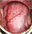
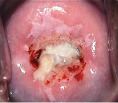
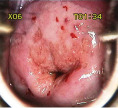
Table 3:
Salient features of various lesions detected on colposcopy.
| Lesion type | Salient features | Colposcopy image |
|---|---|---|
| Leukoplakia | • White patch with shiny, waxy surface • Sharp, raised margin |

|
| Condyloma | • Single or multiple bright white distinct, irregular lesions • Surface irregularity (with pitted or spiky appearance) |

|
| Condyloma/subclinical papillomavirus infection (SPI) | • Thin/milky acetowhite patches • Irregular, geographical margin • Multiple satellite lesions |

|
| Low-grade squamous intraepithelial lesion | • Thin acetowhite epithelium • Irregular, geographical border • Fine mosaic • Fine punctuation |

|
| High-grade squamous intraepithelial lesion | • Dense acetowhite epithelium • Cuffed crypt (gland) openings • Coarse mosaic • Coarse punctation • Sharp border • Inner border sign • Ridge sign |

|
| Invasive carcinoma | • Dense acetowhite area with/without erosion • Surface irregularity • Vascular abnormalities – coarse mosaics/coarse punctations/atypical blood vessels • Exophytic/ulcerative growth |

|
| Adenocarcinoma | • Multiple dense acetowhite areas on columnar epithelium (grated coconut appearance) • Atypical blood vessels |

|
