ABSTRACT
The compartmentalized domains of polarized epithelial cells arise from mutually antagonistic actions between the apical Par complex and the basolateral Scrib module. In Drosophila, the Scrib module proteins Scribble (Scrib) and Discs-large (Dlg) are required to limit Lgl phosphorylation at the basolateral cortex, but how Scrib and Dlg could carry out such a ‘protection’ activity is not clear. We tested Protein Phosphatase 1α (PP1) as a potential mediator of this activity, but demonstrate that a significant component of Scrib and Dlg regulation of Lgl is PP1 independent, and found no evidence for a Scrib-Dlg-PP1 protein complex. However, the Dlg SH3 domain plays a role in Lgl protection and, in combination with the N-terminal region of the Dlg HOOK domain, in recruitment of Scrib to the membrane. We identify a ‘minimal Dlg’ comprised of the SH3 and HOOK domains that is both necessary and sufficient for Scrib localization and epithelial polarity function in vivo.
This article has an associated First Person interview with the first author of the paper.
KEY WORDS: Cell, Polarity, Scrib module, Dlg, Scrib
Summary: A minimal SH3-HOOK fragment of Dlg is sufficient to support epithelial polarity through mechanisms independent of the PP1 phosphatase.
INTRODUCTION
Cell polarity is the fundamental process by which a single cell partitions its plasma membrane into two molecularly distinct, mutually exclusive domains. The ability to polarize is crucial for the development and homeostasis of many cell types, including neurons, stem cells and epithelial cells (St Johnston and Ahringer, 2010). Epithelial cells exhibit apicobasal polarity, a feature critical for their physiological function and morphogenesis of their resident tissues (Buckley and St Johnston, 2022; Rodriguez-Boulan and Macara, 2014). Like many other polarized cells, epithelial cell polarity is often regulated by two highly conserved groups of proteins: the Par complex, composed of Par-3, Par-6 and atypical protein kinase C (aPKC), and the Scrib module, composed of Scribble (Scrib), Discs-large (Dlg) and Lethal giant larvae (Lgl) (Flores-Benitez and Knust, 2016; Goldstein and Macara, 2007). The separation of apical and basolateral domains derives from the mutual antagonism between the apical-defining Par complex and the basolateral-defining Scrib module. Apical aPKC phosphorylates Lgl, which removes it from the plasma membrane, thus excluding Lgl from the apical domain (Bailey and Prehoda, 2015; Betschinger et al., 2003; Dong et al., 2015; Plant et al., 2003). Conversely, basolateral Lgl inhibits aPKC localization to prevent apical domain spread (Hutterer et al., 2004; Wirtz-Peitz et al., 2008; Yamanaka et al., 2003).
For the Par complex, there is now detailed insight into specific functions and molecular interactions for each of its component proteins (Lang and Munro, 2017; Tepass, 2012). By contrast, how the Scrib module determines basolateral polarity is poorly defined (Bonello and Peifer, 2018; Nakajima, 2021; Stephens et al., 2018). The major knowledge gap in Scrib module biology is the molecular mechanism of Scrib and Dlg activity. While Lgl's role as an antagonist of aPKC localization is well known, how Scrib and Dlg act to ensure restriction of the apical domain is not understood. Addressing this question will be essential to a full understanding of cell polarity.
We previously identified several principles of Scrib module protein function, showing that Dlg is required to regulate Scrib cortical localization and providing evidence that Scrib and Dlg are both required to negatively regulate Lgl phosphorylation (Khoury and Bilder, 2020). The data led us to propose a model in which Scrib and Dlg act as molecular switches in the aPKC-Lgl relationship. At the basolateral domain, Scrib and Dlg ‘protect’ Lgl by limiting inhibitory aPKC phosphorylation, allowing Lgl to antagonize aPKC, whereas at the apical domain, where Scrib and Dlg are not present, Lgl is unprotected and can be inhibited by aPKC. Here, we have used a combination of in vivo genetics, biochemistry and an in vitro polarity system to pursue potential molecular bases of this model. We fail to find evidence supporting a plausible mechanism of Lgl protection in which Scrib and Dlg recruit the phosphatase PP1, but we identify a minimal domain of Dlg that is both necessary and sufficient for Scrib recruitment and polarity function.
RESULTS
PP1 is a candidate effector of Scrib and Dlg activity
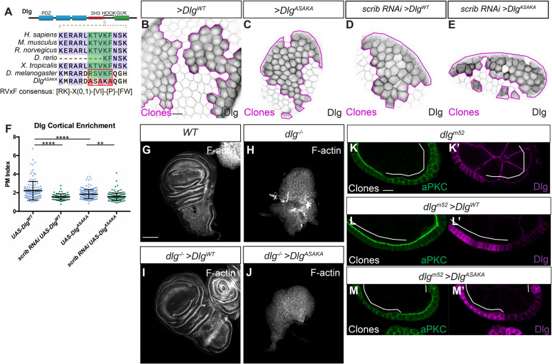
Since Scrib and Dlg are both scaffolding proteins, it is likely that any Lgl protection activity derives from specific binding partners. To search for polarity-relevant Dlg binding partners, we previously carried out proximity proteomics of Dlg in intact epithelial tissue, using a fusion between Dlg and the promiscuous biotin ligase, APEX2, to identify proteins that may interact with or reside near Dlg (Sharp et al., 2021). We mined these data for potential effectors of Lgl protection and uncovered the Drosophila Protein Phosphatase 1α (PP1α) homolog, Pp1-87B (hereafter PP1), in the top 60 most enriched Dlg-proximity hits (log2 fold change=4.8, P=0.001). PP1 is an appealing candidate to mediate Lgl regulation by Scrib and Dlg because PP1 was recently shown to counteract aPKC phosphorylation of Lgl in Drosophila epithelial cells (Moreira et al., 2019). We confirmed that pp1 depletion resulted in decreased cortical Lgl localization in follicle epithelial cells (Fig. S1C-E). Both Scrib and Dlg contain conserved protein sequences that match PP1-binding consensus motifs: Scrib contains SILK and RVxF motifs, and Dlg contains an RVxF motif (Fig. 1A; Fig. S2A) (Hendrickx et al., 2009; Young et al., 2013). These motifs are found in proteins that bind PP1 and can act as substrate specificity factors, recruiting the general PP1 phosphatase to target proteins (Heroes et al., 2013). We therefore hypothesized that Scrib and Dlg could regulate Lgl phosphorylation by scaffolding PP1 at the basolateral cortex.
Fig. 1.
The Dlg RVxF motif is critical for function. (A) Cartoon showing location of the RVxF motif in the Dlg protein and conservation of the motif across species. The resides mutated in DlgASAKA are highlighted in red. The RVxF consensus as defined by Wakula et al. (2003) is shown. (B,C) Like DlgWT (B), DlgASAKA localizes to the basolateral membrane and is enriched at the cell cortex (C). (D-F) DlgWT localization (D) as well as DlgASAKA localization (E) is sensitive to scrib depletion, quantified in F. (G,H) Compared to WT (G), dlg null mutant wing discs form disorganized tumors (H). (I,J) Expression of DlgWT rescues this phenotype (I), while expression of DlgASAKA does not rescue (J). (K,L) In the follicle epithelium, dlgm52 null mutants (K) lose polarity, characterized by lateral aPKC spread, and this is rescued by expressing DlgWT (L). (M) In contrast, polarity loss is not rescued by DlgASAKA expression. Scale bars: 10 µm (B,K), 100 µm (G). Magenta lines in B-E and white lines in K-M indicate clones of given genotype. Clones in B-E are flip-out GAL4 clones and those in K-M are MARCM clones. (F) One-way ANOVA with Tukey's multiple comparisons test. Error bars represent s.d. Data points are PM Index measurements in single cells. PM Index=cortical/cytoplasmic intensity. **P<0.01, ****P<0.0001.
Functional tests of Scrib and Dlg PP1-binding motifs
To test the functional relevance of the putative PP1-binding motifs in Scrib and Dlg, we designed targeted mutations in these sequences. We first generated a UAS construct encoding Dlg with the consensus residues of the RVxF motif mutated to alanine (Fig. 1A). This construct (DlgASAKA) localized to the basolateral membrane when expressed in follicle cells and was enriched at the cell cortex [Plasma Membrane (PM) Index>1] (Fig. 1B,C,F). DlgASAKA localization was slightly less cortical than overexpressed wild-type (WT) Dlg (Fig. 1F). However, DlgASAKA localization was still sensitive to scrib depletion, suggesting that this mutation does not prevent the recently described electrostatic mechanism of Dlg membrane recruitment (Fig. 1D-F) (Lu et al., 2021). DlgASAKA did not rescue the overproliferation or polarity defects when expressed in dlg mutant wing imaginal discs (Fig. 1G-J). Similarly, in dlg mutant follicle cell clones, DlgASAKA had no rescuing activity, and these cells were indistinguishable from dlg null mutants, with ectopic basolateral aPKC localization and epithelial multilayering (Fig. 1K-M′). Thus, the Dlg RVxF motif is required for Dlg's epithelial polarity and growth regulation activities.
Next, we mutated the critical residues in the Scrib SILK and RVxF motifs to alanine in a UAS-driven construct (Fig. S2A). As the SILK motif is located in the Scrib Leucine Rich Repeat (LRR) region, a domain critical for localization and function, we also added an N-terminal myristoylation signal to negate potential complications due to LRR disruption (Zeitler et al., 2004). The resulting protein, myr-ScribTAAA/RAGA, localized to the basolateral membrane in the follicle epithelium and was enriched at the cell cortex (PM Index>1), although myr-ScribTAAA/RAGA localized less well to the cortex than WT myr-Scrib (Fig. S2B-D). When expressed in scrib mutant wing imaginal discs, myr-ScribTAAA/RAGA partially rescued the epithelial architecture defects in scrib mutants, although growth control was not restored (Fig. S2E-H). In follicle cells, myr-ScribTAAA/RAGA was able to partially rescue the polarity loss phenotype. We observed largely normal apical aPKC localization, with incomplete rescue of epithelial multilayering compared to WT myr-Scrib (Fig. S2I-J). These results suggest that myr-ScribTAAA/RAGA retains significant function, and thus that these PP1-interacting consensus motifs are not essential for Scrib's role in polarity.
No evidence for physical interaction between Scrib, Dlg and PP1
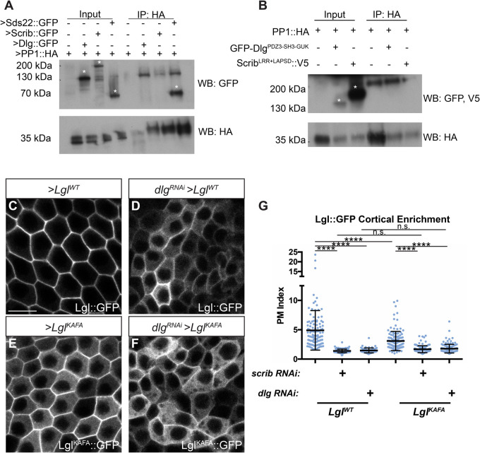
Given the conserved PP1-interaction motifs in both Scrib and Dlg, we tested whether a physical interaction occurs, first using in vivo co-immunoprecipitation (co-IP) assays with transgenic proteins overexpressed in follicle cells. In this assay, we could detect co-purification of transgenic Sds22, a known PP1 binding partner (Fig. 2A) (Ceulemans et al., 2002). However, Scrib or Dlg were not detected co-purifying with PP1 (Fig. 2A). We were also unable to reliably detect interaction between Scrib or Dlg and PP1 when combinations of these proteins were overexpressed in cultured Drosophila S2 cells, even when cells were crosslinked prior to lysis to stabilize weak and transient protein-protein interactions (Fig. 2B).
Fig. 2.
Scrib and Dlg regulate Lgl independently of PP1. (A) Co-IP of transgenic Scrib or Dlg and PP1 from follicle cells fails to detect an interaction, although interaction between PP1 and Sds22 is robustly captured. (B) Co-IP of overexpressed Dlg or Scrib and PP1 from S2 cells following in situ crosslinking also failed to reliably detect interaction between these proteins. Asterisks in A and B indicate relevant bands. (C,D) LglWT membrane localization (C) is severely disrupted by dlg RNAi (D). (E,F) LglKAFA (E) has increased cytoplasmic localization compared to LglWT and is further decreased by dlg RNAi (F). (G) Quantification of Lgl membrane localization. Scale bar: 10 µm. Clones in C-F are flip-out GAL4 clones. (G) One-way ANOVA with Tukey's multiple comparisons test. Error bars represent s.d. PM Index=cortical/cytoplasmic intensity. Data points are measurements from individual cells. n.s. (not significant) P>0.05, ****P<0.0001.
Scrib and Dlg can regulate Lgl independently of PP1
As an additional functional test of the relationship between Lgl regulation by PP1 and its regulation by Scrib and Dlg, we made use of a UAS-driven, mutant Lgl protein that cannot interact with PP1 (LglKAFA) (Moreira et al., 2019). When expressed in the follicle epithelium, LglKAFA exhibits an increased cytoplasmic distribution, presumably resulting from its impaired ability to be dephosphorylated by PP1 and return to the membrane (Fig. 2C,E,G) (Moreira et al., 2019). When expressed in scrib- or dlg-depleted cells rather than WT cells, LglKAFA cortical localization was even further reduced, suggesting that, even in the absence of PP1 regulation, LglKAFA is still dependent on Scrib and Dlg for its localization (Fig. 2F,G). Interestingly, there was no difference between LglKAFA and LglWT cortical levels in scrib- or dlg-depleted cells (Fig. 2D,F,G). Furthermore, overexpression of PP1 in scrib- or dlg-depleted cells did not rescue Lgl mislocalization (Fig. S1F-H). Together, these data suggest that Scrib and Dlg's polarity functions include a PP1-independent component.
A cell culture assay for Scrib recruitment
Although we did not find evidence to functionally implicate PP1 in Scrib/Dlg activity, mutating the Dlg RVxF motif nevertheless caused severe loss of function. We therefore tested other, PP1-independent functions of this protein region. In addition to protecting Lgl, Dlg also stabilizes Scrib at the cell cortex (Khoury and Bilder, 2020; Ventura et al., 2020). We sought to precisely define the regions of Dlg required to recruit Scrib.
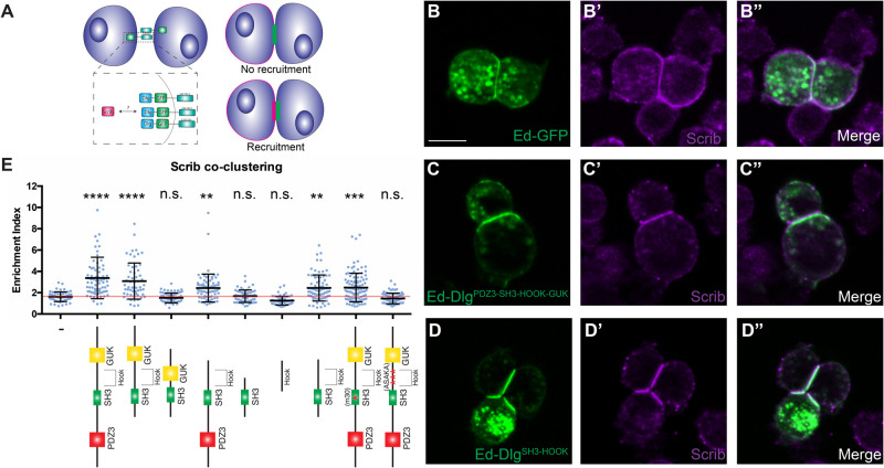
Dlg is a member of the Membrane Associated Guanylate Kinase (MAGUK) family of molecular scaffolds, the members of which are defined by the presence of multiple protein-protein binding domains (Zhu et al., 2016). MAGUK proteins contain one or more PDZ domains (three in the case of Dlg), followed by a non-canonical SH3 domain and a catalytically inactive GUK domain (Fig. 1A). A conserved region of variable length, called the HOOK domain, lies between SH3 and GUK. To identify regions of the Dlg protein required for its Scrib recruitment activity, we adapted a previously described induced polarity assay using cultured Drosophila S2 cells (Johnston, 2020; Johnston et al., 2009). In this method, transgenic expression of the extracellular domain of the homotypic cell adhesion protein Echinoid (Ed) is used to cluster cells due to adhesion between the extracellular domains of Ed on adjacent cells. This adhesion creates a polarized cortical domain at the contact point between the Ed-expressing cells that can be used to study polarity processes in a simplified system. By fusing a protein of interest to the Ed intracellular domain, one can create polarized localization of any target. Importantly, S2 cells do not exhibit native cell-cell adhesion or polarity, although they express a subset of polarity proteins (including Scrib and Dlg) at low to moderate levels. We reasoned that fusing Dlg to Ed would create a discrete cortical domain of polarized Dlg that could recruit endogenous Scrib (Fig. 3A). Indeed, an Ed-fused fragment of Dlg encompassing its PDZ3-SH3-HOOK-GUK domains was able to robustly recruit Scrib to the polarity site, compared to a control construct containing Ed alone (Fig. 3B-C″,E). Because the same Dlg fragment can provide polarity function in vivo (Hough et al., 1997; Lu et al., 2021), the Ed assay provides a useful platform to dissect regions mediating Scrib recruitment by Dlg.
Fig. 3.
DlgSH3-HOOK is sufficient for Scrib localization in an induced polarity system. (A) Cartoon of S2 induced polarity assay. Polarizing Dlg by fusion to Ed enables testing of Scrib recruitment in a minimal synthetic system. (B) S2 cells expressing Ed-GFP can be clustered by adhesion between Ed molecules, but this does not alter Scrib localization. (C) When DlgPDZ3-SH3-HOOK-GUK is fused to Ed, it creates a polarity crescent at the contact site that is able to recruit Scrib. (D) A minimal fragment, DlgSH3-HOOK retains the ability to recruit Scrib to the contact site. (E) Quantification of Scrib recruitment to the polarity site in various Ed-Dlg constructs schematized below. DlgPDZ3-SH3-HOOK-GUK is able to enrich Scrib, while the Ed-GFP negative control cannot. The HOOK and SH3 domains are necessary and, when in combination, sufficient to recruit Scrib. Statistical tests are versus the Ed-GFP negative control construct. Red line denotes the average for the Ed-GFP negative control and indicates no Scrib contact site enrichment. Scale bar: 10 µm. (E) One-way ANOVA with Tukey's multiple comparisons test. Error bars indicate s.d. Data points are individual cell clusters. Enrichment index=contact site/non-contact site intensity. n.s. (not significant) P>0.05, **P<0.01, ***P<0.001, ****P<0.0001.
We tested a series of Ed-Dlg constructs encompassing additional domain truncations and mutations. Consistent with in vivo experiments on Dlg function, we found that PDZ3 and GUK were individually dispensable for Scrib recruitment, although GUK deletion resulted in a mild impairment of Scrib recruitment compared to the full-length construct (Fig. 3E) (Hough et al., 1997; Khoury and Bilder, 2020; Lu et al., 2021). The dlgm30 allele is a missense mutation substituting a leucine for proline in the SH3 domain and results in strong loss of function in vivo. An Ed-Dlg construct mimicking the dlgm30 mutation retained partial ability to recruit Scrib, unlike the in vivo situation (Khoury and Bilder, 2020), although it was significantly worse than the WT construct (Fig. 3E). We generated a second SH3 domain mutation, designed to disrupt conserved residues that would make up the PxxP binding region of a canonical SH3 domain, and found that this construct also disrupted the ability of Ed-Dlg to recruit Scrib (Fig. S3A,B).
Dlg SH3-HOOK is a minimal fragment necessary and sufficient for polarity in vivo
We then turned to the HOOK domain, where the RVxF motif resides. A HOOK-deleted Dlg construct fails to rescue imaginal disc polarity in vivo, but this protein localizes to the nucleus rather than the plasma membrane, limiting interpretation (Hough et al., 1997). In the S2 cell induced polarity assay, the HOOK domain was essential, as a HOOK-deleted construct failed to recruit Scrib (Fig. 3E). Interestingly, the same failure was seen with a construct carrying the DlgASAKA mutation (Fig. 3E). We then tested individual HOOK residues and found that even single amino acid mutations in the RVxF consensus sequence resulted in equivalent disruption of Scrib recruitment activity (Fig. S3A,B). In contrast, mutations in evolutionarily conserved residues at the opposite, C-terminal end of the HOOK domain had no effect, suggesting that the HOOK N-terminal region contains the major functional elements (Fig. S3A,B). Finally, since single amino acid changes in either the HOOK or SH3 domains disrupt Scrib recruitment, we tested the sufficiency of the domains. Neither domain displayed function alone, but strikingly a fragment composed of SH3-HOOK was sufficient to mediate Scrib clustering (Fig. 3D-E).
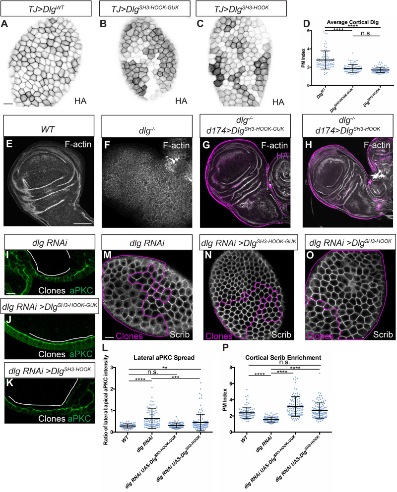
We therefore assessed whether a UAS-driven version of this Dlg construct (DlgSH3-HOOK) was also sufficient for function in vivo. We compared it to a Dlg protein lacking all three PDZ domains, which Lu et al. (2021) recently demonstrated was sufficient to provide polarity and tumor suppressive activity in dlg-deficient follicles and imaginal discs. Our analogous construct (DlgSH3-HOOK-GUK) replicated this result: polarity, architecture, and growth control of discs were also restored when overexpressed in a dlg null mutant background (Fig. 4E-G) (Lu et al., 2021). Importantly, overexpressing the smaller DlgSH3-HOOK also rescued polarity, architecture and growth control in dlg-deficient imaginal discs (Fig. 4E,F,H). We confirmed this result by taking advantage of a validated dlg RNA interference (RNAi) line that targets the PDZ2-encoding sequences, allowing us to deplete the endogenous protein but not our transgenes, which lack this domain. Depletion of dlg in the posterior compartment of wing imaginal discs generates mispolarized tumors, but co-expression of DlgSH3-HOOK efficiently rescued epithelial polarity, architecture and growth, to a degree indistinguishable from the rescue provided by DlgSH3-HOOK-GUK (Fig. S4). We then tested the constructs in the follicle epithelium. Both transgenic proteins localized to the basolateral membrane, albeit at reduced levels compared to WT Dlg (Fig. 4A-D). When expressed in dlg-depleted follicle cells (Fig. 4I), both DlgSH3-HOOK-GUK and DlgSH3-HOOK reduced the basolateral expansion of aPKC to ameliorate polarity and restore monolayer organization (Fig. 4J-L), although the former was more efficient than the latter. Even in cases with altered epithelial architecture, DlgSH3-HOOK restored Scrib recruitment to WT levels, as did DlgSH3-HOOK-GUK (Fig. 4M-P). These data support the conclusion that the SH3 and HOOK domains mediate both Dlg's Lgl protection and Scrib recruitment activities and that they together constitute a minimal functional unit of the protein that can support epithelial polarity, albeit less efficiently in some tissues than others.
Fig. 4.
Dlg SH3 and HOOK domains are sufficient for function in vivo. (A-C) Like WT Dlg (A), DlgSH3-HOOK-GUK (B) and DlgSH3-HOOK (C) localize to the basolateral membrane in follicle cells. All constructs contain an HA epitope tag used for detection. (D) Quantification of cortical localization in A-C. (E-H) Compared to WT (E) and dlg null mutants (F), DlgSH3-HOOK-GUK (G) and DlgSH3-HOOK (H) fully rescue polarity and epithelial architecture in wing imaginal discs. (I-L) In monolayered dlg-depleted follicle cells (I), both DlgSH3-HOOK-GUK (J) and DlgSH3-HOOK (K) provide polarity-rescuing activity, quantitated in L. Full restoration of monolayering is more efficient by DlgSH3-HOOK-GUK than DlgSH3-HOOK: 89.5% (n=38) of DlgSH3-HOOK-GUK show no regions of multilayering in rescued follicles, compared to 20.5% (n=39) of DlgSH3-HOOK rescued follicles and 0% (n=37) of follicles with dlg-depleted clones alone. (M-P) Both DlgSH3-HOOK-GUK (N) and DlgSH3-HOOK (O) fully rescue loss of cortical Scrib seen in dlg-depleted cells (M), quantified in P. Scale bars: 10 µm (A,I,M), 100 µm (E). White or magenta lines indicate clones of given genotypes; Clones in I-K and M-O are flip-out GAL4 clones. (D,L,P) One-way ANOVA with Tukey's multiple comparisons test. Error bars indicate s.d. PM Index=cortical/cytoplasmic intensity. aPKC spread is a ratio of lateral:apical fluorescence intensity. Data points are individual cell measurements. n.s. (not significant) P>0.05, **P<0.01, ***P<0.001, ****P<0.0001.
The Dlg SH3-HOOK unit regulates Scrib localization, and SH3 provides an additional polarity function
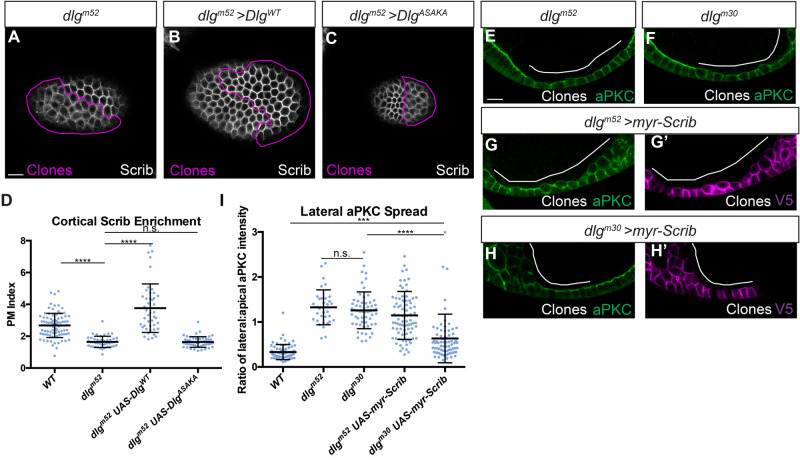
Finally, we investigated the relationship between Dlg's Scrib recruitment and Lgl protection activities. Nuclear localization of previous HOOK deletion constructs prevented conclusions about its role in the former process. We therefore complemented dlg null mutant follicle cells in vivo with our HOOK domain missense mutant construct and found that, as in the S2 cell assays, it fails to rescue Scrib cortical localization (Fig. 5A-D). The SH3 domain is required for Scrib recruitment in vivo, since dlgm30 homozygous cells are defective in recruiting Scrib to the cortex (Khoury and Bilder, 2020). To determine if the SH3 domain is required only for Scrib recruitment, we attempted to bypass its function by expressing a membrane-tethered Scrib protein (myr-Scrib) in dlgm30 homozygous follicle cells. Strikingly, this combination yielded a partial rescue of polarity, as assessed by degree of aPKC mislocalization, compared to myr-Scrib in dlg null cells (Fig. 5E-I). Whereas our previous data show that both SH3 and HOOK domains are required for Scrib localization, this experiment suggests that regions including HOOK cooperate with Scrib to provide Lgl ‘protection’ activity that can be further enhanced by an intact SH3.
Fig. 5.
Dlg SH3-HOOK is primarily required to regulate Scrib localization. (A) dlgm52 null mutant cells show reduced cortical localization of Scrib. (B) Scrib mislocalization is rescued by expression of DlgWT. (C) Scrib mislocalization is not rescued by expression of DlgASAKA. (D) Quantification of Scrib localization in A-C. (E,F) Both dlgm52 null mutants (E) and dlgm30 SH3 point mutant cells (F) lose polarity and mislocalize aPKC. (G,H) Preventing Scrib mislocalization by cortical tethering (myr-Scrib) partially suppresses the polarity loss phenotypes of dlgm30 SH3 mutant cells (H) but not dlgm52 null mutant cells (G). (G′,H′) myr-Scrib contains a V5 epitope tag used for detection. (I) Quantification of aPKC mislocalization in (E-H). Scale bars: 10 µm. Magenta or white lines indicate MARCM clones of given genotypes. (D,I) One-way ANOVA with Tukey's multiple comparisons test. Error bars indicate s.d. PM Index=cortical/cytoplasmic intensity. aPKC spread is a ratio of lateral:apical fluorescence intensity. Data points are individual cell measurements. n.s. (not significant) P>0.05, ***P<0.001, ****P<0.0001.
How does SH3-HOOK regulate Scrib recruitment? In cultured mammalian cells, it was recently shown that the Scrib LRR and LAPSD domains, which are both necessary and sufficient for polarity function in Drosophila (Albertson et al., 2004; Bonello et al., 2019; Choi et al., 2019; Khoury and Bilder, 2020; Zeitler et al., 2004), can co-immunoprecipitate with Dlg1 (Troyanovsky et al., 2021). We tested this Scrib fragment in the S2 induced polarity assay but could not detect recruitment of endogenous Dlg by Ed-ScribLRR+LAPSD (Fig. S5A-C). The ability of Dlg to recruit Scrib in this system, but not vice versa, parallels in vivo data showing Scrib localization to be dependent on Dlg, but Dlg localization to be largely independent of Scrib (Khoury and Bilder, 2020; Lu et al., 2021). We were also unable to co-immunoprecipitate transgenic ScribLRR+LAPSD and DlgPDZ3-SH3-HOOK-GUK from S2 cells (Fig. S5D), even with crosslinking and by increasing the starting material used by severalfold. Combined with our induced polarity and in vivo genetic experiments, these data support the idea that Dlg recruits Scrib via its SH3-HOOK domains, but that this recruitment may not reflect direct physical binding between the two proteins.
DISCUSSION
The molecular mechanism of Scrib module function has been a longstanding challenge in the study of cell polarity. Much work has focused on identifying binding partners of Scrib module proteins (reviewed in Stephens et al., 2018), with less attention given to the relationships that exist within the Scrib module itself. Here, we perform fine-grained functional analysis of the Dlg protein, defining its minimal required domains. These experiments identified a critical SH3-HOOK module that facilitates Scrib localization and is both necessary and sufficient for Dlg's polarity activity in vivo.
Our search for Scrib module effectors yielded PP1 as an appealing candidate for Lgl regulation. Such a role would be consistent with studies from mammalian cell culture, where both Scrib and Dlg have been found to bind to PP1 (Hendrickx et al., 2009; Nagasaka et al., 2013; Troyanovsky et al., 2021; Van Campenhout et al., 2011; Young et al., 2013), and have been proposed to act as targeting factors that direct PP1 to specific substrates. We failed to find evidence for a physical complex between Scrib, Dlg and PP1 in Drosophila, and our data on mutating PP1-binding consensus sequences support alternative functions for these motifs, unrelated to PP1 binding. Although we cannot rule out that PP1-Scrib module interactions occur in Drosophila at a low affinity, we note that a direct role for such interactions in regulating mammalian cell polarity remains to be demonstrated. Moreover, our data demonstrate that Scrib and Dlg influence Lgl localization at least partially independently of PP1, which is consistent with the weak phenotype of pp1 compared to scrib module mutants (Fig. S1A,B) (Moreira et al., 2019).
Our data using LglKAFA are consistent with two possible roles of Scrib and Dlg in polarity. First, Scrib and Dlg could limit Lgl phosphorylation through partners other than PP1, since Lgl mislocalization in dlg-depleted cells can be restored by co-depletion of aPKC or by mutating Lgl phosphorylation sites to alanine (Khoury and Bilder, 2020; Ventura et al., 2020). Second, Scrib and Dlg's regulation of Lgl could involve a phosphorylation-independent component. Consistent with this possibility, we found that a non-phosphorylatable Lgl protein (LglS5A) still exhibited reduced cortical localization in scrib- and dlg-depleted cells (Fig. S6). These findings draw parallels with the Caenorhabditis elegans zygote, where PAR-2 can ‘protect’ PAR-1 both by physical binding as well as competing for aPKC's activity to reduce PAR-1 phosphorylation (Ramanujam et al., 2018). However, evidence for physical binding of Lgl with Scrib or Dlg in Drosophila is currently lacking, outside of a report of binding to the polarity-dispensable GUK domain (Zhu et al., 2014). Thus, although speculative, the analogy of PAR-1 regulation to Lgl ‘protection’ may provide an appealing basis for future experiments. Lastly, although Scrib and Dlg do not require PP1 to regulate Lgl, it is possible that PP1 requires Scrib and Dlg to do so, since the degree of Lgl mislocalization in scrib- and dlg-depleted cells is not enhanced by removing PP1-dependent regulation (LglKAFA, Fig. 2G).
Dlg is required for Scrib recruitment, proximity assays reliably detect Scrib near Dlg (Nakajima et al., 2019; Sharifkhodaei et al., 2019; Sharp et al., 2021), and an optogenetic relocalization experiment showed that either Scrib or Dlg can induce relocation of the other protein (Ventura et al., 2020). However, we were unable to biochemically detect a Scrib-Dlg complex in extracts from follicles or when the proteins were overexpressed in cell culture. A recent mass spectrometry dataset from Drosophila embryos also failed to detect Scrib in Dlg immunoprecipitation (IP) samples and vice versa (Nakajima et al., 2019). In flies, biochemical evidence for such a complex involves co-IP from synapse-containing tissues such as larval muscle and adult brains (Mathew et al., 2002; Rui et al., 2017); in mammalian cells, evidence for co-IP comes from cultured cells (Awadia et al., 2019; Troyanovsky et al., 2021). Several of the above cases involve mutual binding partners, and require that partner for co-IP, such as Gukholder at the neuronal synapse and SGEF in epithelia (Awadia et al., 2019; Mathew et al., 2002). Given the inconsistent evidence for biochemical interaction, we feel that it is prudent to continue to refer to the Scrib proteins as a ‘module’ rather than a complex.
Our studies identify a critical motif in the Dlg N-terminal HOOK domain that is, in combination with the SH3 domain, required for polarity and Scrib localization. In the wing imaginal disc, SH3 and HOOK are alone sufficient to support full polarity function. In follicle cells, SH3 and HOOK are also sufficient to support Scrib recruitment. Polarity activity in this tissue is less efficient, with full architectural rescue that is less penetrant than with the SH3-HOOK-GUK construct. The SH3-HOOK-rescued follicle cell clones resemble clones mutant for GUK-truncated dlg alleles, where polarity in cells retaining epithelial structure is largely normal (Khoury and Bilder, 2020). A follicle-specific role for the GUK domain may involve its known function in spindle orientation, which is required in the follicle but not the wing disc epithelium (Bellaïche et al., 2001; Bergstralh et al., 2013, 2016). However, other domains conserved among MAGUKs that are dispensable in our epithelial overexpression assays play important roles in tissues and contexts not examined here, such as in synaptic organization in neurons (Zhu et al., 2016). Overall, the data demonstrate that SH3 and HOOK domains alone are the minimal elements required for Dlg to recruit cortical Scrib and provide at least partial epithelial polarity function.
How might the SH3 and HOOK domains operate? MAGUK-family SH3 domains are ‘non-canonical’ in that they cannot bind the polyproline ligands bound by typical SH3 domains, because they lack key residues in the PxxP binding pocket (McGee et al., 2001). The HOOK domain is a conserved linker of variable length between the SH3 and GUK domains (Zhang et al., 2013) that is thought to create interdomain allostery, facilitating an intramolecular interaction that enables functions that the individual domains lack in isolation (McCann et al., 2012; McGee and Bredt, 1999; McGee et al., 2001; Rademacher et al., 2019; Zhang et al., 2013). One demonstrated function of HOOK domains is to negatively regulate binding of certain GUK domain ligands, presumably by influencing the SH3-GUK interaction (Golub et al., 2017; Marcette et al., 2009; Qian and Prehoda, 2006). However, the dispensability of the GUK domain for polarity in vivo and in the S2 induced polarity assay reveals that such regulation is not important for Scrib recruitment and polarity function. A second function of HOOK is to mediate electrostatic binding to the membrane (Lu et al., 2021), but our experiments mutating non-polar amino acids and supplying membrane tethering in S2 cells show that additional SH3-dependent functions reside in HOOK. Our single amino acid resolution mutant analysis reveals that the HOOK N-terminus is essential to this function, and an appealing model is that it works with the SH3 domain to bind an additional scaffolding factor to permit Scrib recruitment. Once Scrib has been recruited, SH3-HOOK and Scrib are together competent to protect Lgl through an unknown cooperative activity that defines basolateral identity. In support of this model, we find that constitutively tethering Scrib to the membrane can partially bypass a dlg SH3 mutant allele, demonstrating that a primary function of SH3 is to recruit Scrib. To our knowledge, this is the first case where a Scrib construct can rescue a dlg mutant, providing further evidence for the cooperative nature of Scrib module function in basolateral polarity and pointing to the Dlg SH3-HOOK as a primary mediator of this. Exploring this model will be an important aspect of future studies.
In sum, our in-depth interrogation of the core polarity regulator Dlg defines a minimally sufficient fragment composed of the SH3-HOOK domains, as well as single amino acids in the HOOK domain, that are essential for polarity function. These domains cooperatively recruit Scrib to the cell cortex and supply an additional function that is independent of PP1 that enables Lgl to antagonize aPKC. These data advance our understanding of how basolateral polarity is established and contribute a significant step towards mechanistic understanding of the Scrib module machinery.
MATERIALS AND METHODS
Fly stocks and genetics
Drosophila stocks were raised on cornmeal molasses food at 25°C. Mutant alleles and transgenic lines used are listed in Table S1. Genotypes of Drosophila lines used in the figure panels are listed in Table S3. Follicle cell mutant clones were generated using the MARCM technique (Germani et al., 2018; Lee and Luo, 1999) with hsFLP induction by 37°C heat shock for 1 h on three consecutive days beginning at 120 h after egg deposition (AED) for FRT19A stocks, and two consecutive days for FRT82B stocks. For clonal GAL4 expression (Germani et al., 2018), larvae were heat shocked once for 13 min at 37°C 120 h AED to generate flip out clones. For all clonal experiments, newly eclosed females were fed with yeast and dissected 3 days after eclosion. Unless otherwise noted, pan-follicle cell expression used traffic jam-GAL4 and temperature-sensitive tub-GAL80ts. After 1-2 days on yeast, newly eclosed females were shifted to 29°C for 3 days to induce GAL4 expression before ovary dissection.
Molecular cloning
To generate UAS-DlgASAKA::HA, pUASTattB was digested with EcoRI and XbaI and overlapping fragments amplified from the Dlg cDNA were assembled using Gibson assembly. For the UAS-DlgSH3-HOOK-GUK::HA and UAS-DlgSH3-HOOK::HA constructs, pUASTattB was digested with XhoI and XbaI, and fragments encompassing the appropriate domains were amplified from the pUASTattB-DlgWT vector and assembled via Gibson assemnbly. UAS-myr-ScribTAAA/RAGA::V5 was generated from UAS-myr-Scrib::V5 by first using the NEBuilder Gibson Assembly kit to insert a KpnI site 5′ of the myr signal. Then, the resulting plasmid was digested with KpnI and AgeI, and fragments containing the desired mutations were amplified and assembled using the NEBuilder Gibson Assembly kit. To generate the Ed-Dlg constructs for S2 cell expression, mutations of interest were introduced into the pMT-Ed::GFP::DlgPDZ3-SH3-HOOK-GUK plasmid (Garcia et al., 2014) using the NEBaseChanger site directed mutagenesis kit as directed by the manufacturer (NEB). To generate the cytosolic pMT-GFP:: DlgPDZ3-SH3-HOOK-GUK and pMT-ScribLRR+LAPSD::V5 plasmids, the corresponding regions of the Dlg and Scrib cDNAs were amplified from the pMT-Ed::GFP::DlgPDZ3-SH3-HOOK-GUK and pUASTattB-myr-Scrib::V5 plasmids, respectively. Fragments were then assembled using the NEBuilder HiFi DNA assembly kit (NEB) as instructed into XhoI/EcoRI or AgeI/EcoRI linearized pMT-His-V5 backbone, respectively. The Dlg sequence used in this study is NP_996405.1 and the Scrib sequence is NP_001036761.3. Primers used for cloning are given in Table S1. The deletions made with respect to the Dlg and Scrib reference protein sequences are given in Table S2.
S2 cell culture and induced polarity assay
S2 cells were obtained from the University of California, Berkeley Cell Culture Facility and cultured using standard methods at 25°C in Schneider's medium (Invitrogen) supplemented with 10% fetal bovine serum and 1% penicillin/streptomycin. Transfections were performed using the Effectene kit (Qiagen) according to the manufacturer's instructions. 2×10⁶ cells per well of a six-well plate were transfected with 500 ng of DNA per plasmid. Cells were incubated in transfection complexes for 48 h and then switched into fresh medium containing 0.5 mM CuSO4 to induce expression of the metallothionein promoter for 48 h before experiments. The induced polarity assay was performed essentially as described previously (Johnston, 2020). After 48 h of induction, transfected S2 cells were resuspended in 3 ml of fresh medium containing 0.5 mM CuSO4. The cell suspensions were agitated in an orbital shaker at 150 rpm in six-well plates for 2 h to induce cell clusters. 1 ml per condition of the clustered cell suspension was then allowed to settle on poly-D-lysine-coated coverslips and adhere for 30 min. The cells were then fixed for 20 min in 4% paraformaldehyde (PFA) and processed for immunofluorescence as described below.
Immunofluorescence and microscopy
Ovaries were dissected in PBS, and individual ovarioles were separated prior to fixation in 4% PFA for 20 min. Wing imaginal discs were dissected from wandering L3 larvae in PBS and fixed for 20 min in 4% PFA. Samples were blocked for 30 min to 1 h in 0.1% PBS-T containing 4% normal goat serum and 1% BSA before staining with primary antibodies overnight at 4°C in blocking buffer. Following three washes in PBS-T, samples were incubated in 1:400 fluorescent secondary antibodies (Invitrogen) for 2 h at room temperature. Primary antibodies used are given in Table S1. Imaging was performed on either a Zeiss LSM700 inverted point scanning confocal microscope or an upright Zeiss Axio Imager 2 microscope with Apotome 2 using Plan Apochromat 20×/NA 0.8 or LD C-Apochromat 40×/NA 1.1 W objectives. Uncropped confocal images were 1024×1024 pixels with 2 line averages, and widefield images were 512×512 pixels.
Image analysis and quantification
Image processing and quantification was performed using FIJI software (Schindelin et al., 2012). To quantify Scrib, Dlg and Lgl cortical localization, a 1.17 µm-wide rectangular region of interest (ROI) spanning a single cell-cell boundary and a second identical-width ROI were measured in en face sections. The ratio of membrane:cytoplasmic fluorescence intensity was computed to define the PM Index (Lu et al., 2021). To quantify aPKC localization, lines along the apical and basolateral membranes were measured in FIJI, and the ratio of basolateral:apical intensity was computed to give a measure of lateral mislocalization. To quantify enrichment at S2 cell polarity domains, a 0.39 µm-wide rectangular ROI spanning the contact site between two S2 cells and a second ROI on a non-contacting section of the membrane were measured, and the ratio of contact:non-contact fluorescence intensity was computed to give the Enrichment Index. In all wcases, measurements were taken from single cells, with averages were calculated for each condition. Figures were assembled with Adobe Illustrator.
Co-IP and western blotting
Ovary tissue was lysed in ice cold IP buffer (10 mM Tris, 150 mM NaCl, 0.5 mM EDTA, 0.5% NP-40) (Nakajima et al., 2019) by homogenization. S2 cells were resuspended in ice-cold IP buffer and lysed for 30 min at 4°C by nutation. Lysates were then cleared by centrifugation at 13,400 g for 20 min at 4°C. Following protein concentration determination by BCA assay (Thermo Fisher Scientific), 200 µg of protein per sample was then loaded onto antibody-conjugated Protein G Dynabeads (Thermo Fisher Scientific) and rotated overnight at 4°C. The following day, antibody-bead complexes were washed three times with lysis buffer before eluting the samples by boiling for 10 min in 4× loading dye (Bio-Rad) containing 10% β-mercaptoethanol. 60 µg ‘input’ samples were also prepared in the same way by boiling in β-mercaptoethanol-containing loading dye. Antibodies used for IP are listed in Table S1. To induce crosslinking, cells were washed several times with ice-cold sterile PBS to remove traces of culture media. The cells were then incubated in 2 mM disuccinimidyl suberate (DSS; Thermo Fisher Scientific) in PBS for 30 min at room temperature. The crosslinking reaction was then quenched by adding Tris to a final concentration of 20 mM and incubating for 15 min at room temperature.
Western blotting was performed as previously described (de Vreede et al., 2018). Proteins were separated by SDS-PAGE on 7.5% TGX precast gels (Bio-Rad) before being blotted onto methanol-activated 0.45 µm PVDF membranes (GE Healthcare) at 300 mA for 1 h. Membranes were then blocked for 1 h with TBS-T containing 3% BSA before probing with primary antibodies overnight at 4°C. The following day, membranes were washed three times in TBS-T before being incubated in 1:2000 secondary antibodies in blocking buffer for 2 h at room temperature. Following three more washes, blots were imaged by ECL chemiluminescence (WesternBright) on HyBlot CL autoradiography film (Denville Scientific). Primary antibodies are listed in Table S1.
Multiple sequence alignment
Protein sequence alignments were created with Clustal Omega (Madeira et al., 2019) and visualized with SnapGene Viewer. The UniProt sequences used for Dlg were as follows: Homo sapiens Q12959, Mus musculus Q811D0, Rattus norvegicus Q62696, Danio rerio Q5PYH6, Xenopus tropicalis Q28C55 and Drosophila melanogaster P31007. For Scrib, the sequences used were as follows: H. sapiens Q14160, M. musculus Q80U72, R. norvegicus D3ZWS0, D. rerio A0A1L1QZF0, X. tropicalis XP_031759453.1 and D. melanogaster Q7KRY7.
Statistical analyses
The statistical tests used for each experiment are described in the corresponding figure legends. No data points were excluded. For each experiment, ovaries from at least five females were examined, with at least ten ovarioles being analyzed. All plots show individual data points for all measurements used. All experiments were repeated a minimum of two times. Definitions of significance used are as follows: n.s. (not significant) P>0.05, *P<0.05, **P<0.01, ***P<0.001, ****P<0.0001. Data were analyzed using Microsoft Excel and GraphPad Prism 6.
Supplementary Material
Acknowledgements
We thank E. Morais-de-Sá, C. Johnston and Y. Hong for fly stocks and reagents; L. Mathies for cloning the UAS-Dlg constructs; and K. Prehoda, Y. Hong, M. Kitaoka, and the Hariharan and Bilder laboratories for helpful discussions. We thank the University of California, Berkeley Cell Culture Facility for help with S2 cell experiments. Stocks obtained from the Bloomington Drosophila Stock Center (NIH P40OD018537) and resources from the Drosophila Genomics Resource Center (NIH 2P40OD010949) were used in this study.
Footnotes
Competing interests
The authors declare no competing or financial interests.
Author contributions
Conceptualization: M.J.K., D.B.; Investigation: M.J.K.; Writing - original draft: M.J.K., D.B.; Visualization: M.J.K.; Supervision: D.B.; Funding acquisition: D.B.
Funding
This work was supported by the National Institutes of Health (R35 GM130388 to D.B.). and the American Heart Association (Predoctoral Fellowship 20PRE35120150 to M.J.K.). Open Access funding provided by University of California Libraries. Deposited in PMC for immediate release.
Data availability
The Dlg APEX2 proteomics dataset was previously published (Sharp et al., 2021) and is available from MassIVE proteomics repository and ProteomeXchange using accession numbers MSV000087186 and PXD025378. All other reagents and data will be made available upon request.
References
- Albertson, R., Chabu, C., Sheehan, A. and Doe, C. Q. (2004). Scribble protein domain mapping reveals a multistep localization mechanism and domains necessary for establishing cortical polarity. J. Cell Sci. 117, 6061-6070. 10.1242/jcs.01525 [DOI] [PubMed] [Google Scholar]
- Awadia, S., Huq, F., Arnold, T. R., Goicoechea, S. M., Sun, Y. J., Hou, T., Kreider-Letterman, G., Massimi, P., Banks, L., Fuentes, E. J.et al. (2019). SGEF forms a complex with Scribble and Dlg1 and regulates epithelial junctions and contractility. J. Cell Biol. 218, 2699-2725. 10.1083/jcb.201811114 [DOI] [PMC free article] [PubMed] [Google Scholar]
- Bailey, M. J. and Prehoda, K. E. (2015). Establishment of Par-polarized cortical domains via phosphoregulated membrane motifs. Dev. Cell 35, 199-210. 10.1016/j.devcel.2015.09.016 [DOI] [PMC free article] [PubMed] [Google Scholar]
- Bellaïche, Y., Radovic, A., Woods, D. F., Hough, C. D., Parmentier, M.-L., O'Kane, C. J., Bryant, P. J. and Schweisguth, F. (2001). The partner of inscuteable/discs-large complex is required to establish planar polarity during asymmetric cell division in Drosophila. Cell 106, 355-366. 10.1016/S0092-8674(01)00444-5 [DOI] [PubMed] [Google Scholar]
- Bergstralh, D. T., Lovegrove, H. E. and St Johnston, D. (2013). Discs large links spindle orientation to apical-basal polarity in drosophila epithelia. Curr. Biol. 23, 1707-1712. 10.1016/j.cub.2013.07.017 [DOI] [PMC free article] [PubMed] [Google Scholar]
- Bergstralh, D. T., Lovegrove, H. E., Kujawiak, I., Dawney, N. S., Zhu, J., Cooper, S., Zhang, R. and St Johnston, D. (2016). Pins is not required for spindle orientation in the Drosophila wing disc. Development 143, 2573-–22581.. 10.1242/dev.135475 [DOI] [PMC free article] [PubMed] [Google Scholar]
- Betschinger, J., Mechtler, K. and Knoblich, J. J. A. (2003). The Par complex directs asymmetric cell division by phosphorylating the cytoskeletal protein Lgl. Nature 422, 326-330. 10.1038/nature01486 [DOI] [PubMed] [Google Scholar]
- Bonello, T. T. and Peifer, M. (2018). Scribble: a master scaffold in polarity, adhesion, synaptogenesis, and proliferation. J. Cell Biol. 218, 742-756. 10.1083/jcb.201810103 [DOI] [PMC free article] [PubMed] [Google Scholar]
- Bonello, T. T., Choi, W. and Peifer, M. (2019). Scribble and discs-large direct initial assembly and positioning of adherens junctions during establishment of apical-basal polarity. Development 146, dev180976. 10.1242/dev.180976 [DOI] [PMC free article] [PubMed] [Google Scholar]
- Buckley, C. E. and St Johnston, D. (2022). Apical–basal polarity and the control of epithelial form and function. Nat. Rev. Mol. Cell Biol. 10.1038/s41580-022-00465-y [DOI] [PubMed] [Google Scholar]
- Ceulemans, H., Vulsteke, V., De Maeyer, M., Tatchell, K., Stalmans, W. and Bollen, M. (2002). Binding of the concave surface of the Sds22 superhelix to the α4/α5/α6-triangle of protein phosphatase-1. J. Biol. Chem. 277, 47331-47337. 10.1074/jbc.M206838200 [DOI] [PubMed] [Google Scholar]
- Choi, J., Troyanovsky, R. B., Indra, I., Mitchell, B. J. and Troyanovsky, S. M. (2019). Scribble, Erbin, and Lano redundantly regulate epithelial polarity and apical adhesion complex. J. Cell Biol. 218, 2277-2293. 10.1083/jcb.201804201 [DOI] [PMC free article] [PubMed] [Google Scholar]
- de Vreede, G., Morrison, H. A., Houser, A. M., Boileau, R. M., Andersen, D., Colombani, J. and Bilder, D. (2018). A Drosophila Tumor Suppressor Gene Prevents Tonic TNF Signaling through Receptor N-Glycosylation. Dev. Cell 45, 595-605.e4. 10.1016/j.devcel.2018.05.012 [DOI] [PMC free article] [PubMed] [Google Scholar]
- Dong, W., Zhang, X., Liu, W., Chen, Y.-J., Huang, J., Austin, E., Celotto, A. M., Jiang, W. Z., Palladino, M. J., Jiang, Y.et al. (2015). A conserved polybasic domain mediates plasma membrane targeting of Lgl and its regulation by hypoxia. J. Cell Biol. 211, 273-286. 10.1083/jcb.201503067 [DOI] [PMC free article] [PubMed] [Google Scholar]
- Flores-Benitez, D. and Knust, E. (2016). Dynamics of epithelial cell polarity in Drosophila: how to regulate the regulators? Curr. Opin. Cell Biol. 42, 13-21. 10.1016/j.ceb.2016.03.018 [DOI] [PubMed] [Google Scholar]
- Garcia, J. D., Dewey, E. B. and Johnston, C. A. (2014). Dishevelled binds the Discs large “Hook” domain to activate GukHolder-dependent spindle positioning in Drosophila. PLoS One 9, e114235. 10.1371/journal.pone.0114235 [DOI] [PMC free article] [PubMed] [Google Scholar]
- Germani, F., Bergantinos, C. and Johnston, L. A. (2018). Mosaic analysis in Drosophila. Genetics 208, 473-490. 10.1534/genetics.117.300256 [DOI] [PMC free article] [PubMed] [Google Scholar]
- Goldstein, B. and Macara, I. G. (2007). The PAR proteins: fundamental players in animal cell polarization. Dev. Cell 13, 609-622. 10.1016/j.devcel.2007.10.007 [DOI] [PMC free article] [PubMed] [Google Scholar]
- Golub, O., Wee, B., Newman, R. A., Paterson, N. M. and Prehoda, K. E. (2017). Activation of discs large by aPKC aligns the mitotic spindle to the polarity axis during asymmetric cell division. Elife 6, e32137. 10.7554/eLife.32137 [DOI] [PMC free article] [PubMed] [Google Scholar]
- Hendrickx, A., Beullens, M., Ceulemans, H., Den Abt, T., Van Eynde, A., Nicolaescu, E., Lesage, B. and Bollen, M. (2009). Docking motif-guided mapping of the interactome of protein phosphatase-1. Chem. Biol. 16, 365-371. 10.1016/j.chembiol.2009.02.012 [DOI] [PubMed] [Google Scholar]
- Heroes, E., Lesage, B., Görnemann, J., Beullens, M., Van Meervelt, L. and Bollen, M. (2013). The PP1 binding code: A molecular-lego strategy that governs specificity. FEBS J. 280, 584-595. 10.1111/j.1742-4658.2012.08547.x [DOI] [PubMed] [Google Scholar]
- Hough, C. D., Woods, D. F., Park, S. and Bryant, P. J. (1997). Organizing a functional junctional complex requires specific domains of the Drosophila MAGUK Discs large. Genes Dev. 11, 3242-3253. 10.1101/gad.11.23.3242 [DOI] [PMC free article] [PubMed] [Google Scholar]
- Hutterer, A., Betschinger, J., Petronczki, M. and Knoblich, J. A. (2004). Sequential roles of Cdc42, Par-6, aPKC, and Lgl in the establishment of epithelial polarity during Drosophila embryogenesis. Dev. Cell 6, 845-854. 10.1016/j.devcel.2004.05.003 [DOI] [PubMed] [Google Scholar]
- Johnston, C. A. (2020). A cell adhesion-based reconstitution method for studying cell polarity. Front. Cell Dev. Biol. 8, 598492. 10.3389/fcell.2020.598492 [DOI] [PMC free article] [PubMed] [Google Scholar]
- Johnston, C. A., Hirono, K., Prehoda, K. E. and Doe, C. Q. (2009). Identification of an Aurora-A/PinsLINKER/ Dlg Spindle Orientation Pathway using Induced Cell Polarity in S2 Cells. Cell 138, 1150-1163. [DOI] [PMC free article] [PubMed] [Google Scholar]
- Khoury, M. J. and Bilder, D. (2020). Distinct activities of Scrib module proteins organize epithelial polarity. Proc. Natl. Acad. Sci. USA 117, 11531-11540. 10.1073/pnas.1918462117 [DOI] [PMC free article] [PubMed] [Google Scholar]
- Lang, C. F. and Munro, E. (2017). The PAR proteins: from molecular circuits to dynamic self-stabilizing cell polarity. Development 144, 3405-3416. 10.1242/dev.139063 [DOI] [PMC free article] [PubMed] [Google Scholar]
- Lee, T. and Luo, L. (1999). Mosaic analysis with a repressible cell marker for studies of gene function in neuronal morphogenesis. Neuron 22, 451-461. 10.1016/S0896-6273(00)80701-1 [DOI] [PubMed] [Google Scholar]
- Lu, J., Dong, W., Tao, Y. and Hong, Y. (2021). Electrostatic plasma membrane targeting contributes to Dlg function in cell polarity and tumorigenesis. Development 148, dev196956. 10.1242/dev.196956 [DOI] [PMC free article] [PubMed] [Google Scholar]
- Madeira, F., Park, Y. M., Lee, J., Buso, N., Gur, T., Madhusoodanan, N., Basutkar, P., Tivey, A. R. N., Potter, S. C., Finn, R. D.et al. (2019). The EMBL-EBI search and sequence analysis tools APIs in 2019. Nucleic Acids Res. 47, W636-W641. 10.1093/nar/gkz268 [DOI] [PMC free article] [PubMed] [Google Scholar]
- Marcette, J., Hood, I. V., Johnston, C. A., Doe, C. Q. and Prehoda, K. E. (2009). Allosteric control of regulated scaffolding in membrane-associated guanylate kinases. Biochemistry 48, 10014-10019. 10.1021/bi901160f [DOI] [PMC free article] [PubMed] [Google Scholar]
- Mathew, D., Gramates, L. S., Packard, M., Thomas, U., Bilder, D., Perrimon, N., Gorczyca, M. and Budnik, V. (2002). Recruitment of Scribble to the synaptic scaffolding complex requires GUK-holder, a novel DLG binding protein. Curr. Biol. 12, 531-539. 10.1016/S0960-9822(02)00758-3 [DOI] [PMC free article] [PubMed] [Google Scholar]
- McCann, J. J., Zheng, L., Rohrbeck, D., Felekyan, S., Kühnemuth, R., Sutton, R. B., Seidel, C. A. M. and Bowen, M. E. (2012). Supertertiary structure of the synaptic MAGuK scaffold proteins is conserved. Proc. Natl. Acad. Sci. USA 109, 15775-15780. 10.1073/pnas.1200254109 [DOI] [PMC free article] [PubMed] [Google Scholar]
- McGee, A. W. and Bredt, D. S. (1999). Identification of an intramolecular interaction between the SH3 and guanylate kinase domains of PSD-95. J. Biol. Chem. 274, 17431-17436. 10.1074/jbc.274.25.17431 [DOI] [PubMed] [Google Scholar]
- McGee, A. W., Dakoji, S. R., Olsen, O., Bredt, D. S., Lim, W. A. and Prehoda, K. E. (2001). Structure of the SH3-guanylate kinase module from PSD-95 suggests a mechanism for regulated assembly of MAGUK scaffolding proteins. Mol. Cell 8, 1291-1301. 10.1016/S1097-2765(01)00411-7 [DOI] [PubMed] [Google Scholar]
- Moreira, S., Osswald, M., Ventura, G., Gonçalves, M., Sunkel, C. E. and Morais-de-Sá, E. (2019). PP1-mediated dephosphorylation of Lgl controls apical-basal polarity. Cell Rep. 26, 293-301.e7. 10.1016/j.celrep.2018.12.060 [DOI] [PubMed] [Google Scholar]
- Nagasaka, K., Seiki, T., Yamashita, A., Massimi, P., Subbaiah, V. K., Thomas, M., Kranjec, C., Kawana, K., Nakagawa, S., Yano, T.et al. (2013). A novel interaction between hScrib and PP1γ downregulates ERK signaling and suppresses oncogene-induced cell transformation. PLoS One 8, e53752. 10.1371/journal.pone.0053752 [DOI] [PMC free article] [PubMed] [Google Scholar]
- Nakajima, Y.-I. (2021). Scrib module proteins: control of epithelial architecture and planar spindle orientation. Int. J. Biochem. Cell Biol. 136, 106001. 10.1016/j.biocel.2021.106001 [DOI] [PubMed] [Google Scholar]
- Nakajima, Y., Lee, Z. T., McKinney, S. A., Swanson, S. K., Florens, L. and Gibson, M. C. (2019). Junctional tumor suppressors interact with 14-3-3 proteins to control planar spindle alignment. J. Cell Biol. 218, 1824-1838. 10.1083/jcb.201803116 [DOI] [PMC free article] [PubMed] [Google Scholar]
- Plant, P. J., Fawcett, J. P., Lin, D. C. C., Holdorf, A. D., Binns, K., Kulkarni, S. and Pawson, T. (2003). A polarity complex of mPar-6 and atypical PKC binds, phosphorylates and regulates mammalian Lgl. Nat. Cell Biol. 5, 301-308. 10.1038/ncb948 [DOI] [PubMed] [Google Scholar]
- Qian, Y. and Prehoda, K. E. (2006). Interdomain interactions in the tumor suppressor discs large regulate binding to the synaptic protein GukHolder. J. Biol. Chem. 281, 35757-35763. 10.1074/jbc.M607057200 [DOI] [PMC free article] [PubMed] [Google Scholar]
- Rademacher, N., Kuropka, B., Kunde, S.-A., Wahl, M. C., Freund, C. and Shoichet, S. A. (2019). Intramolecular domain dynamics regulate synaptic MAGUK protein interactions. Elife 8, e41299. 10.7554/eLife.41299 [DOI] [PMC free article] [PubMed] [Google Scholar]
- Ramanujam, R., Han, Z., Zhang, Z., Kanchanawong, P. and Motegi, F. (2018). Establishment of the PAR-1 cortical gradient by the aPKC-PRBH circuit. Nat. Chem. Biol. 14, 917-927. 10.1038/s41589-018-0117-1 [DOI] [PubMed] [Google Scholar]
- Rodriguez-Boulan, E. and Macara, I. G. (2014). Organization and execution of the epithelial polarity programme. Nat. Rev. Mol. Cell Biol. 15, 225-242. 10.1038/nrm3775 [DOI] [PMC free article] [PubMed] [Google Scholar]
- Rui, M., Qian, J., Liu, L., Cai, Y., Lv, H., Han, J., Jia, Z. and Xie, W. (2017). The neuronal protein Neurexin directly interacts with the Scribble–Pix complex to stimulate F-actin assembly for synaptic vesicle clustering. J. Biol. Chem. 292, 14334-14348. 10.1074/jbc.M117.794040 [DOI] [PMC free article] [PubMed] [Google Scholar]
- Schindelin, J., Arganda-Carreras, I., Frise, E., Kaynig, V., Longair, M., Pietzsch, T., Preibisch, S., Rueden, C., Saalfeld, S., Schmid, B.et al. (2012). Fiji: an open-source platform for biological-image analysis. Nat. Methods 9, 676-682. 10.1038/nmeth.2019 [DOI] [PMC free article] [PubMed] [Google Scholar]
- Sharifkhodaei, Z., Gilbert, M. M. and Auld, V. J. (2019). Scribble and Discs-large mediate tricellular junction formation. Development 146, dev174763. 10.1242/dev.174763 [DOI] [PubMed] [Google Scholar]
- Sharp, K. A., Khoury, M. J., Wirtz-Peitz, F. and Bilder, D. (2021). Evidence for a nuclear role for Drosophila Dlg as a regulator of the NURF complex. Mol. Biol. Cell 32, ar23. 10.1091/mbc.E21-04-0187 [DOI] [PMC free article] [PubMed] [Google Scholar]
- St Johnston, D. and Ahringer, J. (2010). Cell polarity in eggs and epithelia: parallels and diversity. Cell 141, 757-774. 10.1016/j.cell.2010.05.011 [DOI] [PubMed] [Google Scholar]
- Stephens, R., Lim, K., Portela, M., Kvansakul, M., Humbert, P. O. and Richardson, H. E. (2018). The scribble cell polarity module in the regulation of cell signaling in tissue development and tumorigenesis. J. Mol. Biol. 430, 3585-3612. 10.1016/j.jmb.2018.01.011 [DOI] [PubMed] [Google Scholar]
- Tepass, U. (2012). The apical polarity protein network in Drosophila epithelial cells: regulation of polarity, junctions, morphogenesis, cell growth, and survival. Annu. Rev. Cell Dev. Biol. 28, 655-685. 10.1146/annurev-cellbio-092910-154033 [DOI] [PubMed] [Google Scholar]
- Troyanovsky, R. B., Indra, I., Kato, R., Mitchell, B. J. and Troyanovsky, S. M. (2021). Basolateral protein Scribble binds phosphatase PP1 to establish a signaling network maintaining apicobasal polarity. J. Biol. Chem. 297, 101289. 10.1016/j.jbc.2021.101289 [DOI] [PMC free article] [PubMed] [Google Scholar]
- Van Campenhout, C. A., Eitelhuber, A., Gloeckner, C. J., Giallonardo, P., Gegg, M., Oller, H., Grant, S. G. N., Krappmann, D., Ueffing, M. and Lickert, H. (2011). Dlg3 trafficking and apical tight junction formation is regulated by Nedd4 and Nedd4-2 E3 Ubiquitin ligases. Dev. Cell 21, 479-491. 10.1016/j.devcel.2011.08.003 [DOI] [PMC free article] [PubMed] [Google Scholar]
- Ventura, G., Moreira, S., Barros-Carvalho, A., Osswald, M. and Morais-de-Sá, E. (2020). Lgl cortical dynamics are independent of binding to the Scrib-Dlg complex but require Dlg-dependent restriction of aPKC. Development 147, dev186593. 10.1242/dev.186593 [DOI] [PubMed] [Google Scholar]
- Wakula, P., Beullens, M., Ceulemans, H., Stalmans, W. and Bollen, M. (2003). Degeneracy and function of the ubiquitous RVXF motif that mediates binding to protein phosphatase-1*. J. Biol. Chem. 278, 18817-18823. 10.1074/jbc.M300175200 [DOI] [PubMed] [Google Scholar]
- Wirtz-Peitz, F., Nishimura, T. and Knoblich, J. A. (2008). Linking cell cycle to asymmetric division: aurora-a phosphorylates the Par complex to regulate numb localization. Cell 135, 161-173. 10.1016/j.cell.2008.07.049 [DOI] [PMC free article] [PubMed] [Google Scholar]
- Yamanaka, T., Horikoshi, Y., Sugiyama, Y., Ishiyama, C., Suzuki, A., Hirose, T., Iwamatsu, A., Shinohara, A. and Ohno, S. (2003). Mammalian Lgl forms a protein complex with PAR-6 and aPKC independently of PAR-3 to regulate epithelial cell polarity. Curr. Biol. 13, 734-743. 10.1016/S0960-9822(03)00244-6 [DOI] [PubMed] [Google Scholar]
- Young, L. C., Hartig, N., Muñoz-Alegre, M., Oses-Prieto, J. A., Durdu, S., Bender, S., Vijayakumar, V., VietriRudan, M., Gewinner, C., Henderson, S.et al. (2013). An MRAS, SHOC2, and SCRIB complex coordinates ERK pathway activation with polarity and tumorigenic growth. Mol. Cell 52, 679-692. 10.1016/j.molcel.2013.10.004 [DOI] [PubMed] [Google Scholar]
- Zeitler, J., Hsu, C. P., Dionne, H. and Bilder, D. (2004). Domains controlling cell polarity and proliferation in the Drosophila tumor suppressor scribble. J. Cell Biol. 167, 1137-1146. 10.1083/jcb.200407158 [DOI] [PMC free article] [PubMed] [Google Scholar]
- Zhang, J., Lewis, S. M., Kuhlman, B. and Lee, A. L. (2013). Supertertiary structure of the MAGUK core from PSD-95. Structure 21, 402-413. 10.1016/j.str.2012.12.014 [DOI] [PubMed] [Google Scholar]
- Zhu, J., Shang, Y., Wan, Q., Xia, Y., Chen, J., Du, Q. and Zhang, M. (2014). Phosphorylation-dependent interaction between tumor suppressors Dlg and Lgl. Cell Res. 24, 451-463. 10.1038/cr.2014.16 [DOI] [PMC free article] [PubMed] [Google Scholar]
- Zhu, J., Shang, Y. and Zhang, M. (2016). Mechanistic basis of MAGUK-organized complexes in synaptic development and signalling. Nat. Rev. Neurosci. 17, 209-223. 10.1038/nrn.2016.18 [DOI] [PubMed] [Google Scholar]
Associated Data
This section collects any data citations, data availability statements, or supplementary materials included in this article.


