

1. INTRODUCTION
Oral lichen planus (OLP) and lichenoid mucositis (LM) are immune‐mediated mucosal reactions (Alrashdan et al., 2016). Multiple factors were proposed to cause OLP and LM, including autoimmunity, medications, dental restorations, stress, and hepatitis C. OLP/LM have been associated with infectious agents such as hepatitis C and human papillomavirus (Lodi et al., 2010; Shang et al., 2020).
Following the description of coronavirus disease 2019 (COVID‐19) and its causative agent severe acute respiratory syndrome coronavirus 2 (SARS‐CoV‐2), case reports of oral mucosal reactions to SARS‐CoV‐2 infection have been published (Amorim Dos Santos et al., 2021; Brandão et al., 2021; da Mota Santana et al., 2022; Fidan et al., 2021; Iranmanesh et al., 2021), including new onset or exacerbation of OLP/LM following SARS‐CoV‐2 infection or vaccination (Burgos‐Blasco et al., 2021; Diaz‐Guimaraens et al., 2021; Fidan et al., 2021; Sharda et al., 2021; Troeltzsch et al., 2021). Here, we report OLP/LM in three categories (Table 1): 1) new‐onset OLP/LM following COVID‐19 infection (case 1), 2) new‐onset OLP/LM following COVID‐19 vaccination (cases 2 and 3), and 3) exacerbation of known OLP/LM following vaccination (cases 4–7).
TABLE 1.
Demographics and clinical features of oral lichenoid reactions to SARS‐CoV‐2 infection and/or vaccination
| Case | Age | Sex | COVID‐19 infection or vaccination | Latency period | New onset or exacerbation | Manifestation | Location | Follow up |
|---|---|---|---|---|---|---|---|---|
| 1 | 41 | M | Infection | 2 weeks | New onset | Lichenoid striations and erythema |
Buccal mucosa Gingiva |
Resolution in 4 weeks with topical steroid therapy |
| 2 | 56 | F | Infection and vaccination | None | New onset | Lichenoid striations and erythema | Buccal mucosa | Not available |
| 3 | 72 | M | Vaccination | 4 weeks | New onset | Erythema | Gingiva and upper lip | Not available |
| 4 | 61 | M | Vaccination | 4 weeks | Exacerabtion and new location | Lichenoid striations and erythema | Gingiva and tongue | Not available |
| 5 | 65 | F | Vaccination | 1 week | Exacerbation | Lichenoid striations and ulceration | Buccal mucosa and tongue | Resolution in 4 weeks |
| 6 | 65 | F | Vaccination | 24 h | Exacerbation |
Lichenoid striations and erythema Dry mouth |
Gingiva and vestibular mucosa | Significant improvement at 1‐month follow up |
| 7 | 51 | M | Vaccination | 2 weeks | Exacerbation | Lichenoid striations, erythema, and erosions | Buccal mucosa | Not available |
2. CASE REPORTS
2.1. Case 1
A 41‐year‐old male was diagnosed with COVID‐19 in December 2020. Two weeks after his diagnosis, he developed oral sensitivity. He had no history of prior mucosal lesions. Lichenoid striations with erythema were present on the buccal mucosa and gingival margins (Figure 1A–C). Fluocinonide 0.05% gel was prescribed, and he experienced symptomatic resolution within 1 month (Figure 1D–F).
FIGURE 1.

New‐onset lichenoid reaction following SARS‐CoV‐2 infection (Case 1). (a–c) Bilateral reticulations of the buccal mucosae and erythema of the soft palatal mucosa. (d–f) Resolution of oral changes following 1‐month treatment with fluocinonide gel (0.05%)
2.2. Case 2
A 56‐year‐old female was diagnosed with COVID‐19 in March 2021. She was hospitalized and received hydroxychloroquine. She received two Pfizer‐BioNTech vaccines in April 2021 and experienced oral sensitivity beginning in April 2021. She had no history of oral mucosal lesions. Striations on the bilateral buccal mucosa with erythema were noted. The assessment included a reaction to hydroxychloroquine or OLP/LM secondary to COVID‐19. Fluocinonide 0.05% gel was prescribed.
2.3. Case 3
A 72‐year‐old male received Moderna vaccines in February and March 2021. He developed new onset of symptoms in April 2021. He had no prior symptoms or mucosal changes. Erythema of the gingiva and upper lip were noted. A biopsy was performed which was consistent with lichenoid mucositis. He was begun on high potency topical steroids and his symptoms improved within 3 months.
2.4. Case 4
A 61‐year‐old male with gingival OLP (Figure 2A–D), which was previously controlled with topical pimecrolimus 1% cream once daily, received Pfizer‐BioNTech vaccine in April 2021, and a second dose of Moderna vaccine in June 2021. He experienced a flare of oral lesions in mid‐July 2021 presenting with erythema and striations involving the gingiva and tongue (Figure 2E–H). His signs and symptoms recovered to baseline 12 weeks after the second vaccine.
FIGURE 2.
Exacerbation of oral lichen planus following vaccination against SARS‐CoV‐2 virus (Case 4). (a–d) Mild reticulations of the marginal gingiva prior to vaccination. (e–h) Erythema and reticulations of the marginal gingiva 4 weeks following vaccination
2.5. Case 5
A 65‐year‐old female with a history of carcinoma‐in‐situ of the left lateral border of tongue, had ongoing follow‐up in the oral medicine clinic for lichenoid changes. She had been originally managed with topical vitamin A 0.025% gel and clobetasol 0.05% gel twice daily with excellent response. One week after receiving the second dose of the Pfizer‐BioNTech vaccine, she developed sensitivity and ulceration of the buccal mucosa and tongue. Oral findings resolved after 4 weeks of the original topical therapy at an increased frequency to 4 times daily.
2.6. Case 6
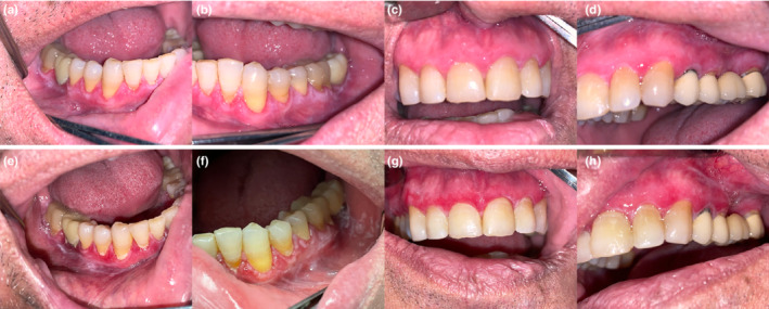
A 65‐year‐old female with a history of oral and cutaneous lichen planus managed with topical steroids received the Pfizer‐BioNTech vaccine in February and March 2021. Twenty‐four hours following the second dose, she reported a feeling of “mouth on fire” which continued for 2 weeks. She was seen in July 2021; she had gingival erythema and striations extending into vestibular mucosa (Figure 3A–C). Saliva production was reduced (resting 0.3 mg/3 min and stimulated 2.1 mg/3 min). She has prescribed topical clobetasol and bethanechol for dry mouth. At one month follow‐up, a significant reduction in erythema was seen (Figure 3D–F).
FIGURE 3.

Exacerbation of oral lichen planus immediately after receiving Pfizer‐BioNTech vaccine (case 6). (a–c) Initial presentation after vaccination showing erythema of the marginal gingiva. (d–f) One month after using topical clobetasol with nearly complete resolution
2.7. Case 7
A 51‐year‐old male with a history of OLP and oral squamous cell carcinoma of the tongue received the second dose of the Pfizer‐BioNTech vaccine in May 2021. He developed increasing oral sensitivity in the buccal mucosa bilaterally 2 weeks following the second dose. Clinical findings include erythema and striations of the posterior buccal mucosal bilaterally. Topical pimecrolimus cream with turmeric supplementation was prescribed. At the 2‐month follow‐up, clinical signs and symptoms returned to baseline.
3. DISCUSSION
We present a case series of oral lichenoid reactions following vaccination or infection by SARS‐CoV‐2. New onset of OLP/LM was noted in 1 case following infection and 2 cases following vaccination. Exacerbation of existing OLP/LM was noted in 4 cases following vaccination. Although a biopsy was performed on one case only, the diagnosis of OLP/LM is generally based on history and clinical features (Alrashdan et al., 2016).
OLP/LM are common conditions with a prevalence of 0.2%–5.0% (Gorouhi et al., 2014). Although the etiopathogenesis of OLP/LM is not fully understood, type 1 immune response appears to play a major role (Wang et al., 2016). Autoimmunity has been proposed to be the underlying mechanism of OLP/LM development (Ebrahimi et al., 2010). This series suggests that OLP/LM may be aggravated due to immune response to COVID‐19/vaccination. Both SARS‐CoV‐2 infection and vaccination elicit type 1 immune response (Alter et al., 2021; Prompetchara et al., 2020). Therefore, it is not surprising for OLP/LM to ensue or be exacerbated by COVID‐19 infection/vaccination.
Lichen planus is reported to follow vaccinations for other viral pathogens, such as hepatitis B, influenza, and herpes zoster (Lai & Yew, 2017). Furthermore, autoimmunity induced by vaccination is not limited to the COVID‐19 vaccine but also in hepatitis B and H1N1 vaccines (Segal & Shoenfeld, 2018). Two theories were proposed to explain this phenomenon, immune hyperstimulation, and molecular mimicry. Immune hyperstimulation following COVID‐19 infection as evidenced by the increase of inflammatory cytokines, including IL‐6 and IL‐10 (Wang et al., 2020). In addition, previous work has shown structural homology between SARS‐CoV‐2 surface peptides and human proteomes, further justifying the occurrence or exacerbation of immune‐mediated conditions following SARS‐CoV‐2 infection (Kanduc & Shoenfeld, 2020).
This case series suggests that COVID‐19 infection and host response to SARS‐CoV‐2 vaccination not only can aggravate known oral immune‐mediated reactions but also can lead to de novo presentation of oral immune‐mediated conditions. While it is not possible to establish causality between COVID‐19 infection/vaccination and OLP/LM development/exacerbation, the timing of events is an important consideration. Whether COVID‐19 infection/vaccination and OLP/LM development/exacerbation are directly or indirectly related, it is important to be aware of the occurrence of such events for diagnosis and management.
AUTHOR CONTRIBUTIONS
Lama Alabdulaaly: Writing – original draft; writing – review and editing. Herve Sroussi: Supervision; writing – review and editing. Joel B. Epstein: Conceptualization; data curation; supervision; writing – review and editing.
CONFLICT OF INTEREST
None.
PEER REVIEW
The peer review history for this article is available at https://publons.com/publon/10.1111/odi.14257.
ACKNOWLEDGMENTS
None.
Alabdulaaly, L. , Sroussi, H. , & Epstein, J. B. (2022). New onset and exacerbation of oral lichenoid mucositis following SARS‐CoV‐2 infection or vaccination. Oral Diseases, 00, 1–5. 10.1111/odi.14257
REFERENCES
- Alrashdan, M. S. , Cirillo, N. , & McCullough, M. (2016). Oral lichen planus: A literature review and update. Archives of Dermatological Research, 308(8), 539–551. [DOI] [PubMed] [Google Scholar]
- Alter, G. , Yu, J. , Liu, J. , Chandrashekar, A. , Borducchi, E. N. , Tostanoski, L. H. , McMahan, K. , Jacob‐Dolan, C. , Martinez, D. R. , Chang, A. , Anioke, T. , Lifton, M. , Nkolola, J. , Stephenson, K. E. , Atyeo, C. , Shin, S. , Fields, P. , Kaplan, I. , Robins, H. , … Barouch, D. H. (2021). Immunogenicity of Ad26.COV2.S vaccine against SARS‐CoV‐2 variants in humans. Nature, 596(7871), 268–272. [DOI] [PMC free article] [PubMed] [Google Scholar]
- Amorim Dos Santos, J. , AGC, N. , da Carvalho Silva, R. L. , Acevedo, A. C. , De Luca Canto, G. , Sugaya, N. , Santos‐Silva, A. R. , & ENS, G. (2021). Oral manifestations in patients with COVID‐19: A living systematic review. Journal of Dental Research, 100(2), 141–154. [DOI] [PubMed] [Google Scholar]
- Brandão, T. B. , Gueiros, L. A. , Melo, T. S. , Prado‐Ribeiro, A. C. , ACFA, N. , GVB, P. , Santos‐Silva, A. R. , & Migliorati, C. A. (2021). Oral lesions in patients with SARS‐CoV‐2 infection: Could the oral cavity be a target organ? Oral Surgery, Oral Medicine, Oral Pathology, Oral Radiology, 131(2), e45–e51. [DOI] [PMC free article] [PubMed] [Google Scholar]
- Burgos‐Blasco, P. , Fernandez‐Nieto, D. , Selda‐Enriquez, G. , Melian‐Olivera, A. , de Perosanz‐Lobo, D. , Dominguez‐Santas, M. , & Alonso‐Castro, L. (2021). COVID‐19: A possible trigger for oral lichen planus? International Journal of Dermatology, 60(7), 882–883. [DOI] [PMC free article] [PubMed] [Google Scholar]
- da Mota Santana, L. A. , Vieira, W. A. , RIC, G. , Lima Dos Santos, M. A. , Takeshita, W. M. , & Miguita, L. (2022). Oral and dermatologic lesions observed in mild COVID‐19 patients infected after 3(rd) vaccine dose. Oral Diseases. Online ahead of print. [DOI] [PMC free article] [PubMed] [Google Scholar]
- Diaz‐Guimaraens, B. , Dominguez‐Santas, M. , Suarez‐Valle, A. , Fernandez‐Nieto, D. , Jimenez‐Cauhe, J. , & Ballester, A. (2021). Annular lichen planus associated with coronavirus SARS‐CoV‐2 disease (COVID‐19). International Journal of Dermatology, 60(2), 246–247. [DOI] [PubMed] [Google Scholar]
- Ebrahimi, M. , Nylander, E. , Bäcklund, B. , Wahlin, Y. B. , Coates, P. J. , & Nylander, K. (2010). The use of a novel ELISA method for detection of antibodies against p63 in sera from patients diagnosed with oral and/or genital and skin lichen planus. Journal of Oral Pathology & Medicine, 39(6), 486–490. [DOI] [PubMed] [Google Scholar]
- Fidan, V. , Koyuncu, H. , & Akin, O. (2021). Oral lesions in Covid 19 positive patients. American Journal of Otolaryngology, 42(3), 102905. [DOI] [PMC free article] [PubMed] [Google Scholar]
- Gorouhi, F. , Davari, P. , & Fazel, N. (2014). Cutaneous and mucosal lichen planus: A comprehensive review of clinical subtypes, risk factors, diagnosis, and prognosis. ScientificWorldJournal, 2014, 742826. [DOI] [PMC free article] [PubMed] [Google Scholar]
- Iranmanesh, B. , Khalili, M. , Amiri, R. , Zartab, H. , & Aflatoonian, M. (2021). Oral manifestations of COVID‐19 disease: A review article. Dermatologic Therapy, 34(1), e14578. [DOI] [PMC free article] [PubMed] [Google Scholar]
- Kanduc, D. , & Shoenfeld, Y. (2020). Molecular mimicry between SARS‐CoV‐2 spike glycoprotein and mammalian proteomes: Implications for the vaccine. Immunologic Research, 68(5), 310–313. [DOI] [PMC free article] [PubMed] [Google Scholar]
- Lai, Y. C. , & Yew, Y. W. (2017). Lichen planus and lichenoid drug eruption after vaccination. Cutis, 100(6), e6–e20. [PubMed] [Google Scholar]
- Lodi, G. , Pellicano, R. , & Carrozzo, M. (2010). Hepatitis C virus infection and lichen planus: A systematic review with meta‐analysis. Oral Diseases, 16(7), 601–612. [DOI] [PubMed] [Google Scholar]
- Prompetchara, E. , Ketloy, C. , & Palaga, T. (2020). Immune responses in COVID‐19 and potential vaccines: Lessons learned from SARS and MERS epidemic. Asian Pacific Journal of Allergy and Immunology, 38(1), 1–9. [DOI] [PubMed] [Google Scholar]
- Segal, Y. , & Shoenfeld, Y. (2018). Vaccine‐induced autoimmunity: The role of molecular mimicry and immune crossreaction. Cellular & Molecular Immunology, 15(6), 586–594. [DOI] [PMC free article] [PubMed] [Google Scholar]
- Shang, Q. , Peng, J. , Zhou, Y. , Chen, Q. , & Xu, H. (2020). Association of Human Papillomavirus with Oral Lichen Planus and Oral Leukoplakia: A meta‐analysis. The Journal of Evidence‐Based Dental Practice, 20(4), 101485. [DOI] [PubMed] [Google Scholar]
- Sharda, P. , Mohta, A. , Ghiya, B. C. , & Mehta, R. D. (2021). Development of oral lichen planus after COVID‐19 vaccination – A rare case report. Journal of the European Academy of Dermatology and Venereology, 36, e82–e83. [DOI] [PMC free article] [PubMed] [Google Scholar]
- Troeltzsch, M. , Gogl, M. , Berndt, R. , & Troeltzsch, M. (2021). Oral lichen planus following the administration of vector‐based COVID‐19 vaccine (Ad26.COV2.S). Oral Diseases. Online ahead of print. [DOI] [PMC free article] [PubMed] [Google Scholar]
- Wang, H. , Zhang, D. , Han, Q. , Zhao, X. , Zeng, X. , Xu, Y. , Sun, Z. , & Chen, Q. (2016). Role of distinct CD4(+) T helper subset in pathogenesis of oral lichen planus. Journal of Oral Pathology & Medicine, 45(6), 385–393. [DOI] [PubMed] [Google Scholar]
- Wang, J. , Jiang, M. , Chen, X. , & Montaner, L. J. (2020). Cytokine storm and leukocyte changes in mild versus severe SARS‐CoV‐2 infection: Review of 3939 COVID‐19 patients in China and emerging pathogenesis and therapy concepts. Journal of Leukocyte Biology, 108(1), 17–41. [DOI] [PMC free article] [PubMed] [Google Scholar]