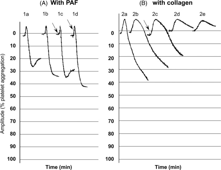
FIGURE 3.

Effect of spike protein on PAF and collagen‐induced WRP aggregation. Platelet aggregation with PAF was defined as the difference between the 0% (PRP) baseline and the 100% (PPP) baseline: (A). PAF‐induced WRP aggregation. (1A) PAF, 1.2 × 10⁻¹¹ M. (1B) 1 min pre‐incubation with 2 μg/ml spike protein and addition of PAF 1.2 × 10⁻¹¹ M. (1C) PAF 1.2 × 10⁻¹¹ M, 2 μg/ml spike protein added 3 s after PAF (see arrow). (1D) PAF 1.2 × 10⁻¹¹ M, 2 μg/ml spike protein added 10 s after PAF (see arrow). (B). Collagen‐induced WRP aggregation. (2A) Collagen 0.08 μg/ml. (2B) 1 min pre‐incubation with 2 μg/ml spike protein and addition of collagen 0.08 μg/ml. (2C) Collagen 0.08 μg/ml, 2 μg/ml spike protein added 30 s after collagen (see arrow). (2D) 1 min pre‐incubation with 20 μg/ml spike protein and addition of collagen 0.08 μg/ml. (2E) 10 min pre‐incubation with 20 μg/ml spike protein and addition of collagen 0.08 μg/ml
