Abstract
Case series summary
Three domestic shorthair cats from California presented to veterinary ophthalmologists with immature cataracts. Other presenting clinical signs included corneal edema, anisocoria, anterior uveitis, elevated intraocular pressure, blepharospasm and/or lethargy. All patients were immunocompromised due to concurrent diseases and/or immunomodulatory drugs. Diagnostics included serial comprehensive ophthalmic examinations with tonometry, ocular ultrasound, electroretinogram and testing for other causes of feline uveitis. Testing for Encephalitozoon cuniculi included serology, histopathology and/or PCR of aqueous humor, lens material or paraffin-embedded whole eye. Treatments included antiparasitic medication, anti-inflammatory medication and supportive care in all three cases. Surgical treatment included enucleation (one case), bilateral phacoemulsification and unilateral intraocular lens placement (one case) and bilateral phacoemulsification with bilateral endolaser ciliary body ablation and bilateral intraocular lens implantation (one case). Both cats for which serologic testing for E cuniculi was performed were positive (1:64–1:4096). In all cats, diagnosis of intraocular E cuniculi was based on at least one of the following: lens histopathology or PCR of aqueous humor, lens material or paraffin-embedded ocular tissue. The clinical visual outcome was best in the patient undergoing phacoemulsification at the earliest stage of the cataract.
Relevance and novel information
Encephalitozoon cuniculi should be considered as a differential cause of cataracts and uveitis in cats in California, the rest of the USA and likely worldwide.
Keywords: Encephalitozoon cuniculi, uveitis, cataracts, phacoemulsification
Introduction
Encephalitozoon cuniculi is a worldwide microsporidian.1,2 Spores can be transmitted via respiratory, oral, 3 conjunctival, 4 intranasal, intraovarial or transplacental routes. 5 In veterinary ophthalmology, E cuniculi is predominantly known as a cause of cataracts and phacoclastic uveitis in rabbits;6,7 after transplacental transmission, spores are speculated to enter the lens via the lenticular blood supply while it is still present. 8 However, a recent study with immunohistochemical evidence of intralenticular E cuniculi after oral infection in 4-month-old, immunocompetent, specific pathogen-free rabbits 9 suggested that alternative mechanisms for lens infections are possible. Reports of ocular involvement in other species are limited and include cataract and uveitis in a snow leopard in France, 10 cataract, uveitis and chorioretinal lesions in dogs in Europe, 11 keratitis and uveitis in an American cat, 12 polyarteritis nodosa and cataract in a blue fox, 13 cataract and neurologic lesions in mink in Norway 14 and keratoconjunctivitis in an American cockatoo. 15 In addition, E cuniculi has been thoroughly investigated as a cause of feline cataract in Austria. 16 This report includes the clinical data from three feline cases of intralenticular E cuniculi in California, USA; one case is discussed at length (case 1), while the other two cases (cases 2 and 3) are summarized in Table 1.
Table 1.
Case summaries
| Case 1 | Case 2 | Case 3 | |
|---|---|---|---|
| Signalment | 3-year-old MN DSH | 15-year-old FS DSH | 1.75-year-old FS DSH |
| Presenting clinical signs | Blepharospasm, anisocoria, lethargy | Rapid-onset cataracts, 6 months after diagnosis of intestinal lymphoma | Upper respiratory signs, ocular discharge, cloudy opacity OS |
| Description of initial cataract | Focal anterior cortical cataracts OU (see Figure 1) | Immature cataracts OU | Incipient peripheral cortical cataracts OU (see Figure 2) |
| Degree of uveitis at presentation | Mild corneal edema OU, moderate keratic precipitates OU, aqueous flare OU (trace OD and 1/4+ OS) | No flare, rubeosis or episcleral injection OU | • OD: no aqueous flare, mild keratic precipitates • OS: rubeosis iridis, 3–4/4+ aqueous flare, keratic precipitates |
| IOP, lowest to highest | • 16–37 mmHg OD • 8–44 mmHg OS |
• 9–70 mmHg OD • 3–56 mmHg OS |
• 11–30 mmHg OD • 15–62 mmHg OS |
| Systemic testing: negative, normal results | • CBC and serum chemistry • Seronegative: T gondii IgG/IgM, FIV, FeLV antigen (IDEXX Reference Laboratory) • Upper respiratory PCR panel (IDEXX Reference Laboratory): C felis, feline calicivirus, M felis and influenza A • Thoracic radiographs • Aqueous humor PCR was negative: FHV-1, FCoV, FeLV, Bartonella species, C neoformans, T gondii, FIV • Normal cytology of a mesenteric lymph node • FIP PCR (blood): negative |
• Seronegative: FeLV/FIV/T gondii
• FCoV IFA <1:400 |
• Seronegative: FIV, coronavirus (T gondii), C neoformans (Antech Diagnostics) |
| Systemic testing positive results | • FCoV titer 1:3200 • C neoformans titer 1:8 • B henselae and B clarridgeiae titer =1:128 • Fecal testing positive: Giardia (ELISA), T foetus, Cryptosporidium species, Giardia species, FCoV and C perfringens alpha toxin gene (PCR; IDEXX Reference Laboratory) • Upper respiratory PCR panel, FHV-1 positive at 0.160 thousands/swab (latent infection; IDEXX Reference Laboratory) • Abdominal ultrasound (splenomegaly and mild mesenteric lymphadenopathy) • Repeat coronavirus titer 1:1600 |
NA | • Bartonella species = 4+ strong positive
(Western blot, National Veterinary
Laboratory) • FeLV-positive (Antech Diagnostics) |
| Systemic diagnoses | • FCoV • Bartonella species serologic positive • Cryptococcosis • FHV-1 • Giardiasis • T foetus infection • Cryptosporidiosis |
• Intestinal lymphoma • FHV-1 (suspected, not confirmed) |
• FeLV • Bartonella species serologic positive |
| Medical treatment for E cuniculi | Fenbendazole 50 mg/kg PO q24h for 3 weeks, repeated twice | Fenbendazole 70 mg/kg PO q24h for 3 weeks | Fenbendazole 50 mg/kg PO q24h for 10 days (multiple courses) |
| Surgical treatment | • Phacoemulsification OU • Intraocular lens OS |
• Phacoemulsification OU • Intraocular lens OU • Endoscopic cyclophotocoagulation OU |
• Enucleation OS |
| Glaucoma treatment | • 2% dorzolamide/0.5% timolol OU q12h • 2% dorzolamide OU q8h |
• 2% dorzolamide OU q12h–q6h • Methazolamide 7.5 mg PO q24h • 0.5% timolol OU q12h |
• Methazolamide (Wedgewood Compounding Pharmacy) 15 mg PO q24h–q12h |
| Uveitis treatment | • Onsior (robenacoxib; Elanco) • Diclofenac 0.1% ophthalmic solution (Bausch and Lomb) OU q12h • Topical 1% prednisolone acetate suspension (Pacific Pharma) OU q24h–q12h • 0.1% nepafenac ophthalmic suspension (Nevanac Alcon) OU q24h–q6h |
• Neomycin polymyxin B sulfates and dexamethasone OU three times
weekly • 1% prednisolone acetate OU q8h |
• Dexamethasone 0.1% (Bausch and Lomb) OS q24h–q12h |
| Keratitis treatment | • Bacitracin neomycin gentamicin ophthalmic ointment (AC
Pharmaceuticals) OU q8h • Famciclovir (Neogen) 250 mg PO q6h • Ofloxacin 0.3% OU q24h • Optixcare (Optixcare Eye Lube Plus; Aventix) OU q12h • 0.5% cidofovir (Wedgewood Compounding Pharmacy) OU q12h |
• 0.5% cidofovir OU q12h • Famciclovir 125 mg PO q12h-q8h • Remend corneal repair gel (Elanco) OU q8h • Ofloxacin 0.3% OU q6h • 5% NaCl ophthalmic ointment OU q6h • Autologous serum OU q6h • 2% ciclosporin aqueous (for stromal keratitis; Stokes Compounding Pharmacy) OU q24h • Buprenorphine 0.005–0.01 mg/kg transbuccal q8h |
NA |
| Medical treatment for other conditions | • Fluconazole 10.7 mg/kg PO q12h • Doxycycline 4.3 mg/kg PO q12h for 3 weeks • Ronidazole (for diarrhea) • Buprenorphine (0.5 mg/ml; Hikma) |
• Prednisolone 5 mg PO q24h • Chlorambucil 2 mg PO q12h for four doses q2weeks • Vitamin B12/cobalamin 250 µg monthly • SC fluids for hyporexia |
• Doxycycline (Road Runner Compounding Pharmacy) 6 mg/kg PO q12h
for 25 days (multiple courses) • Oral pradofloxacin (Veraflox; Elanco) • Prednisolone 1 mg PO EOD |
| Duration of follow-up | 1.4 years post-phacoemulsification | 1 year post-phacoemulsification | 6 years after initial examination, 5.5 years post-enucleation OS |
| Outcome | • OS: pseudophakic • OD: aphakic • OU: menace and PLR positive, comfortable, no aqueous flare, mild capsular opacity, numerous, punctate, gray, slightly hyporeflective retinal lesions • IOP 18/17 mmHg OD/OS |
• OU: pseudophakic, menace negative but patient navigated the
room well, PLR and dazzle positive, comfortable, no flare, mild
retinal degeneration • IOP 24/25 mmHg OD/OS |
• OS: enucleation • OD: mature cataract menace negative, positive PLR and dazzle, mild keratic precipitates and rubeosis iridis, no aqueous flare, fluorescein negative • IOP 21 mmHg |
MN = male neutered; DSH = domestic shorthair; FS = female spayed; IOP = intraocular pressure; CBC = complete blood count; T gondii = Toxoplasma gondii; FIV = feline immunodeficiency virus; FeLV = feline leukemia virus; C felis = Chlamydophila felis; M felis = Mycoplasma felis; FHV-1 = feline herpesvirus-1; FCoV = feline coronavirus; C neoformans = Cryptococcus neoformans; FIP = feline infectious peritonitis; IFA = immunofluorescence; B henselae = Bartonella henselae; B clarridgeiae = Bartonella clarridgeiae; T foetus = Tritrichomonas foetus; C perfringens = Clostridium perfringens; NA = not available; E cuniculi = Encephalitozoon cuniculi; SC = subcutaneous; EOD = every other day; PLR = pupillary light reflex
Case series description
Case 1
A 3-year-old male castrated domestic shorthair cat presented to an emergency service for blepharospasm, anisocoria and lethargy. The patient had a history of chronic upper respiratory infections and diarrhea. It had been rescued from a southern Californian shelter and lived indoors in San Francisco.
On initial emergency examination, the patient’s intraocular pressure was 26 mmHg OD and 33 mmHg OS, with mild corneal edema OU. Treatment included robenacoxib (2 mg/kg SC once [Onsior; Elanco]), ofloxacin (OU q8h) and 2% dorzolamide/0.5% timolol (OU q12h). See Table 1 for the diagnostic testing results (testing for E cuniculi was not immediately performed).
Initial ophthalmic examination revealed menace response and pupillary light reflex (PLR) positive OU, mild corneal edema with moderate keratic precipitates OU, aqueous flare OU (trace OD and 1/4+ OS), focal posterior synechiae OS and focal anterior cortical cataracts OU (Keeler PSLClassic). Intraocular pressure was 20 mmHg OD and 12 mmHg OS (Icare Tonovet; Icare Finland). Retinal examination (Keeler Vantage Indirect) was normal except for numerous, punctate, gray, slightly hyporeflective lesions in the dorsal retina OU. Treatment with topical diclofenac 0.1% ophthalmic solution OU q12h (Bausch and Lomb) was initiated. Treatment with fluconazole (10.7 mg/kg PO q12h long-term for Cryptococcus neoformans) and doxycycline (4.3 mg/kg PO q12h for 21 days for Bartonella species) was initiated.
One week after initial presentation, aqueous flare was unimproved. Treatment with topical 1% prednisolone acetate suspension (OU q12h; Pacific Pharma) and topical 0.5% cidofovir (OU q12h; Wedgewood Compounding Pharmacy) were added.
One month after initial presentation, the patient developed a large superficial corneal ulcer OS, suspected to be related to herpes exacerbated by topical steroids. Prednisolone acetate was discontinued, and bacitracin neomycin gentamicin ophthalmic ointment (AC Pharmaceuticals, Arroyo Grande CA) was added OU q8h.
Approximately 2 months after the initial examination, the corneal ulcer OS persisted. Aqueous flare was trace OD and 1/4+ OS, with an intraocular pressure (IOP) of 19 mmHg OD and 44 mmHg OS. Aqueocentesis was performed OS, and aqueous humor was submitted for PCR to determine if C neoformans, Bartonella species or feline coronavirus (FCoV) were the cause of uveitis. Because the patient’s cataracts appeared to be similar to those described in a previous report, 16 a special request was made to IDEXX to add an E cuniculi PCR test. A contact lens (PureVision BC 8.6; Bausch and Lomb) was placed, and a partial lateral temporary tarsorrhaphy was performed for 2 weeks. Medication administered immediately after the procedures included bacitracin neomycin gentamicin ophthalmic ointment (OU q8h) and dorzolamide HCl/timolol maleate (OS q8h; Bausch and Lomb). Oral medications included ronidazole (for diarrhea), buprenorphine (Hikma 0.5 mg/ml), robenacoxib (6 mg PO q24h for 3 days [Onsior; Elanco]) and famciclovir (250 mg PO q12h; Neogen). Aqueous humor cytology (Veterinary Diagnostics) showed increased cellularity, with 68% mixed (mostly mature) lymphocytes, 15% quiescent to vacuolated macrophages and 17% non-degenerate to slightly poorly preserved neutrophils (see Tables 1 and 2). Given the positive aqueous humor PCR result for E cuniculi, treatment with fenbendazole (50 mg/kg PO q24h for 3 weeks) was initiated.
Table 2.
Encephalitozoon cuniculi testing
| Case 1 | Case 2 | Case 3 | |
|---|---|---|---|
| Serology (IgG) | 1:64* | NA | 1:4096 (2014) then 1:256 (2018)* |
| PCR | • Aqueocentesis fluid, positive
†
• Lens material, positive, strain II ‡ |
• Phacoemulsified lens fluid, positive
§
• Urine negative ¶ |
Paraffin scrolls of enucleated eye (OS), positive ¶ |
| Histopathology | Lens capsule: Gram-positive, Ziehl–Neelsen acid-fast positive ∞ | NA | Globe: intralenticular organisms, Gram-positive, variably acid-fast, Luna stain positive (see Figure 3) ¶ |
University of Miami Avian & Wildlife Laboratory
IDEXX Reference Laboratories
Department for Pathobiology, Veterinary University Vienna
Athens Veterinary Diagnostic Laboratory, University of Georgia
Comparative Pathology Laboratory, University of California, Davis
Comparative Ocular Pathology Laboratory of Wisconsin
NA = not available
Three months after initial presentation, ophthalmic examination indicated similar signs of uveitis with punctate fluorescein positivity OS only, and IOP was 16 mmHg OD and 8 mmHg OS. Topical 0.1% nepafenac ophthalmic suspension (OU q12h; Nevanac Alcon) was initiated.
Four months after initial presentation, aqueous flare had resolved with normal IOP without any dorzolamide/timolol in the previous 3 days. Given the anticipated difficulty in controlling uveitis medically and the likelihood that cataracts would progress in the long term, cataract surgery was considered. Owing to the patient’s positive FCoV status, thoracic radiographs (unremarkable), abdominal ultrasound (splenomegaly and mild mesenteric lymphadenopathy) and ultrasound-guided aspiration of a mesenteric lymph node (cytologically normal) were performed (see also Table 1).
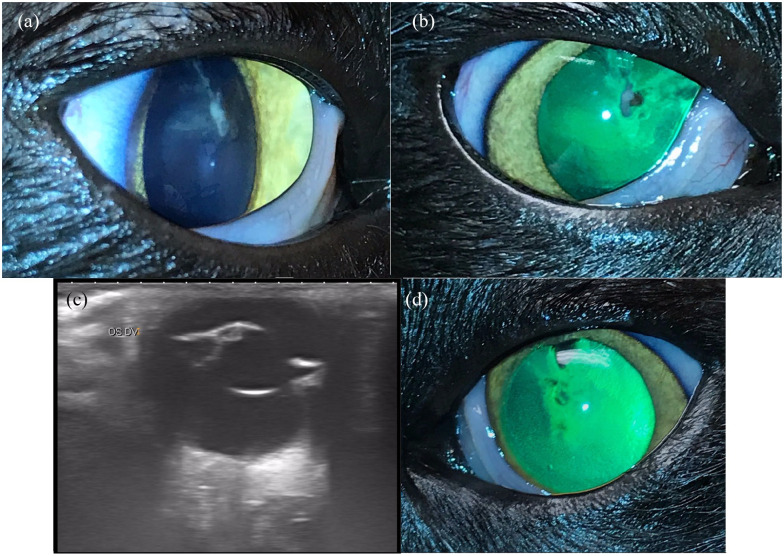
Five months after initial presentation, E cuniculi serology was 1:64 (University of Miami Avian & Wildlife Laboratory; see Table 2 for the full list of E cuniculi tests). Electroretinogram (ERG Retinographics BNP200) was normal with b-wave amplitudes >300 µV OU. Ocular ultrasound (Toshiba AplioMX) was normal OU except for multifocal capsular/cortical lens irregularities OU (Figure 1). Phacoemulsification was performed OU (Acrivet Alexos). An intraocular lens was placed OS only (An-lens MC1-13) due to excision of a peripheral capsular plaque necessitating excess capsule removal OD. Immediate postoperative medications included 0.3% ofloxacin ophthalmic solution (OU q6h; Bausch and Lomb), 0.5% cidofovir (OU q12h), 0.1% Nevanac (OU q6h), 1% prednisolone acetate (OU q6h), 2% dorzolamide ophthalmic solution (OU q8h; Micro Labs) and Optixcare (OU q12h; Optixcare Eye Lube Plus Aventix). Oral medications included fenbendazole (50 mg/kg PO q24h for 3 weeks), fluconazole, amoxicillin trihydrate/clavulanate potassium (62.5 mg PO q12h; Zoetis), transmucosal buprenorphine (0.02 mg/kg q8h; Wedgewood Compounding Pharmacy) and robenacoxib (6 mg PO q24h for three doses [Onsior; Elanco]). One day postoperatively, IOP was 37 mmHg OD and 43 mmHg OS but normalized after two extra doses of 2% dorzolamide/0.5% timolol OU. Perincisional superficial corneal ulcers and 1/4+ aqueous flare were present OU.
Figure 1.
Case 1: (a,b,d) clinical photos and (c) ultrasound image. Pupils were pharmacologically dilated with 1% tropicamide ophthalmic in (a), (b) and (d) (Akorn). (a,b) OD focal anterior subcapsular to anterior cortical cataract and focal pigment on lens capsule. The lens capsule appeared focally wrinkled at the site of the cataract clinically, but no capsular tears were visible on slit-lamp examination. (c) OS: vertical ultrasound image showing echoic dorsal anterior subcapsular cataract with anterior cortical to nuclear extension. The lens capsule was interpreted to be intact via ultrasound. Other than lens abnormalities, anterior and posterior segments were within normal limits. (d) OS: clinical photo showing retro-illumination of focal subcapsular to anterior cortical cataract (dark lenticular opacities). Images courtesy of Dr Mitzi Zarfoss
Lens capsule plaques were submitted to the Comparative Ocular Pathology Laboratory of Wisconsin. Histopathology of the right lens capsule presented moderate numbers of foamy-to-epithelioid macrophages with numerous 1–3 µm, rod-shaped microsporidia consistent with E cuniculi. The organisms were strongly Gram positive and lightly Ziehl–Neelsen acid-fast positive (see Table 2).
Postoperatively, topical anti-inflammatories were tapered over several months, and dorzolamide/timolol was eventually discontinued. On ophthalmic recheck 17 months postoperatively, the patient was visual, comfortable, normotensive and PLR positive OU. There was mild anisocoria, dyscoria and mydriasis OD, aphakia OD, pseudophakia OS, no aqueous flare OU, minimal capsular opacity and an unchanged retinal examination with numerous punctate gray lesions in the dorsal retina OU. Medications consisted of 0.1% Nevanac (OU q24h). At home, vision was reportedly very good.
Discussion
This case series demonstrates that intralenticular E cuniculi is a potential cause of cataracts, uveitis and secondary glaucoma in domestic cats in California, USA.
Although E cuniculi is found worldwide, feline ocular encephalitozoonosis has only been reported in Austria, 16 France 10 and the USA (feline cornea). 12 Factors including climate and animal reservoirs may affect E cuniculi’s prevalence and risk to cats. Specifically, environmental spore viability varies by temperature. 3 Given that encephalitozoonosis in rodents has been documented worldwide,5,17–22 rodents likely spread disease, as corroborated by case 1 and several Austrian cases that tested positive for the mouse strain (strain II). 16
Although E cuniculi is an opportunistic pathogen in immunocompromised people, 23 the role of immunosuppression in feline ocular encephalitozoonosis remains unclear. The cases in this study were immunocompromised due to concurrent diseases (see Table 1) and immunomodulatory drugs (prednisolone and chlorambucil in case 2). This aligns with the current understanding that immunosuppression exacerbates rabbit encephalitozoonosis. 4 However, in the 2011 study published by Benz et al, 16 11 systemically healthy European Shorthair cats also developed cataracts and uveitis from E cuniculi, though 4/11 cats had positive titers for Toxoplasma gondii (IgG 1:4000). In the same study, 2/100 ophthalmologically healthy cats had a positive antibody titer for E cuniculi. Research conducted in North America,24,25 Europe18,26,27 and Asia28–30 has found that E cuniculi prevalence range from 0% to 26.8%, with one paper demonstrating a seroprevalence of 6.1% (18/295) 30 in healthy, asymptomatic cats.
The mechanism by which E cuniculi causes uveitis is unknown. E cuniculi antigens may contribute to the inflammatory response; 31 this is supported by Nell et al 11 and cases 1 and 3, which suggest that focal anterior cataracts due to E cuniculi may be more inflammatory relative to focal cataracts due to other etiologies. Alternatively, E cuniculi may replicate and physically disrupt the lens, leading to lens-induced uveitis. 7 In case 1, aqueous humor PCR screening failed to show any evidence of other intraocular infections and supported E cuniculi being the causative agent for uveitis.
Currently, phacoemulsification surgery, antiparasitic medication and symptomatic treatment are employed to treat intralenticular E cuniculi infections. Phacoemulsification treats cataracts, removes microsporidia and minimizes further pathogen replication and intraocular inflammation. Fenbendazole, often prescribed at ranges of 20–50 mg/kg q24h for 3 weeks (extra-label), targets various pathogen stages. 32 Symptomatic treatment often includes oral and ophthalmic anti-inflammatories to address anterior uveitis. Since systemic immunosuppression facilitates E cuniculi, 33 corticosteroids should be employed at anti-inflammatory doses. The literature and this report suggest that surgical management of intraocular E cuniculi via phacoemulsification, especially early phacoemulsification, 34 can successfully maintain vision and comfort, while medical management alone may more commonly lead to blindness, discomfort and enucleation. 16
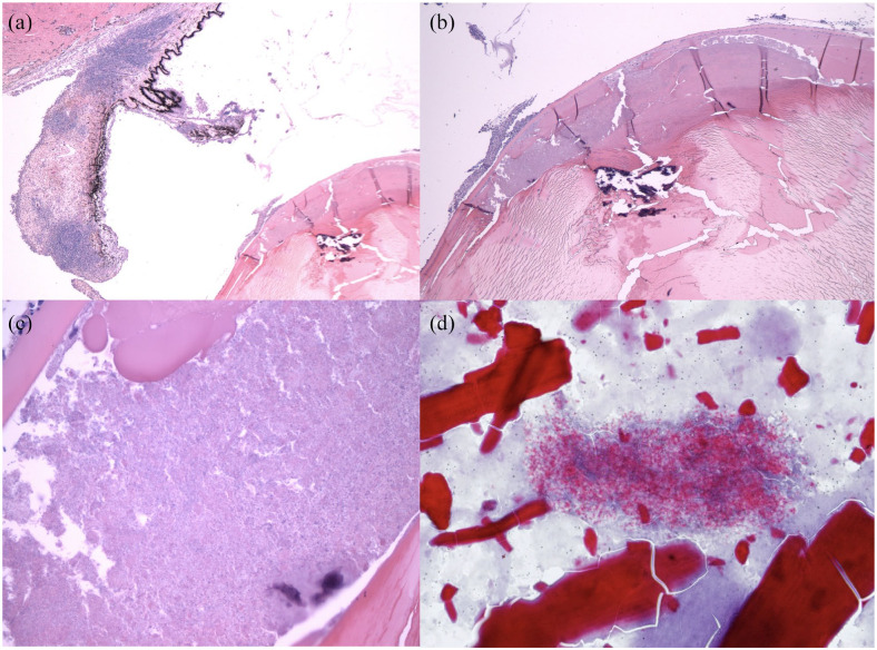
Various diagnostic testing is available for E cuniculi (see Table 2). Serology is a non-invasive, low-risk screening tool that is expected to be weakly or strongly positive for E cuniculi in cats with intraocular E cuniculi; however, PCR positivity of ocular fluid/tissues provides more definitive evidence of intraocular involvement. PCR detection of E cuniculi varies based on sample location. In Benz et al, aqueous humor from 10/19 affected cats was PCR positive, whereas lens material was PCR positive in one or both eyes in 11/11 of these cats. 16 Histopathology with hematoxylin and eosin stains can help guide the diagnosis of E cuniculi (see Figure 3a–c); however, the preferred histologic stains for E cuniculi spore detection are modified trichrome and Gram stain with light microscopy and calcofluor white stain with ultraviolet light microscopy, 35 though acid fast trichome can be effective (see Figure 3d); 16 in case 3, Luna stain was helpful.
Figure 3.
Histopathology. (a) Case 3, hematoxylin and eosin, × 2 magnification showing lymphoplasmacytic iritis. (b) Case 3, hematoxylin and eosin, × 4 magnification showing regionally severe equatorial lens fiber degeneration. (c) Case 3, hematoxylin and eosin, × 40 magnification showing innumerable Encephalitozoon cuniculi organisms within the lens, with swollen lens fibers/Morgagnian globules (top center) and a few neutrophils outside the capsule (upper left). (d) Case 1, histopathology. Ziehl–Neelsen acid fast stain, × 60 magnification, lens material and E cuniculi organisms. Images (a), (b) and (c) courtesy of Dr Christopher Reilly, DACVP. Image (d) courtesy of Dr Barbara Nell
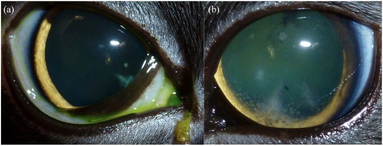
When feline cataracts are identified, possible causes include chronic uveitis (most common), trauma (especially penetrating trauma), E cuniculi, secondary to glaucoma or lens luxation, congenital, possibly hereditary, nutritional or uncommonly metabolic (hypocalcemia, hyperphosphatemia, diabetes). 36 The cause of feline cataracts can be very difficult to determine, particularly since chronic uveitis commonly causes cataracts and vice versa. Cataracts caused by chronic lens-induced uveitis and those caused by E cuniculi can be very similar in appearance and size (ranging anywhere from incipient to mature in this report). However, in the experience of the authors, E cuniculi cataracts typically originate as focal lesions in the anterior cortex and spread from there to the whole lens. As cases are presented at different stages, the appearance of E cuniculi cataracts can differ in size and stage of maturity. We suspect that smaller (incipient) E cuniculi cataracts can cause disproportionately severe and acute uveitis and/or may progress somewhat more quickly relative to incipient cataracts of other etiologies (with the possible exception of penetrating trauma where an obvious corneal lesion would be expected). In two cases in this report (cases 1 and 3), incipient cataracts were associated with 1/4 or 3/4+ aqueous flare, which is unusual in cataracts not associated with traumatic intralenticular bacterial implantation or long-standing uveitis. However, inflammation caused by E cuniculi cataracts can be variable; in case 2, cataracts were immature and uveitis was initially minimal (although this patient was also on oral prednisolone for intestinal lymphoma). Speed of progression of E cuniculi cataracts can also be variable. In case 2, cataracts were reported to be rapidly progressive, whereas in case 3 the cataract progressed from incipient to mature over 6 years. Furthermore, features of chronic uveitis that may have led to these cataracts (such as chronic iris discoloration or large areas of posterior synechiation) were generally lacking in the cases presented here, except for mild rubeosis in case 3 and very focal synechiation in case 1 (see Figures 1 and 2). Ultimately, serology for E cuniculi is recommended as a screening tool in all cases of feline cataracts for which an alternative underlying cause is not apparent. If E cuniculi serology is positive, then referral to an ophthalmologist for additional (PCR) testing of ocular tissues and more intensive medical and surgical treatment should be considered, as this would be expected to improve the clinical outcome.
Figure 2.
Case 3. Both pupils were dilated with 1% tropicamide (Akorn). (a) OD (initial examination): no aqueous flare, mild keratic precipitates, incipient peripheral cortical cataracts, fluorescein negative. (b) OS (first recheck after 3 weeks): mild iris thickening, trace aqueous humor cells, keratic precipitates, incipient peripheral cortical cataract, fluorescein negative. Images courtesy of Dr Holly Hamilton
Conclusions
This study highlights E cuniculi as a cause of feline cataracts in the USA (and likely worldwide). Study limitations include low case numbers and heterogeneous, incomplete patient data with limited follow-up. Although this series provides clinically relevant information, it does not necessarily represent optimal treatment of feline ocular E cuniculi. Because the literature on feline encephalitozoonosis is somewhat lacking, future studies should more thoroughly evaluate systemic involvement, pathophysiology and/or best treatment practices. E cuniculi should be considered in cats presenting with cataracts, especially those with concurrent anterior uveitis. The authors hope that increased awareness and testing will lead to earlier diagnosis of feline intraocular E cuniculi and improved clinical outcomes.
Acknowledgments
The authors would like to recognize the work of Christopher M Reilly DVM, DACVP of SpecialtyVETPATH for his pathology expertise and work in characterizing case 3, as well as significant contributions to the manuscript. In addition, Leandro BC Teixeira DVM, DACVP (Comparative Ocular Pathology Laboratory of Wisconsin) provided histopathology expertise. Holly Hamilton DVM, DACVO primarily managed case 3 and provided valuable feedback on the manuscript. The authors thank Dr Carolyn Cray for providing valuable expert consultation in case 1 and in preparation of the manuscript. The authors thank Dr Patty Smith for her clinical assistance with case 3, Drs Lana Linton and Kristina Gronkiewicz for their support in treatment of case 2 and Dr Marcella Harb-Hauser for her clinical support with case 1. The authors would also like to thank Dr Klaas-Ole Blohm for assistance with PCR testing for case 1.
Footnotes
Accepted: 25 May 2022
Author note: Case 3 of this series was presented at a specialty conference in 2015. 37
Conflict of interest: The authors declared no potential conflicts of interest with respect to the research, authorship, and/or publication of this article.
Funding: The authors received no financial support for the research, authorship, and/or publication of this article.
Ethical approval: The work described in this manuscript involved the use of non-experimental (owned or unowned) animals. Established internationally recognized high standards (‘best practice’) of veterinary clinical care for the individual patient were always followed and/or this work involved the use of cadavers. Ethical approval from a committee was therefore not specifically required for publication in JFMS Open Reports. Although not required, where ethical approval was still obtained it is stated in the manuscript.
Informed consent: Informed consent (verbal or written) was obtained from the owner or legal custodian of all animal(s) described in this work (experimental or non-experimental animals, including cadavers) for all procedure(s) undertaken (prospective or retrospective studies). For any animals or people individually identifiable within this publication, informed consent (verbal or written) for their use in the publication was obtained from the people involved.
ORCID iD: Joie Lin  https://orcid.org/0000-0001-7803-5121
Taemi Horikawa  https://orcid.org/0000-0002-7388-0999
References
- 1. Bohne W, Böttcher K, Groß U. The parasitophorous vacuole of Encephalitozoon cuniculi: biogenesis and characteristics of the host cell-pathogen interface. Int J Med Microbiol 2011; 301: 395–399. [DOI] [PubMed] [Google Scholar]
- 2. Ashton N, Cook C, Clegg F. Encephalitozoonosis (nosematosis) causing bilateral cataract in a rabbit. Br J Ophthalmol 1976; 60: 618–631. [DOI] [PMC free article] [PubMed] [Google Scholar]
- 3. Harcourt-Brown F. Infectious diseases of domestic rabbits. In: Harcourt-Brown F. (ed). Textbook of rabbit medicine. St Louis, MO: 2002, pp 361–385. [Google Scholar]
- 4. Jeklova E, Leva L, Kovarcik K, et al. Experimental oral and ocular Encephalitozoon cuniculi infection in rabbits. Parasitology 2010; 137: 1749–1757. [DOI] [PubMed] [Google Scholar]
- 5. Hinney B, Sak B, Joachim A, et al. More than a rabbit’s tale – Encephalitozoon spp. in wild mammals and birds. Int J Parasitol Parasites Wildl 2016; 5: 76–87. [DOI] [PMC free article] [PubMed] [Google Scholar]
- 6. Künzel F, Fisher PG. Clinical signs, diagnosis, and treatment of Encephalitozoon cuniculi infection in rabbits. Vet Clin North Am Exot Anim Pract 2018; 21: 69–82. [DOI] [PubMed] [Google Scholar]
- 7. Giordano C, Weigt A, Vercelli A, et al. Immunohistochemical identification of Encephalitozoon cuniculi in phacoclastic uveitis in four rabbits. Vet Ophthalmol 2005; 8: 271–275. [DOI] [PubMed] [Google Scholar]
- 8. Ozkan O, Karagoz A, Kocak N. First molecular evidence of ocular transmission of Encephalitozoonosis during the intrauterine period in rabbits. Parasitol Int 2019; 71: 1–4. DOI: 10.1016/j.parint.2019.03.006. [DOI] [PubMed] [Google Scholar]
- 9. Jeklová E, Levá L, Kummer V, et al. Immunohistochemical detection of Encephalitozoon cuniculi in ocular structures of immunocompetent rabbits. Animals 2019; 9: 988. DOI: 10.3390/ani9110988. [DOI] [PMC free article] [PubMed] [Google Scholar]
- 10. Scurrell EJ, Holding E, Hopper J, et al. Bilateral lenticular Encephalitozoon cuniculi infection in a snow leopard (Panthera uncia). Vet Ophthalmol 2015; 18 Suppl 1: 143–147. [DOI] [PubMed] [Google Scholar]
- 11. Nell B, Csokai J, Fuchs-Baumgartinger A, et al. Encephalitozoon cuniculi causes focal anterior cataract and uveitis in dogs. Tierarztl Prax Ausg K Kleintiere Heimtiere 2015; 43: 337–344. [DOI] [PubMed] [Google Scholar]
- 12. Buyukmihci N, Bellhorn R, Hunziker J, et al. Encephalitozoon (Nosema) infection of cornea in a cat. J Am Vet Med Assoc 1977; 171: 355–357. [PubMed] [Google Scholar]
- 13. Arnesen K, Nordstoga K. Ocular encephalitozoonosis (nosematosis) in blue foxes: polyarteritis nodosa and cataract. Acta Ophthalmol 1977; 55: 641–651. [DOI] [PubMed] [Google Scholar]
- 14. Bjerkås I. Brain and spinal cord lesions in encephalitozoonosis in mink. Acta Vet Scand 1990; 31: 423–432. [DOI] [PMC free article] [PubMed] [Google Scholar]
- 15. Phalen DN, Logan KS, Snowden KF. Encephalitozoon hellem infection as the cause of a unilateral chronic keratoconjunctivitis in an umbrella cockatoo (Cacatua alba). Vet Ophthalmol 2006; 9: 59–63. [DOI] [PubMed] [Google Scholar]
- 16. Benz P, Maaß G, Csokai J, et al. Detection of Encephalitozoon cuniculi in the feline cataractous lens. Vet Ophthalmol 2011: 14 Suppl 1: 37–47. [DOI] [PubMed] [Google Scholar]
- 17. Hofmannová L, Sak B, Jekl V, et al. Lethal Encephalitozoon cuniculi genotype III infection in Steppe lemmings (Lagurus lagurus). Vet Parasitol 2014; 205: 357–360. [DOI] [PubMed] [Google Scholar]
- 18. Meredith AL, Cleaveland SC, Brown J, et al. Seroprevalence of Encephalitozoon cuniculi in wild rodents, foxes and domestic cats in three sites in the United Kingdom. Transbound Emerg Dis 2015; 62: 148–156. [DOI] [PubMed] [Google Scholar]
- 19. Kitz S, Grimm F, Wenger S, et al. Encephalitozoon cuniculi infection in Barbary striped grass mice (Lemniscomys barbarus). Schweiz Arch Tierheilkd 2018; 160: 394–400. [DOI] [PubMed] [Google Scholar]
- 20. Sak B, Kváč M, Květoňová D, et al. The first report on natural Enterocytozoon bieneusi and Encephalitozoon spp. infections in wild East-European house mice (Mus musculus musculus) and West-European house mice (M. m. domesticus) in a hybrid zone across the Czech Republic–Germany border. Vet Parasitol 2011; 178: 246–250. [DOI] [PubMed] [Google Scholar]
- 21. Perec-Matysiak A, Leśniańska K, Buńkowska-Gawlik K, et al. The opportunistic pathogen Encephalitozoon cuniculi in wild living Murinae and Arvicolinae in Central Europe. Eur J Protistol 2019; 69: 14–19. [DOI] [PubMed] [Google Scholar]
- 22. Tsukada R, Tsuchiyama A, Sasaki M, et al. Encephalitozoon infections in Rodentia and Soricomorpha in Japan. Vet Parasitol 2013; 198: 193–196. [DOI] [PubMed] [Google Scholar]
- 23. Mathis A, Weber R, Deplazes P. Zoonotic potential of the microsporidia. Clin Microbiol Rev 2005; 18: 423–445. [DOI] [PMC free article] [PubMed] [Google Scholar]
- 24. Kourgelis C, Reilly C, Von Roedern M, et al. Serological survey for antibodies to Encephalitozoon cuniculi in cats within the United States. Vet Parasitol Reg Stud Rep 2017; 9: 122–124. [DOI] [PubMed] [Google Scholar]
- 25. Hsu V, Grant DC, Zajac AM, et al. Prevalence of IgG antibodies to Encephalitozoon cuniculi and Toxoplasma gondii in cats with and without chronic kidney disease from Virginia. Vet Parasitol 2011; 176: 23–26. [DOI] [PubMed] [Google Scholar]
- 26. Piekarska J, Kicia M, Wesołowska M, et al. Zoonotic microsporidia in dogs and cats in Poland. Vet Parasitol 2017; 246: 108–111. [DOI] [PubMed] [Google Scholar]
- 27. Lores B, del Aguila C, Arias C. Enterocytozoon bieneusi (Microsporidia) in faecal samples from domestic animals from Galicia, Spain. Mem Int Oswaldo Cruz 2002; 97: 941–945. [DOI] [PubMed] [Google Scholar]
- 28. Jamshidi S, Tabrizi AS, Bahrami M, et al. Microsporidia in household dogs and cats in Iran; a zoonotic concern. Vet Parasitol 2012; 185: 121–123. [DOI] [PubMed] [Google Scholar]
- 29. Askari Z, Mirjalali H, Mohebali M, et al. Molecular detection and identification of zoonotic Microsporidia spore in fecal samples of some animals with close-contact to human. Iran J Parasitol 2015; 10: 381–388. [PMC free article] [PubMed] [Google Scholar]
- 30. Tsukada R, Osaka Y, Takano T, et al. Serological survey of Encephalitozoon cuniculi infection in cats in Japan. J Vet Med Sci 2016; 78: 1615–1617. [DOI] [PMC free article] [PubMed] [Google Scholar]
- 31. Wolfer J, Grahn B, Wilcock B, et al. Phacoclastic uveitis in the rabbit. Prog Vet Comp Ophthalmol 1993; 3: 92–97. [Google Scholar]
- 32. Addie DD, Tasker S, Boucraut-Baralon C, et al. Encephalitozoon cuniculi infection in cats: European guidelines from the ABCD on prevention and management. J Feline Med Surg 2020; 22: 1084–1088. [DOI] [PMC free article] [PubMed] [Google Scholar]
- 33. Kotkova M, Sak B, Kvetonova D, et al. Latent microsporidiosis caused by Encephalitozoon cuniculi in immunocompetent hosts: a murine model demonstrating the ineffectiveness of the immune system and treatment with albendazole. PLoS One 2013; 8. DOI: 10.1371/journal.pone.0060941. [DOI] [PMC free article] [PubMed] [Google Scholar]
- 34. Felchle LM, Sigler RL. Phacoemulsification for the management of Encephalitozoon cuniculi-induced phacoclastic uveitis in a rabbit. Vet Ophthalmol 2002; 5: 211–215. [DOI] [PubMed] [Google Scholar]
- 35. Rodríguez-Tovar LE, Villarreal-Marroquín A, Nevárez-Garza AM, et al. Histochemical study of Encephalitozoon cuniculi spores in the kidneys of naturally infected New Zealand rabbits. J Vet Diagn Investig 2017; 29: 269–277. [DOI] [PubMed] [Google Scholar]
- 36. Stiles J. Feline opthalmology. In: Gelatt KN. (ed). Veterinary ophthalmology. 5th ed. Chichester: Wiley-Blackwell, 2014, pp 1477–1559. [Google Scholar]
- 37. Reilly CM, Hamilton HL, Cray C, et al. Naturally occurring lenticular encephalitozoonosis in a domestic cat [abstract]. Vet Ophthalmol 2015; 18: E17–E34. [Google Scholar]


