Pemphigus vulgaris (PV) is an autoimmune blistering disease that causes intraepidermal blisters on the skin and mucous membranes. Autoantibodies targeting desmoglein 1 and desmoglein 3, which are cell adhesion molecules connecting epidermal cells, play an essential role in the development of PV. 1 It has been suggested that one mechanism for the production of autoantibodies is molecular mimicry, i.e. a phenomenon in which different molecules have similar structures. For example, if antigens of pathogenic microorganisms such as bacteria and viruses are in a molecular mimicry relationship with self‐antigens, immune responses to infections may cause autoimmune diseases. 2 , 3 Autoimmune diseases caused by molecular mimicry could also be caused by vaccines. However, cases of PV occurring after vaccination are rare.
The mechanism by which PV is triggered by vaccination is unknown. Here, we report a case of PV with latent hypopharyngeal and gastric cancer that manifested after SARS‐CoV‐2 vaccination.
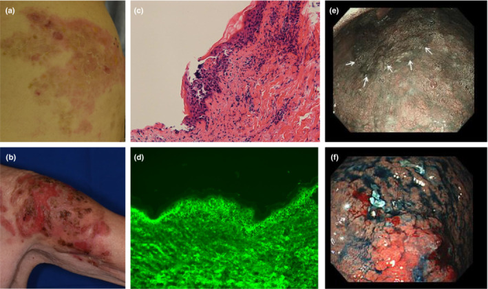
An 86‐year‐old Japanese man received the SARS‐CoV‐2 vaccine (BNT162b2) on days 0 and 21. On day 22, flaccid bullae appeared mainly in the lumbar region. PV was suspected, and the patient was referred to our hospital on day 33. On arrival at our hospital, we found flaccid bullae scattered mainly in the right lumbar region (Figure 1a) and left upper arm (Figure 1b). The skin biopsy showed an intraepidermal blister with a single basal layer remaining (Figure 1c). Direct immunofluorescent staining showed IgG deposition on the keratinocytes of the lesional epidermis (Figure 1d). Blood tests showed high levels of anti‐desmoglein 1 antibody (962.3 U/mL, normal <19.9) and anti‐desmoglein 3 antibody (131.1 U/mL, normal <19.9). After further systematic examination, upper gastrointestinal endoscopy found abnormal conditions in the hypopharynx (Figure 1e) and stomach (Figure 1f). These lesions were biopsied by a gastroenterologist, who confirmed that they were hypopharyngeal and gastric cancers. Gastric cancer was pathologically confirmed to be adenocarcinoma. Subsequent positron emission tomography showed distant metastasis. In addition to these findings, the absence of intraoral blisters, the lack of keratinocyte necrosis on histopathological examination, no deposition of C3 on the keratinocytes by direct immunofluorescent staining, and the fact that the malignant tumor in the patient was not a hematological malignancy led us to conclude that the patient had PV associated with malignant tumors rather than paraneoplastic pemphigus. 1 , 4 Although topical steroids were used, there was no improvement in the skin rash on the lumbar region, and the blisters expanded further to the face. Therefore, intravenous steroid pulse therapy (1000 mg/day) was administered on days 40–42. Subsequently, the skin symptoms improved. The patient was still on maintenance therapy with oral steroids at 30 mg/day. As a matter of preference, the patient opted out of specific therapy for the malignant tumor. Recently, a number of studies have shown that autoimmune abnormalities appear simultaneously with COVID19 infection. 5 In the same way, vaccines against COVID19 have been described to cause autoimmune abnormalities as an adverse effect. 2 , 3 These indicate that there is a molecular mimicry between COVID19 antigens and self‐antigens, possibly triggering autoimmune abnormalities in the immune response to COVID19.
FIGURE 1.

Skin symptoms, histopathological findings of skin blister lesions and gastrointestinal endoscopic findings. Flaccid bullae with erythema on the right lumbar region (a) and left upper arm (b) at the time of our hospital visit. The histopathological sections, stained with hematoxylin and eosin, revealed intraepidermal blisters (magnification × 200) (c). Direct immunofluorescent staining of tissue sections from blister lesions demonstrated IgG deposition on keratinocytes (magnification × 400) (d). Hypopharyngeal cancer (e) and gastric cancer (f). The white arrows indicate the margin of hypopharyngeal cancer.
CONFLICT OF INTEREST
None declared.
ACKNOWLEDGMENTS
None.
REFERENCES
- 1. Committee for Guidelines for the Management of Pemphigus Disease , Amagai M, Tanikawa A, Shimizu T, Hashimoto T, Ikeda S, et al. Japanese guidelines for the management of pemphigus. J Dermatol. 2014;41:471–86. [DOI] [PubMed] [Google Scholar]
- 2. Segal Y, Shoenfeld Y. Vaccine‐induced autoimmunity: the role of molecular mimicry and immune crossreaction. Cell Mol Immunol. 2018;15:586–94. [DOI] [PMC free article] [PubMed] [Google Scholar]
- 3. Li N, Aoki V, Liu Z, Prisayanh P, Valenzuela JG, Diaz LA. From insect bites to a skin autoimmune disease: a conceivable pathway to endemic pemphigus foliaceus. Front Immunol. 2022;13:907424. [DOI] [PMC free article] [PubMed] [Google Scholar]
- 4. Wade MS, Black MM. Paraneoplastic pemphigus: a brief update. Australas J Dermatol. 2005;46:1–8; quiz 9‐10. [DOI] [PubMed] [Google Scholar]
- 5. Wang EY, Mao T, Klein J, Dai Y, Huck JD, Jaycox JR, et al. Diverse functional autoantibodies in patients with COVID‐19. Nature. 2021;595:283–8. [DOI] [PubMed] [Google Scholar]


