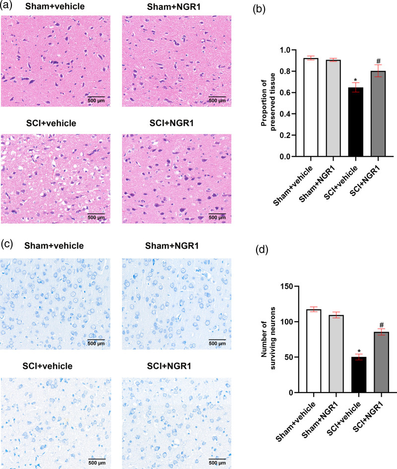
Fig. 2.
NGR1 administration attenuates tissue damage and motor neurons loss after SCI. (a) The representative image of HE staining in the Sham + vehicle, Sham + NGR1, SCI + vehicle, and SCI+NGR1 groups at 21 days after SCI. Bars = 500 μm (200×). (b) Quantitative analysis of the proportion of preserved tissue in each group. N = 6. Data are represented as mean ± SEM. *P < 0.05 vs. the Sham + vehicle group; #P < 0.05 vs. the SCI+NGR1 group. (c) The representative image of Nissl staining in the Sham + vehicle, Sham + NGR1, SCI + vehicle, and SCI + NGR1 groups at 14 days after SCI. Bars = 500 μm (200×). (d) Quantitative analysis of the number of surviving neurons in each group. N = 6. Data are represented as mean ± SEM. One-way analysis of ANOVA with Turkey’s post hoc tests was used. *P < 0.05 vs. the Sham + vehicle group; #P < 0.05 vs. the SCI+NGR1 group. ANOVA, analysis of variance; HE, hematoxylin-eosin; NGR1, notoginsenoside R1; SCI, spinal cord injury.

