Abstract
Objectives: The aim of this study was to evaluate the influence of varying dentin and enamel layer thicknesses of two nano-composite resins on color match of composite resins and lithium disilicate dental ceramic.
Materials and Methods: Twenty-six specimens of two types of nano-composite resins, Opallis and Vittra, were fabricated using the two-layered technique with different thickness ratios of enamel and dentin composites (A2 shade) with a total thickness of 1.2mm. Thirteen discs of the same shade and thickness of IPS e.max Press LT (low translucency) lithium disilicate dental ceramic were also fabricated. Specimen color was measured with a spectrophotometer. The difference in color (ΔE00) of composite and ceramic specimens, and the translucency parameter (TP) of all specimens were calculated. Data were analyzed using multi-factor ANOVA (P<0.05).
Results: The color difference (ΔE00) values of composites and ceramic were not clinically acceptable in any areas of either of the two composites (ΔE00>2.25). But ΔE00 between the two composite resins was in the clinically acceptable range (ΔE00<2.25). The mean TP value of IPS e.max Press was greater than that of Vittra and lower than that of Opallis.
Conclusion: In similar thicknesses, composite resins with any enamel/dentin thickness ratio could not successfully simulate the color and translucency of IPS e.max Press LT ceramic.
Key Words: Ceramics, Color, Composite Resins, Lithia Disilicate
Introduction
Today, the increasing demand for esthetic dental restorations has challenged clinicians to gain extra clinical skills and knowledge in the field of dental materials [ 1 ]. Despite the wide range of available options for indirect restorative materials, clinicians often prefer the use of direct restorative materials such as composite resins due to numerous advantages like lower cost, desirable clinical outcomes, bonding capability to dental structures, and conservative tooth preparation [2,3].
Full-ceramic dental restorations are among the commonly used esthetic restorations that are fabricated indirectly. IPS e.max Press (Ivoclar Vivadent AG, Schaan, Liechtenstein), introduced in 2005, is a lithium disilicate pressed glass ceramic with satisfactory physical properties, and improved translucency compared with its previous generations [4].
The shade selection process is one of the most challenging treatment steps in esthetic dentistry. The difference in color of the same designated Vita shade materials among various commercial brands makes this procedure even more challenging [5]. In fact, because of the complexity of the color and translucency of natural teeth, it is usually difficult to imitate the natural tooth appearance by using only one single Vita shade for the restoration, and multiple layers of different shades and opacities of composite resins are often needed, using the incremental or layering technique [2, 6]. The two-layered technique is one of the simplest techniques using two layers of dentin and enamel composites [7,8]. However, since variations in opacity, color, and thickness of different layers are all influential factors on the final results of restorations, implementing this technique is highly complicated and it does not guarantee an ideal color match in all clinical situations, and the selection of suitable color and translucency for optimal color match of various restorations remains problematic. The process of shade matching gets even more complicated when the clinician needs to match the color and translucency of the restorations made up of different dental materials, as when the clinician has to match the color of a new composite restoration with an existing ceramic restoration in the patient’s mouth [5-7,9-11].
The information available regarding the color match between various dental materials is limited. Considering the importance of acceptable esthetic clinical outcomes, the aim of this study was to evaluate the influence of varying dentin and enamel layer thicknesses of two nano-composite resins on color match of composite resins and lithium disilicate dental ceramics. The null hypothesis was that there would be no differences in color and translucency parameter (TP) of different layering areas of two types of composite resins and IPS e.max Press ceramic equal in thickness and of the same Vita shade.
MATERIALS AND METHODS
In this study, a comparison of color and translucency was made between two compo-sites using the layering technique, and a lithium disilicate dental ceramic, IPS e.max Press, of the same Vita shade. For this purpose, an evaluation of different combinations of enamel and dentin composite layer thicknesses was made to determine the influence of different thickness combinations on the final color and translucency, and their matching degree with ceramic restorations of the same thickness.
Preparation of composite specimens:
To standardize the composite specimen thickness and to simulate a two-layered restoration with different thicknesses of enamel and dentin composites, a special Teflon mold (internal dimensions: 1.2×15×10mm) was used to produce dentin and enamel composite layered specimens (Figure 1) [1].
Fig. 1.
Schematic illustration of specimen layers and dimensions
Thirteen specimens of each type of composite and 26 total composite specimens were fabricated as below:
Opallis Enamel A2 shade + Opallis Dentin A2 shade (FGM; Joinville, SC, Brazil)
Vittra APS Enamel A2 shade + Vittra APS Dentin A2 shade (FGM; Joinville, SC, Brazil)
Both composites match the standard VITAPAN® Classical shade scale as claimed by the manufacturer [12,13].
The composites were slightly heated in warm water prior to application to decrease their viscosity and enhance their application into the molds [1]. The Teflon mold was placed on a glass slab, and then the dentin composite was applied into the mold and pressed against the bottom with another glass slab covering it. While holding under pressure, the composite was light-cured with a curing unit (LITEX 680A Curing Light; Dentamerica Inc., CA, USA) for 40 s at 500 mW/cm2 [13]. Then, the glass cover was removed and light curing was repeated. In the second step, the enamel composite was directly applied on the previously cured dentin composite layer without any medium between the layers. A Mylar strip (Henry Schein; Melville, NY, USA) was placed on top of the enamel composite, and a glass slide was pressed against it to extrude the excess composite resin and to form a flat surface without any voids. After 10s of photo-polymerization, the glass slide was removed and the distal end of the light guide was placed against the surface of the matrix strip and the material was light-cured at 500 mW/cm2 for 20s according to the manufacturer’s recom-mendations [13]. The thicknesses of the samples were carefully measured with a digital caliper (Shoka Gulf; Malaga, Spain). The output of the curing unit was checked periodically by using a light-meter (SDS; Kerr, Orange, CA, USA). The specimens were then kept in a dark and humid environment at room temperature for 24h [1,6].
Preparation of ceramic specimens:
Thirteen ceramic specimens of IPS e.max Press (Ivoclar Vivadent AG, Schaan, Liechtenstein) with A2 shade and low translucency (LT) were fabricated in the form of discs with a diameter of 10mm and a thickness of 1.2mm, to simulate monolithic ceramic restorations. The spec-imens were initially fabricated in wax with 10mm diameter and 1.5mm thickness. The discs were fabricated according to the manufacturer’s instructions. The surfaces of the discs were then ground and polished under water spray until the thickness of 1.2±0.02mm was achieved, which was measured and controlled by a digital caliper [14].
Color measurement:
The color of the specimens was measured by a trained operator using a spectrophotometer (Micro SpectroShade; MHT, Verona, Italy) against a black and a white background. The device has a built-in aiming mode that produces a reproducible position per-pendicular to the surface of the specimens, and it was calibrated according to the manufacturer’s instructions, so that all the measurements were made under equal standardized conditions.
For each composite specimen, the color values were measured in three areas by the SpectroShade software: area 1 (thicker dentin composite), area 2 (middle), and area 3 (thicker enamel composite) (Figure 1). For each specimen area, the color measurement was repeated 3 times and the mean value was recorded [1].
Color measurements for the ceramic specimens were made in 3 random spots on the surface of the discs against a black and a white background, and the mean value was considered as the color of the specimen.
The color difference between the two types of composites, and between different areas of the composite specimens and the ceramic specimens, was calculated using CIEDE2000 system (ΔE00) with the following equation [15]:
 |
Where ΔL’ , ΔC’ , and ΔL’ are the differences in lightness, chroma, and hue, respectively, between two specimens in CIEDE2000, and RT is a rotation function accounting for the interaction between the chroma and hue differences in the blue region; SL, SC, SH are weighting functions that adjust the total color difference for variation in the location of the color difference in L’ , a’ , b’ coordinates between two color readings, and the parametric factors, KL, KC, KH, are correction terms for experimental conditions. In this study, the parametric factors were set to 1 and the clinical acceptability threshold was set at 2.25 ΔE00 units [15].
The TP of different areas of composite specimens and ceramic specimens was calculated using the following formula [1, 15]:
 |
Where the B and W subscripts refer to the color coordinates measured against the black and white backgrounds, respectively.
The data were analyzed using descriptive statistics (mean ± standard deviation). Normal distribution was verified with the Kolmogorov-Smirnov test. To analyze the effect of different thickness areas and the composite type on color match and TP of composite resin, two-factor ANOVA and the Tukey’s post-hoc test were used. Between-group translucency comparison was done by one-way ANOVA and Tukey’s post-hoc test. Statistical significance level for all the tests was set at 0.05. All the statistical analyses were conducted using SPSS 25.0 (SPSS, Chicago, IL, USA).
Results
Color difference:
The color difference (ΔE00 (composite-ceramic)) between the ceramic discs and various areas of composites is presented as mean ± standard deviation in Table 1.
Table 1.
Comparison of translucency parameter and ΔE00 (composite-ceramic) between composite resins and different areas using two-way ANOVA
| Variables | Composite | Area of different thicknesses | P* | P# | ||
|---|---|---|---|---|---|---|
| 1 | 2 | 3 | ||||
| ΔE00 (composite-ceramic) | Vittra | 5.92±1.05 | 6.83±1.38 | 6.74±0.73 | 0.21 | 0.29 |
| Opalis | 5.96±1.85 | 5.82±1.98 | 6.54±1.13 | |||
| Translucency parameter | Vittra | 12.46±0.39 | 14.63±0.58 | 15.03±0.49 | <0.001 | <0.001 |
| Opalis | 17.04±0.46 | 18.67±0.56 | 18.52±0.55 | |||
Between groups;
Within groups
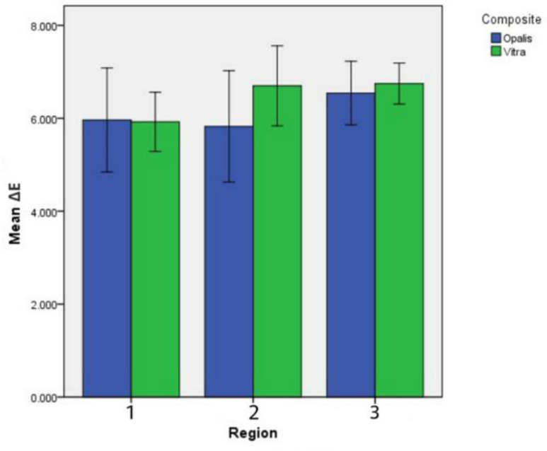
ΔE00 (composite-ceramic) values for various areas of composite resins showed that the color difference was not clinically acceptable in any area of either of the two composites (ΔE00>2.25).
Two-way ANOVA (Table 1) revealed no significant difference between composite resin types and different areas. Also, the interaction effect of these two factors was not significant (P=0.5). Figure 2 illustrates the ΔE00 of the two composite resins in different areas.
Fig. 2.
ΔE00 (composite-ceramic) of the two composite resins in different areas
The results showed that the mean±standard deviation value of ΔE00 between the Opallis and Vittra composite resins was 2.20±0.58, which was within the clinically acceptable range (ΔE00<2.25). Also, the mean ±standard deviation color difference between similar areas of the two composites in areas 1, 2, and 3 was 2.48±0.39, 1.94±0.67, and 2.20±0.55, respectively; thus, Opallis and Vittra had the least color difference in their middle area. Moreover, in areas 2 and 3, the color difference between the two composites was clinically acceptable (ΔE00<2.25), but in the area 1, the difference was not within the clinically acceptable range (ΔE00>2.25).
One-way ANOVA and Tukey’s post-hoc tests were used to compare the color difference of Opallis and Vittra in different areas. The results revealed that the difference between areas 1 and 2 was statistically significant (P=0.044), but the difference was not significant between areas 2 and 3 (P=0.417).
TP:
Two-way ANOVA (Table 1) revealed significant effects of composite resin type, different areas, and also the interaction effect of these two factors on TP (P<0.001(.
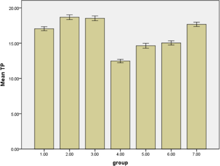
Opallis was more translucent than Vittra. Also, TP increased from area 1 towards area 3 (as the enamel layer thickened), except in Opallis in which the TP of areas 2 and 3 was equal.Comparison of the TP of different areas of the two composites showed significant differences between them (P<0.001), except for areas 2 and 3 of Vittra (p=0.43) and areas 2 and 3 of Opallis (p=0.98), where there were no significant differences.
According to one-way ANOVA, the TP value of ceramic (17.66±0.48) and different areas of both composites was significantly different (P<0.001). The bar chart (Figure 3) illustrates the mean TP of various areas of the two composites and ceramic.
Fig. 3.
Bar chart of TP of different areas of composites and ceramic. Numbers 1 to 3 refer to different areas of Opallis (from thicker to thinner dentin), 4 to 6 refer to different areas of Vittra (from thicker to thinner dentin), and number 7 refers to ceramic
Discussion
In our study, the color difference between the two types of composites was in the clinically acceptable range. This might be due to the fact that both of them were of the same commercial brand (FGM), and the color standardization and calibrations might have been done similarly for both composites by the manufacturer. However, Paravina et al. [16] reported that 75% of the composites of the same shade did not match in color. Also, Da Costa et al. [7] showed that the composite shades did not well match the Vita Shade guide, even when the layering technique was applied. In addition, statistically significant differences were found between the areas 1 and 2 of both composites; but the difference was not significant between areas 2 and 3. This may indicate the more prominent effect of dentin composite on defining the final color, in comparison with the enamel composite. These findings comply with the previous studies concluding that the dentin is considered the dental tissue of higher relevance to tooth color, and that the covering enamel plays a minor role of modulating the underlying dentinal color [17, 18]. Also, Friebel et al. [8] reported that the final color perception of layered composite restorations depended on the thickness of each layer of dentin and enamel composites, and also on the degree of translucency of each layer. Our results were in agreement with those of Vichi et al, [6] who concluded that both the layer thickness and thickness ratios of dentin and enamel composites highly affect the final appearance of restoration. Moreover, Khashayar et al. [1] found that the final color of restoration was affected by even small changes in the thickness of layers.
Despite the clinically acceptable ΔE00 values between the two composites in our study, the color difference with ceramic was not acceptable in any area of either of the two composites. Thus, the first null hypothesis of the study stating that “there would be no difference in color of different areas in equal thicknesses of composites and ceramic with similar shade (A2)”, was rejected. Therefore, a similarity in Vita shade selection, cannot be a reliable criterion for the clinician to match the shades of esthetic restorations of different materials. This color mismatch might be due to the fact that ceramic materials possess crystalline phases similar to the tooth enamel structure, unlike composites which have an amorphous structure including a resin matrix and scattered fillers; as a result, they probably present dissimilar optical behaviors.
In accordance with our study, Kim et al. [19] showed a significant color difference between a composite and a porcelain with the same shade. According to them, the probable reason for this difference might be the compositional and structural differences between the two materials. They added that the brand of composite resin had a greater effect than its shade on the color difference between porcelain and composite, and hybrid composites showed smaller color difference with porcelain in comparison with nanofilled composites [19]. In addition, Seghi et al. [20] reported that different porcelain systems also exhibited significant color differences despite having identical shades.
On the other hand, our results revealed significant differences between the TP of ceramic and various areas of both composites. Therefore, in similar thicknesses, composites in any enamel/dentin thickness ratio, could not successfully simulate the translucency of an IPS e.max (LT) ceramic; thus, the second null hypothesis was rejected as well.
Another result was that composite type, area, and the interaction between these two factors significantly affected the TP, and Opallis was more translucent than Vittra. Various factors such as the organic matrix, pigments, and the amount, size, shape, and organization of filler particles directly affect the light transmission and opacity of composites and their final color [21-25].
The Vittra APS composite formula is free of Bis-GMA and Bis-EMA (Bisphenol-A free) and contains nanospheres of zirconia silicate with an average particle size of 200 nm, and total inorganic load of 72%-82% in weight (52%-60% in volume). The advanced polymerization system technology allows polymerization with no visually noticeable change in color and opacity and longer working time under ambient light [13]. Opallis, is a nanohybrid composite resin composed of a monomeric matrix containing Bis-GMA, Bis-EMA, UDMA, and TEGDMA and glass filler particles, including barium-aluminum, silicate and nanoparticles of silicone dioxide, with particle size range of 40 nm to 3.0 µm and average size of 0.5 µm (78.5%-79.8% in weight, 57- 58% in volume) [12]. Haas et al. [26] investigated the effect of different metal oxide opacifiers on the translucency of composites and concluded that TiO2, ZrO2, and Al2O3 opacifiers decreased the translucency of UDMA-based experimental composite resins, with Al2O3 having the least effect. Accordingly, presence of spheroidal zirconia silicate filler particles in Vittra might be the reason for its lower light transmittance and higher opacity in comparison with Opallis. On the other hand, it is reported that the amount of Bis-GMA significantly affects the translucency and the refractive index of dental composite resins containing silica fillers, and it is considered as a way of adjusting the translucency of composite resins [22]. Considering the fact that Vittra is a Bis-GMA free composite, while opallis contains Bis-GMA, the difference in the composition of these two composites could be the cause of the difference in their translucencies, and the higher translucency of Opallis. In composite specimens, TP increased from area 1 to area 3, as the enamel composite layer thickness increased except for areas 2 and 3 in Opallis, which had equal TPs. Regarding the fact that enamel composite is more translucent than dentin composite, these results were expected. Our results were in agreement with the results of Rocha Maia et al, [21] who concluded that the composite layer thickness affected the light transmittance properties.
Our results revealed that in equal thicknesses (1.2mm), the mean value of TP in IPS e.max Press (LT) was greater than that of Vittra and lower than that of Opallis composite. In fact, in all proportions of dentin and enamel layer thicknesses, Vittra was opaquer than IPS e.max Press (LT) ceramic, while Opallis was more translucent. As explained before, higher opacity of Vittra might be due to the spheroidal zirconia silicate filler particles [26]. But the reason for higher opacity of IPS e.max Press ceramic compared with Opallis might be due to its crystalline structure and the presence of needle-shaped crystals of lithium disilicate that comprise about two-thirds of the volume of the glass ceramic [27].
Studies have shown that various all-ceramic systems have different translucencies compared with each other [28-30]. The translucency of materials for all-ceramic restorations varies depending on the nature of their reinforcing crystalline phase, and the more the crystalline phase, the lower the translucency would be [31]. If the crystalline size is smaller than the visible light wavelength (400-700 nm), the light will be transmitted and the ceramic will appear transparent, but if the crystalline size is greater, the light will be scattered and the material will appear more opaque [32]. Additionally, the more a ceramic material contains voids and porosities, the more light dispersion and the less light transmission will occur [33].
In the clinical situations, the final color of restorations is not only influenced by the color and optical properties of the material, but also by the background color, surface texture, and the degree of polishing of restoration [34-38]. Therefore, it is essential for the clinicians to consider all these factors.
It should be pointed out that the results of our study are only attributed to the studied composites and ceramic, and cannot be generalized to other materials. Also, in this study, the thickness of all specimens was 1.2mm while in clinical situations, a wide range of thicknesses are applied for esthetic restorations. Thus, more studies are recommended to evaluate the color match of other types of ceramics and composites and also different thicknesses of these materials.
CONCLUSION
The total mean color difference between the two composites, Opallis and Vittra, was in the clinically acceptable range, but the color difference between IPS e.max Press (LT) ceramic and different areas of the two composites was not clinically acceptable.
The type of composite affected the color of the composite only in areas with thicker dentin composite. The TP of ceramic and different areas of both composites was significantly different. In similar thicknesses, Vittra was opaquer than IPS e.max Press (LT) in all enamel/dentin thickness ratios, but Opallis was more translucent than IPS e.max Press (LT). In similar thicknesses, composites in any enamel/dentin thickness ratios could not successfully simulate the color and translucency of an IPS e.max Press (LT) ceramic.
ACKNOWLEDGMENTS
The authors would like to thank Tabriz University of Medical Sciences for the financial support provided.
Notes:
Cite this article as: Saati Khosroshahi E, Jafari Navimipour E, Pournaghi Azar F, Abed-Kahnamoui M, Bahari M. Influence of Varying Dentin and Enamel Layer Thicknesses of Nano-Composite Resins on Color Match between Lithium Disilicate Dental Ceramic and Composite Resins. Front Dent. 2021:18:12
CONFLICT OF INTEREST STATEMENT
None declared.
References
- 1.Khashayar G, Dozic A, Kleverlaan C, Feilzer A, Roeters J. The influence of varying layer thicknesses on the color predictability of two different composite layering concepts. Dent Mater. 2014 May;30(5):493–8. doi: 10.1016/j.dental.2014.02.002. [DOI] [PubMed] [Google Scholar]
- 2.Blank JT. Simplified techniques for the placement of stratified polychromatic anterior and posterior direct composite restorations. Compend Contin Educ Dent. 2003 Feb;24(2 Suppl):19–25. [PubMed] [Google Scholar]
- 3.Pontons-Melo JC, Furuse AY, Mondelli J. A direct composite resin stratification technique for restoration of the smile. Quintessence Int. 2011 Mar;42(3):205–11. [PubMed] [Google Scholar]
- 4.Stappert CF, Att W, Gerds T, Strub JR. Fracture resistance of different partial-coverage ceramic molar restorations: An in vitro investigation. J Am Dent Assoc. 2006 Apr;137(4):514–22. doi: 10.14219/jada.archive.2006.0224. [DOI] [PubMed] [Google Scholar]
- 5.Carney MN, Johnston WM. Appearance differences between lots and brands of similar shade designations of dental composite resins. J Esthet Restor Dent. 2017 Apr;29(2):E6–E14. doi: 10.1111/jerd.12263. [DOI] [PMC free article] [PubMed] [Google Scholar]
- 6.Vichi A, Fraioli A, Davidson CL, Ferrari M. Influence of thickness on color in multi-layering technique. Dent Mater. 2007 Dec;23(12):1584–9. doi: 10.1016/j.dental.2007.06.026. [DOI] [PubMed] [Google Scholar]
- 7.Da Costa J, Fox P, Ferracane J. Comparison of various resin composite shades and layering technique with a shade guide. J Esthet Restor Dent. 2010 Apr;22(2):114–24. doi: 10.1111/j.1708-8240.2010.00322.x. [DOI] [PubMed] [Google Scholar]
- 8.Friebel M, Pernell O, Cappius H-J, Helfmann J, Meinke MC. Simulation of color perception of layered dental composites using optical properties to evaluate the benefit of esthetic layer preparation technique. Dent Mater. 2012 Apr;28(4):424–32. doi: 10.1016/j.dental.2011.11.017. [DOI] [PubMed] [Google Scholar]
- 9.Mikhail SS, Schricker SR, Azer SS, Brantley WA, Johnston WM. Optical characteristics of contemporary dental composite resin materials. J Dent. 2013 Sep;41(9):771–8. doi: 10.1016/j.jdent.2013.07.001. [DOI] [PubMed] [Google Scholar]
- 10.Mikhail SS, Johnston WM. Confirmation of theoretical colour predictions for layering dental composite materials. J Dent. 2014 Apr;42(4):419–24. doi: 10.1016/j.jdent.2014.01.008. [DOI] [PubMed] [Google Scholar]
- 11.Devoto W, Saracinelli M, Manauta J. Composite in everyday practice: how to choose the right material and simplify application techniques in the anterior teeth. Eur J Esthet Dent. 2010 Spring;5(1):102–24. [PubMed] [Google Scholar]
- 12.Opallis composite resin. Available at: https://fgmdentalgroup.com/en/pages/opallis.
- 13.Vittra composite resin. Available at: https://dentaum.com.ua/image/data/FGM%20materials/ARTICLES/Mate%CC%81ria%20Vittra%20APS.pdf.
- 14.Pires LA, Novais PM, Araújo VD, Pegoraro LF. Effects of the type and thickness of ceramic, substrate, and cement on the optical color of a lithium disilicate ceramic. J Prosthet Dent. 2017 Jan;117(1):144–9. doi: 10.1016/j.prosdent.2016.04.003. [DOI] [PubMed] [Google Scholar]
- 15.Kürklü D, Azer SS, Yilmaz B, Johnston WM. Porcelain thickness and cement shade effects on the colour and translucency of porcelain veneering materials. J Dent. 2013 Nov;41(11):1043–50. doi: 10.1016/j.jdent.2013.08.017. [DOI] [PubMed] [Google Scholar]
- 16.Paravina RD, Kimura M, Powers JM. Color compatibility of resin composites of identical shade designation. Quintessence Int. 2006 Oct;37(9):713–9. [PubMed] [Google Scholar]
- 17.Dietschi D. Layering concepts in anterior composite restorations. J Adhes Dent. 2001 Spring;3(1):71–80. [PubMed] [Google Scholar]
- 18.Magne P, Holz J. Stratification of composite restorations: systematic and durable replication of natural aesthetics. Pract Periodontics Aesthet Dent. 1996 Jan-Feb;8(1):61–8. [PubMed] [Google Scholar]
- 19.Kim SH, Lee YK, Lim BS, Rhee SH, Yang HC. Difference in color and color parameters between dental porcelain and porcelain‐repairing resin composite. J Biomed Mater Res B Appl Biomater. 2006 Jan;76(1):149–54. doi: 10.1002/jbm.b.30344. [DOI] [PubMed] [Google Scholar]
- 20.Seghi RR, Johnston WM, O'brien W. Spectrophotometric analysis of color differences between porcelain systems. J Prosthet Dent. 1986 Jul;56(1):35–40. doi: 10.1016/0022-3913(86)90279-9. [DOI] [PubMed] [Google Scholar]
- 21.Rocha Maia R, Oliveira D, D'antonio T, Qian F, Skiff F. Comparison of light-transmittance in dental tissues and dental composite restorations using incremental layering build-up with varying enamel resin layer thickness. Restor Dent Endod. 2018 Apr 16;43(2):e22. doi: 10.5395/rde.2018.43.e22. [DOI] [PMC free article] [PubMed] [Google Scholar]
- 22.Azzopardi N, Moharamzadeh K, Wood DJ, Martin N, van Noort R. Effect of resin matrix composition on the translucency of experimental dental composite resins. Dent Mater. 2009 Dec;25(12):1564–8. doi: 10.1016/j.dental.2009.07.011. [DOI] [PubMed] [Google Scholar]
- 23.Lee YK. Translucency of human teeth and dental restorative materials and its clinical relevance. J Biomed Opt. 2015 Apr;20(4):045002. doi: 10.1117/1.JBO.20.4.045002. [DOI] [PubMed] [Google Scholar]
- 24.Lim YK, Lee YK, Lim BS, Rhee SH, Yang HC. Influence of filler distribution on the color parameters of experimental resin composites. Dent Mater. 2008 Jan;24(1):67–73. doi: 10.1016/j.dental.2007.02.007. [DOI] [PubMed] [Google Scholar]
- 25.Arikawa H, Kanie T, Fujii K, Takahashi H, Ban S. Effect of filler properties in composite resins on light transmittance characteristics and color. Dent Mater J. 2007 Jan;26(1):38–44. doi: 10.4012/dmj.26.38. [DOI] [PubMed] [Google Scholar]
- 26.Haas K, Azhar G, Wood DJ, Moharamzadeh K, van Noort R. The effects of different opacifiers on the translucency of experimental dental composite resins. Dent Mater. 2017 Aug;33(8):e310–6. doi: 10.1016/j.dental.2017.04.026. [DOI] [PubMed] [Google Scholar]
- 27.McLaren EA, Cao PT. Ceramics in dentistry—part I: classes of materials. Inside Dent. 2009 Oct;5(9):94–103. [Google Scholar]
- 28.Jurišić S, Jurišić G, Zlatarić DK. In vitro evaluation and comparison of the translucency of two different all-ceramic systems. Acta Stomatol Croat. 2015 Sep;49(3):195–203. doi: 10.15644/asc49/3/1. [DOI] [PMC free article] [PubMed] [Google Scholar]
- 29.Heffernan MJ, Aquilino SA, Diaz-Arnold AM, Haselton DR, Stanford CM, Vargas MA. Relative translucency of six all-ceramic systems. Part II: core and veneer materials. J Prosthet Dent. 2002 Jul;88(1):10–5. [PubMed] [Google Scholar]
- 30.Barizon KT, Bergeron C, Vargas MA, Qian F, Cobb DS, Gratton DG, et al. Ceramic materials for porcelain veneers: part II. Effect of material, shade, and thickness on translucency. J Prosthet Dent. 2014 Oct;112(4):864–70. doi: 10.1016/j.prosdent.2014.05.016. [DOI] [PubMed] [Google Scholar]
- 31.Powers JM, Sakaguchi RL, Craig RG. Craig's restorative dental materials/edited by Ronald L. In: Ronald L. Sakaguchi, John M., editors. Powers. 13th ed. . Philadelphia: Elsevier/Mosby; 2012. 255 pp. [Google Scholar]
- 32.Van Noort R. 4th ed. Philadelphia: Elsevier/Mosby; 2013. Introduction to dental materials; 212 pp. [Google Scholar]
- 33.O'Keefe K, Pease P, Herrin H. Variables affecting the spectral transmittance of light through porcelain veneer samples. J Prosthet Dent. 1991 Oct;66(4):434–8. doi: 10.1016/0022-3913(91)90501-m. [DOI] [PubMed] [Google Scholar]
- 34.Barath VS, Faber FJ, Westland S, Niedermeier W. Spectrophotometric analysis of all-ceramic materials and their interaction with luting agents and different backgrounds. Adv Dent Res. 2003 Dec;17:55–60. doi: 10.1177/154407370301700113. [DOI] [PubMed] [Google Scholar]
- 35.Li Q, Yu H, Wang Y. Spectrophotometric evaluation of the optical influence of core build-up composites on all-ceramic materials. Dent Mater. 2009 Feb;25(2):158–65. doi: 10.1016/j.dental.2008.05.008. [DOI] [PubMed] [Google Scholar]
- 36.Spyropoulou P-E, Giroux EC, Razzoog ME, Duff RE. Translucency of shaded zirconia core material. J Prosthet Dent. 2011 May;105(5):304–7. doi: 10.1016/S0022-3913(11)60056-5. [DOI] [PubMed] [Google Scholar]
- 37.Wang H, Xiong F, Zhenhua L. Influence of varied surface texture of dentin porcelain on optical properties of porcelain specimens. J Prosthet Dent. 2011 Apr;105(4):242–8. doi: 10.1016/S0022-3913(11)60039-5. [DOI] [PubMed] [Google Scholar]
- 38.Peyton JH. Finishing and polishing techniques: direct composite resin restorations. Pract Proced Aesthet Dent. 2004 May;16(4):293–8. [PubMed] [Google Scholar]


