Abstract
Objectives: This study aimed to evaluate the efficacy of ProTaper, Mtwo, and WaveOne retreatment files and Hedstrom files for removal of gutta-percha from the straight root canals using cone-beam computed tomography (CBCT).
Materials and Methods: Forty freshly extracted single-rooted and single-canal teeth were selected for this study. The teeth were decoronated, and biomechanical preparation was performed up to #30 K-file. The root canals were obturated using lateral compaction technique with gutta-percha and Resilon sealer. The teeth were then randomly divided into 4 groups, and CBCT images were obtained. All the canals were then retreated with either ProTaper retreatment files, Mtwo retreatment files, WaveOne files, or Hedstrom files. The surface area of the remaining filling material after the retreatment procedure was quantified by CBCT. Statistical analysis was performed via one-way ANOVA and the Tukey-Kramer multiple comparisons test.
Results: None of the file systems could completely remove the filling material from the canals. Data analysis revealed significant differences between the groups in the apical and middle thirds (P<0.05).
Conclusion: All the file systems left some filling material in the canals. Mtwo retreatment files had maximum efficacy for removal of filling materials in comparison with other files. WaveOne files can also be used for root canal retreatment.
Key Words: Gutta-Percha, Resilon Sealer, Cone-Beam Computed Tomography, Root Canal Preparation, Root Canal Therapy
Introduction
Endodontic treatment failure may occur due to the residual bacteria remaining in the root canal system as a result of inadequate biomechanical preparation, imperfect obturation, or improper post-endodontic restoration, leading to coronal/apical leakage [1]. The endodontic treatment outcome can be evaluated by radiographic examination and also based on clinical signs and symptoms of the treated teeth [2].
Preservation of a healthy periapical tissue is one objective of non-surgical removal of gutta-percha from the root canal system. Success of retreatment procedures depends on complete removal of the root filling material and persistent bacteria, as well as appropriate re-obturation [3]. Gutta-percha and root canal sealer are most commonly used as root filling materials, and provide a hermetic seal [4,5].
Use of hand files for the retreatment procedure of the root canals with well-condensed root filling material with or without solvent is tiresome and time consuming [6]. Therefore, it is important to find the proper rotary file system for easy and effective removal of remnants from the root canal system [7]. One major advantage of using rotary instruments for gutta-percha removal is their fast action [8].
The ProTaper Universal retreatment system is composed of three files (D1, D2 and D3) with the taper and tip size of 0.09/0.30mm, 0.08/0.25mm and 0.07/0.20mm, respectively. D1, D2 and D3 have been exclusively designed for gutta-percha removal from the coronal third, middle third, and apical third of the root canal system, respectively. The Mtwo retreat-ment system is composed of two retreatment files (size 15 with 0.05 taper and size 25 with 0.05 taper) with cutting tips for effective removal of root canal filling material [9]. Also, biomechanical preparation of the root canal system using nickel-titanium files with M-wire alloy with one single file from the start to finish is a newly introduced concept [10].
Reciprocating files are manufactured from the M-wire NiTi alloy, which allows for higher flexibility and greater resistance to cyclic fatigue in comparison with the conventional NiTi files [11]. Currently, Reciproc and WaveOne are two reciprocating files available in the market. Reciproc has three file sizes (R25, R40 and R50) while WaveOne contains a small (21.06), a primary (25.08) and a large (40.08) file [12,13]. The procedure and technique of endodontic retreatment with rotary files are the same as the conventional procedure, and the gutta-percha is retrieved by the brushing motion of a file against the lateral root canal walls [13].
Cone-beam computed tomography (CBCT) is commonly used in endodontics, and has higher efficacy than conventional radiography for diagnosis of periapical pathologies and internal and external root resorption defects, evaluation of root canal morphology, and management of endodontic surgery [9,14]. The aim of the present study was to compare the gutta-percha removal efficacy of ProTaper, Mtwo, and WaveOne retreatment files and Hedstrom files from the root canals using CBCT. The null hypothesis was that there would be no statistically significant difference in gutta-percha removal efficacy of ProTaper, Mtwo and WaveOne retreatment files and Hedstrom files.
MATERIALS AND METHODS
Root canal treatment:
Forty freshly extracted human mandibular single-rooted and single-canal premolars were collected after gaining ethical approval. Preoperative mesiodistal and buccolingual radiographs were obtained from each root to confirm the canal configuration and complete root formation, and ensure absence of root fillings, pins, internal resorptions or localized/ diffuse calcifications.
Access cavity was prepared using high speed handpiece and diamond burs under continuous water irrigation. To ensure canal patency, a #10 K-file was placed in the canal. Roots were decoronated using a diamond disc operated at low-speed and standardized at 16mm length. The root canals were prepared using K-files with the step-back technique. Instrumentation was standardized with a #30 K-file reaching to the working length. Subsequently #35, #40, #45, #50, and #55 K-files were used for step-back canal preparation, and final coronal flaring was performed with the Gates Glidden drills. During shaping, each canal was irrigated with 17% EDTA (for smear layer removal) and 2.5% NaOCl.
Canal obturation:
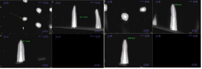
The root canals were dried with paper points and obturated by lateral compaction, using gutta-percha cones (Dentsply Maillefer, Ballaigues, Switzerland) and Resilon sealer (Research LLC, Madison, CT, USA). The coronal access cavity was sealed with a temporary filling material (Cavit G; 3M ESPE, Seefeld, Germany). The specimens were stored at 37ºC in 100% humidity for 2 weeks. At this stage, all primary CBCT images were taken using i-CAT CBCT scanner with 120kVp and 3-8mA (Imaging Sciences International Inc., Hatfield, PA, USA) (Fig. 1).
Fig. 1.
Preoperative Images of obturated extracted premolars
Retreatment technique:
The samples were randomly allocated to groups 1 to 4 (n=20) according to the retreatment technique. A drop of xylene solvent was introduced into each canal and left in the canal for 2 min.
Group I: Hedstrom files
Hedstrom files (H type; Dentsply Maillefer, Ballaigues, Switzerland) #45, #40, #35, #30 and #25 were used in the crown-down technique using circumferential quarter turn push-pull filing motion to remove gutta-percha and sealer from the canal until the working length was reached with a #25 Hedstrom file. Sizes 15 and 20 Hedstrom files were used for deep penetration down into the canal until the working length was reached. A step-back procedure was performed with 1mm increments to #55 file. After withdrawal, the files were cleansed of any filling material before being reintroduced again into the root canal. Each file was discarded after instrumentation of five canals. Deformed files were discarded. Retreatment was considered complete when no filling material was observed on the file, and the canal walls were smooth and free of visible debris (Fig. 2).
Fig. 2.
Postoperative CBCT images of the samples treated with Hedstrom file, ProTaper, Mtwo, and WaveOne retreatment files
Group II: ProTaper retreatment system
All the 3 ProTaper Universal System retreatment files were used sequentially with the crown-down technique, until the working length was reached using a brushing action with lateral pressing movements. The D1 ProTaper file was used to remove the filling material from the cervical third of the root canal. A D2 ProTaper file was used in the coronal two thirds of the root canal. The D3 ProTaper file was used with light apical pressure until the working length was reached and no further filling material could be removed.
Group III: Mtwo retreatment system
Mtwo retreatment file was used according to the manufacturer’s instructions. Retreatment was initiated by placing the tip of R2 size 25, 0.05 taper retreatment file on the gutta-percha. The canals were instrumented to the working length using Mtwo R2 file with circumferential filing and a lateral pressing movement. Progression of the rotary files was performed by applying slight apical pressure and frequently removing the files to inspect the blade and clean the flutes from the debris.
Group IV: WaveOne system:
WaveOne single file (Dentsply Maillefer, Ballaigues, Switzerland) was used with a WaveOne motor (Dentsply Maillefer, Ballaigues, Switzerland) and operated with a reciprocating handpiece. The files were used with a progressive up and down movement no more than four times with minimal apical pressure. The files were then removed and wiped clean.
CBCT evaluation:
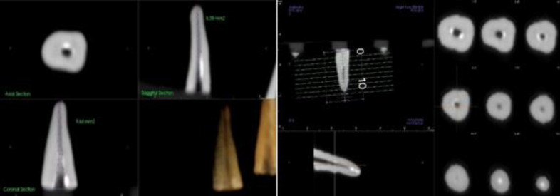
Removal of the filing material from the canal walls was evaluated by CBCT using INVIVO-5.1 View software (Imaging Sciences Inter-national Inc., Hatfield, PA, USA). Forty teeth were fixed in 1-cm thick wax plates and placed on the desk of the i-CAT tomography device (120kVp, 3-8mA; Imaging Sciences Inter-national Inc., Hatfield, PA, USA) for image acquisition. The longitudinal axis of each tooth was determined by the rotation tool. The area with maximum filling material was measured on axial, coronal and sagittal sections after adjusting the appropriate parameters for scanning, with 0.2mm voxel size (Fig. 2).
The surface area of the canal and the residual filling material were calculated on the coronal and sagittal sections, and the percentage of the remaining filling material on the canal walls was calculated with the following equation:
APRFM= × 100
APRFM represents area percentage of the remaining filling material. A grading system was used to score the amount of residual filling material in the coronal, middle and apical portion of each canal as follows [15]:
1. No or slight presence (0-25%) of debris on the dentinal surface
2. Some (25-50%) debris on the dentinal surface
3. Moderate (50-75%) amount of debris on the dentinal surface
4. Heavy presence (>75%) of debris on the dentinal surface. No attempt was made to distinguish between the filling material and sealer remnants.
Next, the coronal, middle and apical thirds of each tooth were evaluated on axial sections (Fig. 3).
Fig. 3.
Axial, coronal and sagittal sections of the coronal, middle and apical thirds
Statistical analysis:
Statistical analysis was carried out using SPSS version 16 software. Descriptive statistics such as mean and standard deviation were used to report the data. Comparisons between the groups were performed using ANOVA followed by Tukey-Kramer multiple comparisons test. Intergroup comparison was made using t-test.
Results
The results showed that all the files used in the present study had optimal gutta-percha removal efficacy, but none of the files could completely remove the gutta-percha from the canals. There was a significant difference between Mtwo and Hedstrom files, and also ProTaper and Hedstrom files in the coronal and sagittal sections. The largest area of filling material remnants in the coronal, middle and apical thirds was seen in the Hedstrom file group followed by the WaveOne and ProTaper retreatment files. The least filling material remnants were seen in Mtwo retreatment file group. The percentage of remaining debris in each treatment group was calculated by means of i-CAT.
Tables 1-4 show the comparison of files in the coronal and sagittal sections. Figure 4 shows the residual filling material in the apical, middle and coronal third of the roots after using different files. There was no significant difference between Mtwo file, ProTaper retreatment file and WaveOne file, but there was a significant difference between Mtwo and Hedstrom files (P<0.05). There was a significant difference between ProTaper and Hedstrom files as well (P<0.05). There was no significant difference between WaveOne and Hedstrom files. As shown in Table 3, there was a significant difference within groups between the apical and middle sections. The groups were not significantly different in the coronal section (P=0.7).
Table 1.
Comparison of residual filling material in Mtwo with ProTaper, WaveOne and Hedstrom file in the coronal and sagittal sections (mean±standard deviation)
| Section | Mtwo | ProTaper | Mean difference | 95% CI of difference | t-value | P |
|---|---|---|---|---|---|---|
| Coronal | 36.76±8.6 | 40.54±14.2 | 3.78 | -7.2–14.8 | 0.72 | 0.48 |
| Sagittal | 40.28±10.2 | 41.56±7.1 | 1.28 | -6.9–9.5 | 0.32 | 0.75 |
| WaveOne | ||||||
| Coronal | 36.76±8.6 | 47.94±17.9 | 11.18 | -2.03–24.38 | 1.78 | 0.09 |
| Sagittal | 40.28±10.2 | 43.76±6.34 | 3.48 | -4.48–11.44 | 0.92 | 0.37 |
| Hedstrom | ||||||
| Coronal | 36.76±8.6 | 55.16±14.4 | 14.62 | 1.21–28.03 | 2.29 | 0.03 |
| Sagittal | 40.28±10.2 | 52.58±12.66 | 11.02 | 1.36–20.68 | 2.39 | 0.03 |
Table 4.
Mean values and standard deviations of residual filling material in each group
| Area | Mtwo | ProTaper | WaveOne | Hedstrom | F-value | P |
|---|---|---|---|---|---|---|
| Apical | 12.76±1.52 | 13.69±1.31 | 13.80±1.20 | 15.13±1.87 | 4.24 | 0.01 |
| Middle | 8.56±0.89 | 9.17±1.32 | 9.30±1.20 | 10.13±1.13 | 3.17 | 0.04 |
| Coronal | 4.18±0.17 | 4.67±1.32 | 4.74±1.31 | 5.39±0.69 | 2.48 | 0.07 |
Fig. 4.
Residual filling material in the apical, middle and coronal thirds of the roots after using different files
Table 3.
Comparison of residual filling material between WaveOne and Hedstrom file (mean±standard deviation)
| Section | Wave One | Hedstrom | Mean difference | 95% CI of difference | t-value | P |
|---|---|---|---|---|---|---|
| Coronal | 47.94±17.9 | 55.16±14.4 | 7.22 | -8.04–22.48 | 0.99 | 0.33 |
| Sagittal | 43.76±6.34 | 52.58±12.66 | 8.82 | -0.58–18.23 | 1.97 | 0.064 |
There was no significant difference between Mtwo and ProTaper in the apical, middle or coronal third (P=0.16, P=0.24, P=0.47, respectively). There was a significant difference between Mtwo and Hedstrom files in the apical, middle and coronal thirds (P=0.00 for all).
Table 4 shows the mean values and standard deviation of residual filling material in each group. Analyzing the data showed that there was a significant difference between the groups in the apical (P=0.01) and middle thirds (P=0.04). In the coronal section, the groups did not have a significant difference (P>0.05).
Table 2.
Comparison of residual filling material in ProTaper, with WaveOne and Hedstrom file in the coronal and sagittal sections (mean±standard deviation)
| Section | ProTaper | WaveOne | Mean difference |
95% CI of
difference |
t-value | P |
|---|---|---|---|---|---|---|
| Coronal | 40.54±14.2 | 47.94±17.9 | 7.40 | -7.77–22.58 | 1.02 | 0.32 |
| Sagittal | 41.56±7.1 | 43.76±6.34 | 2.20 | -8.55–4.15 | 0.73 | 0.47 |
| Hedstrom | ||||||
| Coronal | 40.54±14.2 | 55.16±14.4 | 14.62 | 1.21–28.03 | 2.29 | 0.03 |
| Sagittal | 41.56±7.1 | 52.58±12.66 | 11.02 | 1.36–20.68 | 2.39 | 0.03 |
Discussion
Persistent residual bacteria are the main reason for failure of root canal treatment [16]. Success of non-surgical root canal retreatment depends on complete elimination of root filling materials [8]. Complete removal of pre-existing filling materials from the canals is a prerequisite for successful nonsurgical root canal retreatment. This procedure can also remove the residual necrotic tissues or bacteria that may be responsible for persistent periapical inflammation, and allow further cleaning and refilling of the root canal system. Different methodologies have been reported to evaluate the amount of filling material remaining in the root canal after the retreatment procedure [8]. It can be assessed by radiography [17], longitudinal root splitting and linear measurement of the remaining gutta-percha and sealer, or by using a scoring system [18] or a clearing technique. Computed tomography [19] and microscopes have also been used for this purpose. Ideally, three-dimensional visualization of the root canal system would provide a better understanding of the distribution of debris after retreatment.
Several methods and instruments can be used for retreatment of the root canal system such as stainless steel hand files, Gate Glidden drills, nickel titanium rotary instruments, ultrasonic devices, heat-bearing instruments, lasers, and solvents [20].
In the current study, all teeth were decoronated to ensure standardization of the teeth [21]. Several obturation techniques are available for root canal treatment. The choice depends on the canal anatomy and the unique objectives of treatment in each case. Newer methods include the use of injectable, thermoplasticized gutta-percha, carriers coated with an alpha phase gutta-percha, cold flowable filling materials that combine gutta-percha and sealer in one product, and glass ionomer-embedded gutta-percha points [22]. Two basic obturation procedures include the lateral compaction and warm vertical condensation. The lateral compaction technique has been the gold standard to which other techniques have been compared.
The lateral cold compaction technique has the advantage of excellent length control. Also, it can be used with any acceptable sealer. The lateral compaction technique is extensively used for root canal obturation [15].
Rotary and Hedstrom files with solvents were used in this study because they decrease patient and operator fatigue [11]. Conventional radiography cannot provide accurate information in all dimensions; whereas, CBCT provides three-dimensional information. CBCT has been designed to represent three-dimensional views of the maxillomandibular region, with a lower patient radiation dose compared with computed tomography. This technique gives detailed information about the morphological features without damaging the teeth or their surrounding tissue [23].
CBCT eliminates the shortcomings of two-dimensional imaging, has lower radiation dose than medical computed tomography, and enables the clinicians to make more accurate decisions and design more accurate treatment plans. Three important parameters of CBCT include voxel size, field of view and slice thickness. The best voxel size would be 0.2mm, with a shorter scanning time and lower radiation dose of patient [24]. Smaller voxel size increases the noise and the resolution. Images in smaller voxel sizes are sharper. For suspected root fractures, the used voxel size should be <0.2mm. For evaluation of internal root resorption, the voxel size should be 0.16mm. Voxel size used for the detection of depth of proximal carious lesions is 0.125mm. The voxel size of 0.2mm is more accurate than 0.4mm. Voxel size is important in terms of image quality and has a direct correlation with scanning and image reconstruction time as well as the field of view, amperage and voltage [25]. In this study, 0.2mm voxel size was used.
In the present study, all teeth showed residual filling materials, and none of the file systems could completely remove the filling material. Among the tested file systems, Mtwo retreatment files had maximum efficacy for removal of the filling material followed by ProTaper retreatment files and WaveOne.
The maximum amount of residual filling material was recorded in Hedstrom file group. Mtwo Retreatment files had maximum efficacy for removal of filling material, because of the design and characteristic features of these files. Mtwo retreatment files have positive rake angle, two cutting edges, increasing pitch length from the apical towards the coronal region, and S-shaped cross section. The cutting blades form a vertical spiral, which provides better control and precise cutting through the canal [9].
The results of the present study were in agreement with those of Yadav et al, [9] who found that Mtwo and ProTaper retreatment files left less filling material. Contrary to the results of the present study, Khedmat et al. [20] reported that ProTaper retreatment files had maximum removal efficacy of filling materials compared with Mtwo retreatment files. After Mtwo retreatment files, ProTaper retreatment files showed maximum gutta-percha removal efficacy in this study. Superior performance of gutta-percha removal by ProTaper retreatment files was attributed to its progressive taper and length [26]. In the present study, WaveOne files also efficiently removed gutta-percha from the canals, as it has a reciprocating motion as well as marked taper, which makes greater contact area between the instrument and gutta-percha [27]. Silva et al. [27] showed that WaveOne files have faster rate of removal of gutta-percha than ProTaper retreatment files.
CONCLUSION
None of the systems tested in this study could completely remove gutta-percha from the canals. Further in vivo studies are required to evaluate post-instrumentation pain and prognosis in use of these systems.
Notes:
Cite this article as: Madhu K, Karade P, Chopade R, Jadhav Y, Chodankar K, Alane U. CBCT Evaluation of Gutta-Percha Removal Using Protaper and Mtwo Retreatment Files, Wave One, and Hedstrom Files: An Ex Vivo Study. Front Dent. 2021;18:19.
CONFLICT OF INTEREST STATEMENT
None declared.
References
- 1.Giuliani V, Cocchetti R, Pagavino G. Efficacy of ProTaper universal retreatment files in removing filling materials during root canal retreatment. J Endod. 2008 Nov;34(11):1381–4. doi: 10.1016/j.joen.2008.08.002. [DOI] [PubMed] [Google Scholar]
- 2.Lin LM, Skribner JE, Gaengler P. Factors associated with endodontic treatment failures. J Endod. 1992 Dec;18(12):625–7. doi: 10.1016/S0099-2399(06)81335-X. [DOI] [PubMed] [Google Scholar]
- 3.Hammad M, Qualtrough A, Silikas N. Three-dimensional evaluation of effectiveness of hand and rotary instrumentation for retreatment of canals filled with different materials. J Endod. 2008 Nov;34(11):1370–3. doi: 10.1016/j.joen.2008.07.024. [DOI] [PubMed] [Google Scholar]
- 4.Imura N, Zuolo ML, Ferreira MO, Novo NF. Effectiveness of the Canal Finder and hand instrumentation in removal of gutta percha root fillings during root canal retreatment. Int Endod J. 1996 Nov;29(6):382–6. doi: 10.1111/j.1365-2591.1996.tb01402.x. [DOI] [PubMed] [Google Scholar]
- 5.Keles A, Koseoglu M. Dissolution of root canal sealers in EDTA and NaOCl solutions. J Am Dent Assoc. 2009 Jan;140(1):74–9. doi: 10.14219/jada.archive.2009.0021. [DOI] [PubMed] [Google Scholar]
- 6.Pinto de Oliveira D, Vicente Baroni Barbizam J, Trope M, Teixeira FB. Comparison between gutta-percha and Resilon removal using two different techniques in endodontic retreatment. J Endod. 2006 Apr;32(4):362–4. doi: 10.1016/j.joen.2005.12.006. [DOI] [PubMed] [Google Scholar]
- 7.Ring J, Murray PE, Namerow KN, Moldauer BI, Garcia-Godoy F. Removing root canal obturation materials: a comparison of rotary file systems and re-treatment agents. J Am Dent Assoc. 2009 Jun;140(6):680–8. doi: 10.14219/jada.archive.2009.0254. [DOI] [PubMed] [Google Scholar]
- 8.Schirrmeister JF, Wrbas KT, Schneider FH, Altenburger MJ, Hellwig E. Effectiveness of a hand file and three nickel titanium rotary instruments for removing gutta-percha in curved root canals during retreatment. Oral Surg Oral Med Oral Pathol Oral Radiol Endod. 2006 Apr;101(4):542–7. doi: 10.1016/j.tripleo.2005.03.003. [DOI] [PubMed] [Google Scholar]
- 9.Yadav P, Bharath MJ, Sahadev CK, Ramachandra PK, Rao Y, Ali A, et al. An in vitro CT comparison of gutta-percha removal with two rotary systems and Hedstrom files. Iran Endod J. 2013;8(2):59–64. [PMC free article] [PubMed] [Google Scholar]
- 10.Prichard J. Rotation or reciprocation: A contemporary look at NiTi instruments. Br Dent J. 2012;212:345–6. doi: 10.1038/sj.bdj.2012.268. [DOI] [PubMed] [Google Scholar]
- 11.Koçak MM, Koçak M, Türker S, Sağlam BC. Cleaning efficacy of reciprocal and rotary systemsin the removal of root canal filling material. J Conserv Dent. 2016 Mar;19(2):184–6. doi: 10.4103/0972-0707.178706. [DOI] [PMC free article] [PubMed] [Google Scholar]
- 12.Alapati SB, Brantley WA, Iijma M, Clark WA, Kovarik L, Buie C, et al. Metallurgical characterization of a new nickel-titanium wire for rotary endodontic instruments. J Endod. 2009 Nov;35(11):1589–93. doi: 10.1016/j.joen.2009.08.004. [DOI] [PubMed] [Google Scholar]
- 13.Gutmann JL, Gao Y. Alteration in the inherent metallic and surface properties of nickel-titanium root canal instruments to enhance performance, durability and safety: A focused review. Int Endod J. 2012 Feb;45(2):113–28. doi: 10.1111/j.1365-2591.2011.01957.x. [DOI] [PubMed] [Google Scholar]
- 14.Kim S. Endodontic application of cone-beam computed tomography in South Korea. J Endod. 2012 Feb;38(2):153–7. doi: 10.1016/j.joen.2011.09.008. [DOI] [PubMed] [Google Scholar]
- 15.Somma F, Cammarota G, Plotino G, Grande NM, Pameijer CH. The effectiveness of manual and mechanical instrumentation for the retreatment of three different root canal filling materials. J Endod. 2008 Apr;34(4):466–9. doi: 10.1016/j.joen.2008.02.008. [DOI] [PubMed] [Google Scholar]
- 16.Nair PR, Sjögren U, Krey G, Kahnberg KE, Sundqvist G. Intraradicular bacteria and fungi in root-filled, asymptomatic human teeth with therapy-resistant periapical lesions: a long-term light and electron microscopic follow-up study. J Endod. 1990 Dec;16(12):580–8. doi: 10.1016/S0099-2399(07)80201-9. [DOI] [PubMed] [Google Scholar]
- 17.De Carvalho Maciel AC, Zaccaro Scelza MF. Efficacy of automated versus hand instrumentation during root canal retreatment: an ex vivo study. Int Endod J. 2006 Oct;39(10):779–84. doi: 10.1111/j.1365-2591.2006.01148.x. [DOI] [PubMed] [Google Scholar]
- 18.Hülsmann M, Stotz S. Efficacy, cleaning ability and safety of different devices for gutta-percha removal in root canal retreatment. Int Endod J. 1997 Jul;30(4):227–33. doi: 10.1046/j.1365-2591.1997.00036.x. [DOI] [PubMed] [Google Scholar]
- 19.Barletta FB, Rahde ND, Limongi O, Maranhao Moura AA, Zanesco C, Mazocatto G, et al. In vitro comparative analysis of 2 mechanical techniques for removing gutta-percha during retreatment. J Can Dent Assoc. 2007 Feb;73(1):65–65e. [PubMed] [Google Scholar]
- 20.Khedmat S, Azari A, Shamshiri AR, Fadae M, Fakhar HB. Efficacy of ProTaper and Mtwo retreatment files in removal of gutta-percha and GuttaFlow from root canals. Iran Endod J. 2016;11(3):184–7. doi: 10.7508/iej.2016.03.007. [DOI] [PMC free article] [PubMed] [Google Scholar]
- 21.Pooja L, Navneet G, Ravi V. Evaluation of efficiency of three NiTi instruments in removing gutta-percha from root canal during retreatment - An in vitro study. Endodontology. :80–8. [Google Scholar]
- 22.Girish K. New root canal obturation techniques: A review. EC Dent Sci. 2017;11(2):68–76. [Google Scholar]
- 23.Marfisi K, Mercade M, Plotino G, Duran‐Sindreu F, Bueno R, Roig M. Efficacy of three different rotary files to remove gutta-percha and Resilon from root canals. Int Endod J. 2010 Nov;43(11):1022–8. doi: 10.1111/j.1365-2591.2010.01758.x. [DOI] [PubMed] [Google Scholar]
- 24.Özer SY. Detection of vertical root fractures by using cone beam computed tomography with variable voxel sizes in an in vitro model. J Endod. 2011 Jan;37(1):75–9. doi: 10.1016/j.joen.2010.04.021. [DOI] [PubMed] [Google Scholar]
- 25.Spin-Neto R, Gotfredsen E, Wenzel A. Impact of voxel size variation on CBCT base diagnostic outcome in dentistry: a systemic review. J Digit Imaging. 2013;26(4):813–20. doi: 10.1007/s10278-012-9562-7. [DOI] [PMC free article] [PubMed] [Google Scholar]
- 26.Gu LS, Ling JQ, Wei X, Huang XY. Efficacy of ProTaper Universal rotary retreatment system for gutta-percha removal from root canals. Int Endod J. 2008 Apr;41(4):288–95. doi: 10.1111/j.1365-2591.2007.01350.x. [DOI] [PubMed] [Google Scholar]
- 27.Silva EJ, Orlowsky NB, Herrera DR, Machado R, Krebs RL, Countinho-Filho TD. Effectiveness of rotatory and reciprocating movements in root canal filling material removal. Braz Oral Res. 2015;29:1–6. doi: 10.1590/1807-3107bor-2015.vol29.0008. [DOI] [PubMed] [Google Scholar]


