Abstract
Endoscopic lung volume reduction using unidirectional endobronchial valves is a new technique in the treatment of patients with severe emphysema. However, the movements of the thoracic structures after endobronchial valves insertion are still unpredictable We report the unusual outcome of six patients after valves insertion in the left upper lobe. They all developed a complete atelectasis of the target lobe, a pneumothorax and sequential genuine bullae in the treated left lung of unknown etiology. The chest CT scan prior to the valves insertion was unremarkable. Three patients developed an air–liquid level in the bullae the day before a bacterial infection of their left lower lobe. The three other patients had an uneventful spontaneous resolution of their bullae at long-term follow-up. Therefore, a conservative attitude should be followed in this particular setting.
Keywords: endoscopic lung volume reduction, pneumothorax, chest CT scanner, chest tube drainage
Introduction
Endoscopic lung volume reduction (ELVR) using unidirectional endobronchial valves (EBV) is an established guideline technique to improve lung function, exercise capacity and quality of life in patients with severe emphysema.1,2 However, the movements of the thoracic structures (lungs, ribs, mediastinum and diaphragm) after EBV insertion are unpredictable. Additionally, the occurrence of pneumothorax, which is the most frequent adverse event following EBV placement, was found in 4.2–34.4% of the treated patients.3 Because the ipsilateral lobe rapidly expands into the space created in the pleural cavity by atelectasis of the target lobe, some blebs or bullae from the ipsilateral lobe could break up, especially in the presence of pleural adhesions.3,4 Some risk factors were reported in association with a higher rate of pneumothorax such as a high percentage of low attenuation volume of the untreated ipsilateral lobe and a high residual volume. However, a high ratio of the untreated ipsilateral lobe volume compared to the volume of the hemithorax and panlobular emphysema was found to be protective.5
Here, we report a case series of six patients who presented septate bullae inside the left lung after EBV insertion for ELVR in the setting of severe emphysema.
Case Reports
We described the unusual outcome of six patients after Zephyr® EBV (PulmonX, Redwood City, California, USA) insertion, all in the left upper lobe (LUL). All patients fulfilled the key selection criteria for ELVR therapy.6 Careful examination of their chest CT scan performed before valves insertion showed absence of bulla adjacent to the target lobe, paraseptal emphysema, severe scaring, fibrotic lesions and significant pleural adhesions. The Chartis evaluation performed prior to valves insertion showed absence of collateral ventilation in all patients. As previously described, the endoscopic procedures were performed in Erasme Hospital (University Hospital, Brussels, Belgium) for five patients and in CHU Sart-Tilman (University Hospital, Liège, Belgium), for one patient.2,6 The patients’ characteristics are summarized in Table 1. All patients have given consent to participate as well as consent to publish the data. Institutional approval was not required to publish this case series.
Table 1.
Patients’ Characteristics
| Age (years) | Gender | Type of Emphysema | Target Lobe | Baseline Target Lobe Volume (mL) | Number of Valves | Pneumothorax | Infection | Air–Liquid Level in Bulla | Lung Function Evolution at 3 Months | Exercise Capacity at 3 Months | |
|---|---|---|---|---|---|---|---|---|---|---|---|
| Patient 1 | 69 | M | Heterogeneous | LUL | 1855 | 4 | Yes | No | No | ΔFEV1: +90 mL ΔRV: −0.3L |
Δ6MWT: +5m |
| Patient 2 | 66 | M | Homogeneous | LUL | 1928 | 4 | Yes | No | No | ΔFEV1: +250mL ΔRV: −1.71L | Δ6MWT: +67m |
| Patient 3 | 56 | M | Heterogeneous | LUL | 2149 | 5 | Yes | No | No | ΔFEV1: −60mL ΔRV: +0.28L | NA |
| Patient 4 | 74 | M | Homogeneous | LUL | 1985 | 6 | Yes | Yes | Yes | ΔFEV1: +100mL ΔRV: −1. 18L | Δ6MWT: −30m |
| Patient 5 | 57 | F | Homogeneous | LUL | 1339 | 4 | Yes | Yes | Yes | ΔFEV1: +10mL ΔRV: −0. 997L | Δ6MWT: −10m |
| Patient 6 | 65 | M | Homogeneous | LUL | 1671 | 6 | Yes | Yes | Yes | ΔFEV1: +6mL ΔRV: +0.04L | NA |
| Patient 7# | NA | NA | NA | LLL | NA | NA | Yes | NA | Yes | NA | NA |
| Patient 8# | NA | NA | NA | LLL | NA | NA | No | No | No | NA | NA |
| Patient 9* | NA | NA | NA | RLL | NA | NA | Yes | NA | NA | NA | NA |
Notes: Patients 1 to 6 are described in the present study. #Patients 7 and 8 are described in Valipour A et al4 *Patient 9 is described in Van Dijk M et al3
Abbreviations: LUL, left upper lobe; FEV1, forced expiratory volume in 1 second, RV, residual volume; 6MWT, 6-minute walking test; NA, not available; LLL, left lower lobe; RLL, right lower lobe.
These six patients developed septate and isolated bullae in their left lung within 48 hours after EBV insertion in their left upper lobe (LUL) (Figures 1–6).
Figure 1.
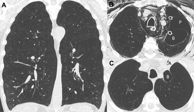
Chest CT evolution (patient 1). (A): Baseline chest CT before endobronchial valves insertion in the left upper lobe (LUL). The circle shows a low attenuation zone next to the fissure. (B): Chest CT performed 35 days after valves insertion. We can see the complete atelectasis of the LUL (A), presence of pneumothorax (P) with subcutaneous emphysema, chest tube drainage and occurrence of a new bulla (B) next to the fissure. (C): Chest CT performed 83 days after valves insertion. The air leak resolved and the bulla (B) size is decreasing. (D): Chest CT performed 508 days after valves insertion with a complete resolution of the bulla.
Figure 2.
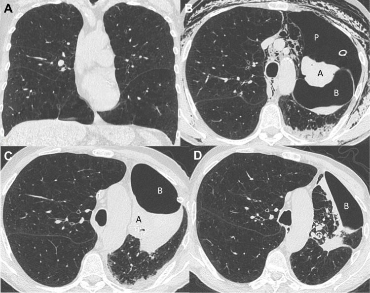
Chest CT evolution (patient 2). (A) Baseline chest CT. Presence of homogeneous emphysema. (B) Chest CT performed 5 days after valves insertion in the left upper lobe. Presence of pneumothorax (P), pneumomediastinum, subcutaneous emphysema and 2 chest tubes. Apparition of a bulla (B) in the left lung fissure. (C) Chest CT performed 84 days after valves insertion with resolution of air leak and decreasing size of the bulla (B) and an atelectasis (A).
Figure 3.
Chest CT evolution (patient 3). (A) Chest CT performed before valves insertion characterized by heterogeneous emphysema. (B) Chest CT performed one day after valves insertion in the left upper lobe (LUL). There is a complete atelectasis (A) of the LUL and occurrence of a bulla (B). (C) Chest CT performed six days after valves insertion with a pneumothorax (P) drained by a chest tube, the bulla (B) and the atelectasis (A) of the LUL. (D) Chest CT performed 30 days after valves insertion with decrease of the size of the pneumothorax (P) and the bulla (B). Only partial atelectasis (A) persisted in the LUL.
Figure 6.
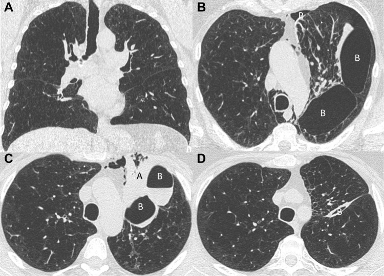
Chest CT evolution (patient 6). (A) Baseline chest CT which is characterized by homogeneous emphysema. (B) Chest CT performed 12 days after valves insertion in the left upper lobe (LUL). We can see occurrence of partial atelectasis (A), pneumothorax (P), chest tube drainage and the presence of a bulla (B) in left fissure with air–liquid level. (C) Chest CT performed 82 days after valves insertion (7 days after valves removal for post-obstructive pneumonia) which is characterized by the loss of the LUL atelectasis and a persistent bulla (B) completely filled with liquid in the left fissure. (D) Chest CT performed 262 days after valves insertion (6 months after valves removal for post-obstructive pneumonia) showing a nearly complete resolution of the bulla (B).
Interestingly, these six patients presented several common characteristics: all had LUL treatment and the appearance of the bullae occurred concomitantly with a rapid loss of volume of the target lobe (complete atelectasis within the first day after valves insertion). They all had early complete atelectasis complicated by a pneumothorax within 2 days after EBV insertion. Chest tube drainage was required in all patients except in patient 5 who presented an ex vacuo pneumothorax spontaneously resolving.
The outcomes of patients 1, 2 and 3 were uneventful with complete resolution of the bullae for patient 1 at 1.5 years of follow-up (Figure 1D).
An air–liquid level in the LUL bullae occurred within 2 days after EBV placement in patients 4, 5 and 6 (Figures 4, 5 and 6) and was associated with a bacterial infection documented by sputum culture, lung infiltrates on chest CT and high CRP level on blood analysis. A 6-week antibiotic treatment (14 days of intravenous piperacillin/tazobactam 4g, four times a day followed by 4 weeks of amoxicillin/clavulanic acid 875 mg 3 times a day) was given. Patients 4 and 5 had a very similar outcome. Despite continuance of two persistent bullae with an air–liquidlevel and infection in their left lower lobe, their outcome was favorable under antibiotic treatment. Patient 6 developed a post-obstructive pneumonia in the treated lobe. Therefore, the valves were removed 4 months later. A bulla completely filled with liquid persisted in the left fissure but nearly completely resolved 6 months after valves removal (Figure 6D). The patient completely recovered with a lung function similar to that of before the valves insertion (Table 1).
Figure 4.
Chest CT evolution (patient 4). (A) Baseline chest CT characterized by homogeneous emphysema. (B) Chest CT performed 7 days after valves insertion in the left upper lobe (LUL). We can see a complete atelectasis (A) of the LUL, a pneumothorax (P) of the left lung, pneumomediastinum, subcutaneous emphysema, chest tube drainage and a bulla (B) with air–liquid level. (C) Chest CT performed 26 days after valves insertion showing resolution of the air leak, bulla (B) with air–liquid level, the atelectasis (A) of the LUL and left lower lobe (LLL) infiltrates. (D) Chest CT performed 100 days after valves insertion characterized by resolution of the air leak and decreased size of the bulla (B) (always with air–liquid level) as well as the LLL infiltrates.
Figure 5.
Chest CT evolution (patient 5). (A) Baseline chest CT before endobronchial valves insertion in the left upper lobe (LUL). (B) Chest CT performed one day after valves insertion. We can see the hypoventilation of the LUL, presence of a pneumothorax (P) which did not require chest tube drainage, pneumomediastinum and the occurrence of two new bullae (B) between the upper and lower lobes. (C) Chest CT performed 5 days after valves insertion. There is a regression of the pneumomediastinum, increasing of the atelectasis (A) and the bullae (B) with an air–liquid level. (D) Chest CT performed 63 days after valves insertion with a partial atelectasis of the LUL and a nearly complete resolution of both bullae (B).
At 3 months after valves insertion, data could suggest a more limited improvement in lung function and exercise capacity in these six patients (average ΔFEV1: +66mL, ΔRV: −0, 644L and average difference from baseline in 6-minute walk distance (Δ6MWD): +8m) compared to 5 other patients from our center who presented an uncomplicated pneumothorax and complete atelectasis after valves insertion (ΔFEV1: +268mL (p=0.053), ΔRV: −1.026L (p=0.372) and Δ6MWD: +39m (p=0.299)).
Discussion
This descriptive case series is the first long-term follow-up report about the occurrence of unusual spontaneously formed septate bullae in the left lung associated with pneumothorax, following EBV insertion in the left upper lobe of the lung in patients with severe emphysema.
Indeed, isolated bullae associated with complete pneumothorax as shown in our six patients had not been reported previously except three incidental cases included in two reports,3,4 whose available characteristics are included in Table 1. The occurrence of interlobar pneumothorax was previously reported in congestive heart failure, asthma or after surgical pleurodesis.7,8 In all previously reported interlobar pneumothorax, the air in the pleural space was restricted in the fissure likely due to pleural adhesions without complete pneumothorax. This was also observed in two patients with only interlobar pneumothorax after valves placement by Valipour et al.4 This is quite different from our observed patients as they had both bullae and complete pneumothorax requiring chest tube drainage except in patient 5. Moreover, chest tube drainage which allowed the expansion of the lung had no influence on the bullae. Patient 5 had an ex-vacuo pneumothorax, bullae with an air–liquid level and a pneumomediastinum. She probably developed an ex vacuo pneumothorax due to a rapid loss of volume in the target lobe and an interlobar pneumothorax in a restricted pleural cavity with an air leak through the mediastinum resulting in a pneumomediastinum.9 Interestingly, nearly all patients who presented bullae after valves insertion (six in the present study and two in a statement published by Valipour et al on nine described patients (Table 1)) occurred in the left lung.4 This could be explained, in part, as more than two-thirds of the valves are placed in the left lung.10
The mechanism responsible for the occurrence of these bullae is still unknown. Moreover, the location of those bullae (inside lung parenchyma or pleural cavity) remains unclear. In our opinion, two hypothetical causes for these clinical situations may be suggested. The first could be a rupture of parenchymal septa leading to the formation of giant parenchymal bulla without air leak into the pleural cavity associated with a “usual” complete pneumothorax complicating valves insertion. This clinical situation may be illustrated by patient 1 as we may see a low attenuation zone next to the interlobar fissure (Figure 1A). In case of rapid expansion of the left lower lobe, rupture of septa in this particular zone may result in the formation of a bulla in the lung parenchyma (Figure 1B and C). The second potential explanation could be the hypothesis of two air leaks; the first air leak in a restricted pleural cavity in the fissure due to pleural adhesions completely isolated from the second one which occurred in an “open” pleural cavity leading to a complete pneumothorax. This clinical situation may be suggested by patient 6. Indeed, we may see on Figure 6C that there is still an interlobar pleural cavity completely fulfilled with liquid after valves removal suggesting two different air leaks in two separated zones of the pleural cavity.
Regarding the follow-up of our patients, different outcomes were observed depending on the presence or not of an air–liquid level in the bulla. In cases of bulla without an air–liquid level (patients 1, 2 and 3), there was spontaneous resolution of the bullae over time (after 1.5 years in patient 1). A “wait-and-see” attitude should be proposed in this particular setting. In cases of bulla with an air–liquid level, which could be considered as a secondary pleural effusion, a close monitoring of those patients should be undertaken as they have an increased risk of bacterial pneumonia.8 Prophylactic antibiotherapy should be discussed.
Regarding the improvement of lung function and exercise capacity at 3 months following endobronchial valves therapy, our results could suggest a decreased improvement, but not statistically significant, of FEV1 (p=0.053), of RV (p=0.372) and 6MWT (p=0.299) compared to patients with uncomplicated pneumothorax of our center. These results are in line with previously reported data by Gompelmann et al on patients who presented a pneumothorax and complete atelectasis after valves insertion (average ΔFEV1: +100mL, ΔRV: −0.400L and Δ6MWT: +15m).10
This case series has some limitations. First, it is a descriptive study of adverse events following endobronchial valves insertion in severe emphysematous patients. Large, worldwide register should be done to increase the number of such cases. Second, longer follow-up of those patients should be undertaken to evaluate the impact of those bullae on lung function and exercise capacity. Third, a majority of these complications (five out of six patients) have been detected in a single center probably because a systematic chest CT scan has been performed in case of pneumothorax, those bullae being hard to diagnose on routine chest X-ray.
Conclusion
Parenchymal or interlobar bulla is a complication after endobronchial valves insertion for endoscopic lung volume reduction in patients with severe emphysema. This complication seems to be associated with a more limited improvement of lung function and exercise capacity compared to patients with an uncomplicated pneumothorax. In the case of bulla without an air–liquid level, a conservative attitude should be taken as spontaneous resolution was described, whereas in the presence of an air–liquid level in the bulla, antibiotic prophylaxis should be discussed to prevent secondary infection.
Acknowledgments
The authors thank Samantha Phillips for the correction of this manuscript.
Disclosure
OT, BB, KC, OM and DL have no conflicts of interest in this work. VH reports grants from Astra Zeneca, GlaxoSmithKline, Chiesi, and Boeringher Ingelheim, outside the submitted work. DJS is a physician-advisor and investigator for Pulmonx, CA, USA. PLS received lecture fees and is a consultant of Pulmonx, CA, USA.
References
- 1.Klooster K, ten Hacken N, Hartman JE, Kerstjens H, van Rikxoort EM, Slebos DJ. Endobronchial valves for emphysema without interlobar collateral ventilation. N Engl J Med. 2015;373:2325–2335. doi: 10.1056/NEJMoa1507807 [DOI] [PubMed] [Google Scholar]
- 2.Klooster K, Slebos DJ. Endobronchial valves for the treatment of advanced emphysema. Chest. 2021;159:1833–1842. doi: 10.1016/j.chest.2020.12.007 [DOI] [PMC free article] [PubMed] [Google Scholar]
- 3.Van Dijk M, Sue R, Criner GJ, et al. Expert statement: pneumothorax associated with one-way valve therapy for emphysema: 2020 Update. Respiration. 2021;100:969–978. doi: 10.1159/000516326 [DOI] [PMC free article] [PubMed] [Google Scholar]
- 4.Valipour A, Slebos DJ, de Oliveira HG, et al. Expert statement: pneumothorax associated with endoscopic valve therapy for emphysema – potential mechanisms, treatment algorithm, and case examples. Respiration. 2014;87:513–521. doi: 10.1159/000360642 [DOI] [PubMed] [Google Scholar]
- 5.Gompelmann D, Lim HJ, Eberhardt R, et al. Predictors of pneumothorax following endoscopic valve therapy in patients with severe emphysema. Int J Chron Obstruct Pulmon Dis. 2016;11:1767–1773. doi: 10.2147/COPD.S106439 [DOI] [PMC free article] [PubMed] [Google Scholar]
- 6.Slebos D-J, Shah PL, Herth F, Valipour A. Endobronchial valves for endoscopic lung volume reduction: best practice recommendations from expert panel on endoscopic lung volume reduction. Respiration. 2017;93:138–150. doi: 10.1159/000453588 [DOI] [PMC free article] [PubMed] [Google Scholar]
- 7.Rabinowitz JG, Kongtawng T. Loculated interlobar air-fluid collection in congestive heart failure. Chest. 1978;74(6):681–683. doi: 10.1378/chest.74.6.681 [DOI] [PubMed] [Google Scholar]
- 8.Gourdon G, Dietemann A, Beigelman C, Sohier B, Pauli G. Recurrent interlobar pneumothorax in an asthmatic patient. Eur Respir J. 1993;6:748–749. [PubMed] [Google Scholar]
- 9.Woodring JH, Baker MD, Stark P. Pneumothorax ex vacuo. Chest. 1996;110(4):1102–1105. doi: 10.1378/chest.110.4.1102 [DOI] [PubMed] [Google Scholar]
- 10.Gompelmann D, Heinhold T, Rötting M, et al. Long-term follow up after endoscopic valve therapy in patients with severe emphysema. Ther Adv Respir Dis. 2019;13:1–11. doi: 10.1177/1753466619866101 [DOI] [PMC free article] [PubMed] [Google Scholar]


