



Abstract
Neuregulin 1 signals are essential for the development and function of Schwann cells, which form the myelin sheath on peripheral axons. Disruption of myelin in the peripheral nervous system (PNS) can lead to peripheral neuropathy, which is characterized by reduced axonal conduction velocity and sensorimotor deficits. Charcot-Marie-Tooth disease is a group of heritable peripheral neuropathies that may be caused by variants in nearly 100 genes. Despite the evidence that Neuregulin 1 is essential for many aspects of Schwann cell development, previous studies have not reported variants in the neuregulin 1 gene (NRG1) in patients with peripheral neuropathy. We have identified a rare missense variant in NRG1 that is homozygous in a patient with sensory and motor deficits consistent with mixed axonal and de-myelinating peripheral neuropathy. Our in vivo functional studies in zebrafish indicate that the patient variant partially reduces NRG1 function. This study tentatively suggests that variants at the NRG1 locus may cause peripheral neuropathy and that NRG1 should be investigated in families with peripheral neuropathy of unknown cause.
Keywords: peripheral neuropathy, NRG1, neuregulin 1, functional genomics, zebrafish
Charcot-Marie-Tooth disease (CMT) encompasses a group of clinically and genetically heterogenous peripheral sensory and motor neuropathies (Lassuthova et al., 2018). CMT occurs in ~1 in 2,500 individuals and has been associated with pathogenic variants in >90 genes (Lassuthova et al., 2018; Ravenscroft et al., 2021; Senderek et al., 2020). There are two main CMT sub-types, which are differentiated based on electrophysiological and neuropathological criteria. CMT1 is a demyelinating neuropathy characterised by slow motor nerve conduction velocity and is caused by abnormalities in the myelin sheath due to cell-autonomous defects in Schwann cells, the glia that make myelin in the peripheral nervous system (PNS). CMT2 is an axonal neuropathy, characterised by normal or mildly reduced nerve conduction velocities and chronic axonal degeneration and regeneration. Differentiation between axonal and demyelinating forms can be complicated by secondary axonal degeneration and demyelination in CMT1 and CMT2 respectively. More than 50% of CMT patients remain without a definitive molecular diagnosis following comprehensive screening using comprehensive gene panels or clinical exomes (Drew et al., 2015; Lassuthova et al., 2018; Ravenscroft et al., 2021; Senderek et al., 2020), suggesting there are additional CMT disease genes to identify. Herein we describe the identification of a rare homozygous missense variant in the neuregulin 1 gene (NRG1) in a consanguineous patient presenting with CMT disease.
Neuregulin signals are essential for Schwann cell development and myelination in vertebrates including mouse and zebrafish (Britsch et al., 1998; Garratt et al., 2000; Glenn & Talbot, 2013; Lyons et al., 2005; Meyer & Birchmeier, 1995; Morris et al., 1999; Perlin et al., 2011). In developing peripheral nerves, Nrg1 on the axonal membrane activates a heterodimeric ErbB2-ErbB3 receptor tyrosine kinase on associated glia, triggering Schwann cell proliferation, migration, and myelination (Nave & Salzer, 2006). Through different promoters and alternative mRNA splicing, the Nrg1 gene encodes more than 30 different isoforms, which are further modified by posttranslational processing (Falls, 2003; Hu et al., 2006; La Marca et al., 2011; Willem et al., 2006). In humans, the NRG1 gene has distinct promoters that generate six main classes of isoforms (types I, II, III, IV, V and VI) that have different N-terminal regions but share a common EGF-related domain that activates ErbB receptors (Chou & Ozaki, 2010). While types IV-VI are not present in all vertebrates, the type I, type II, and type III isoforms are widely conserved among vertebrates, and mutational studies in mouse and zebrafish have identified Nrg1 type III signals as essential regulators of Schwann cell development and myelination (Perlin et al., 2011; Wolpowitz et al., 2000). In mouse and zebrafish, type III isoform-specific mutants lack Schwann cells in peripheral nerves (Perlin et al., 2011; Wolpowitz et al., 2000). The extent of myelination is exquisitely controlled by the level of Nrg1 type III on peripheral axons: overexpression of Nrg1 type III generates thicker myelin, whereas mice heterozygous for a Nrg1 type III specific mutation have thinner myelin (Michailov et al., 2004; Taveggia et al., 2005). In addition, compound heterozygous mice for mutations in Nrg1 and ErbB2 have significantly thinner myelin and slower nerve conduction velocity (Michailov et al., 2004). Although overexpression of soluble Nrg1 signals can modulate the phenotypes of CMT animal models (Fledrich et al., 2014), previous studies have not reported NRG1 loss-of-function variants in patients with peripheral neuropathy. Our analysis provides evidence that partial loss of NRG1 function may impair nerve conduction in humans. We propose that variants in NRG1 may be responsible for cases of peripheral neuropathy with unknown cause.
The proband was a female, born to consanguineous parents. She noticed after the birth of her first child, when she was aged 20 years, that she stumbled when walking on uneven surfaces and that her hands were weak, particularly when manipulating small objects like keys. These symptoms gradually worsened over the ensuing 13 years. When examined at the age of 33 years she had normal strength proximally, but distally had wasting of the thenar muscles and the intrinsic muscles of the hand, and the tibialis anterior of the leg. Strength-wise, she had completely lost all thumb opposition, had 3/5 power in intrinsic muscles of the hand, and 4/5 wrist movements. Unusually, her lower limbs were not as affected as her upper limbs. She could still stand on her toes, although not on the toes of one foot only, but could not walk on her heels. All other lower limb movements were within normal limits. Her deep tendon reflexes were slightly augmented, except she had lost her ankle jerks. Plantar reflexes were flexor. Pinprick sensation was reduced to the knees and the mid-forearms. Nerve conduction studies were performed on the proband at the age of 35 years (Supp. Table S1). Right peroneal and tibial motor and right sural sensory responses were absent. Right median sensory and motor left median sensory and right radial sensory responses were also absent. Bilateral ulnar motor distal latencies were essentially normal. Motor conduction velocities were slowed, and bilateral ulnar F wave latencies were essentially normal. Limited EMG of left arm and leg muscles showed large motor unit potentials in distal muscles, with fibrillation. In conclusion there is electrophysiologic evidence of a mixed axonal and demyelinating distally prominent neuropathy. Over the ensuing ten years, the movements about the ankle have worsened to the extent that she cannot walk on toes or heels and has bilateral foot drop.
Whole genome sequencing identified 14 rare (allele frequency <0.001) coding homozygous variants, including a missense variant in the Neuregulin1 (NRG1) gene (ENST00000287842, NM_013956, c.1652G>A, p.(Arg551Gln), referred to here as R551Q; rs141355195). This variant is present on 23 alleles in gnomAD v2.1.1 and is absent from the Greater Middle East Variome Project (Scott et al., 2016). Multiple in silico predictions suggest this variant to have a detrimental functional impact; SIFT: deleterious, PolyPhen: probably damaging, MutationAssessor: medium, MutationTaster: disease-causing, CADD score 21. The substituted arginine is conserved in human, mouse, and frog; in zebrafish the corresponding residue is a serine.
Bi-directional Sanger sequencing confirmed the NRG1 c.1652G>A variant to be homozygous in the proband and showed that all healthy relatives were either carriers or homozygous for the reference allele (Figure 1A). Limited clinical details are available for the youngest sister (II.7), who resides in Iraq. Prior to genomic testing, she reported cramps and numbness in the legs and feet. She is also homozygous for the variant. We have been unable to obtain further details or formal clinical review of this individual.
Figure 1: Recessively inherited R551Q variant in the cytoplasmic tail region of NRG1 segregates with Charcot-Marie-Tooth neuropathy in an Australian-Iraqi family.
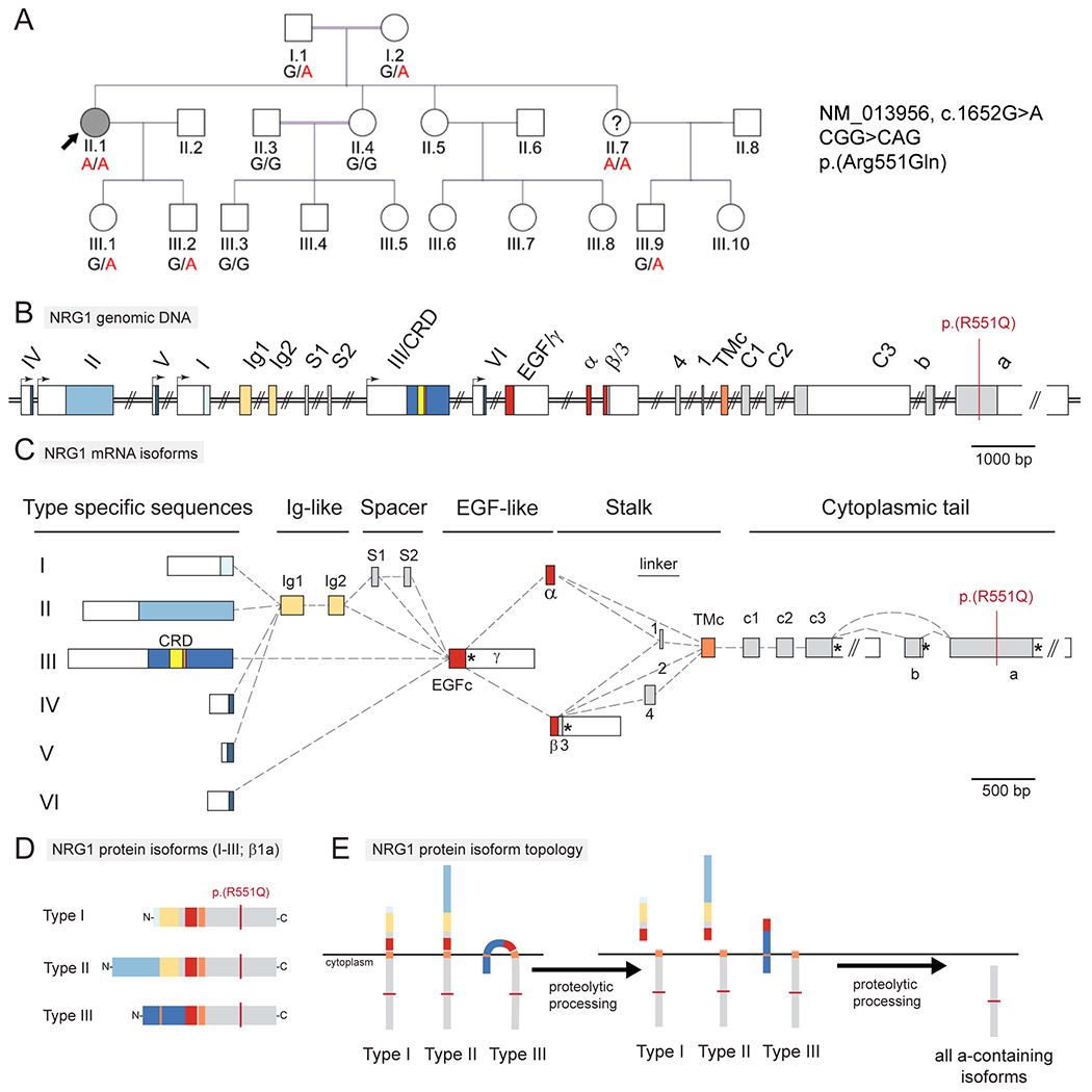
(A) Segregation of the homozygous NRG1 missense variant [NM_013956.5: c.1652G>A p.(Arg551Gln)] with disease is demonstrated. Limited clinical information is available for individual II:7, her current affection status is unknown, although she reported symptoms in keeping with a CMT neuropathy prior to genetic testing. Double lines indicate consanguinity. Proband indicated with arrow. Note II:3 is a first cousin of the proband. (B) Human NRG1 gene structure. Six “types” of NRG1s are generated through use of alternative transcriptional start sites (arrows), giving each type a unique N-terminal exon (exons and introns to scale, unless indicated with slashes; white boxes, 5’- or 3’-UTRs). We have identified an R551Q missense variant (red line) in the last exon (exon “a”), which encodes the cytoplasmic tail. (C) NRG1 mRNA splicing diagram, illustrating the diversity of transcripts generated through alternative starts sites and alternative splicing. The R551Q variant affects transcripts utilizing the “a” exon, for example NRG1 type III beta1a, a type III isoform that includes the EGF beta exon (red), linker 1, and terminates with the “a” exon. Grey dashed lines indicate alternative splicing paths utilized in RefSeq transcripts in NCBI gene database. Asterisks indicate alternative stop codons. (D) Linear protein diagrams, illustrating the location of the R551Q substitution in isoforms types I-III (transmembrane domains, orange). (E) NRG1 protein isoform topology diagram showing the role of proteolytic processing in generating mature signaling proteins. Type III has an N-terminal TM domain and can remain localized to the membrane after proteolysis; axonal Nrg1 type III is required for myelination of peripheral nerves in vertebrates(Morrissey et al., 1995; Perlin et al., 2011; Taveggia et al., 2005). Subsequent proteolysis can release the C-terminal cytoplasmic tail containing sequences encoded by the “a” exon, in which we have identified the R551Q variant.
Of the 13 other rare homozygous variants identified (Supp. Table S2), two had homozygous individuals in gnomAD, and we therefore excluded these. Variants in three other candidate genes (DDX59, GSR and ST3GAL5) cause diseases in humans that do not include neuropathy (Supp. Table S2, Notes) and thus were not regarded as candidates for the disease in this family. We used Sanger sequencing to analyze segregation for the remaining eight variants. By Sanger sequencing six variants did not co-segregate with disease. Two variants co-segregated with disease in the family, including an in-frame indel in ZFHX4 and a missense variant in DMTN. Previously, a de novo balanced translocation of ZFXH4 was identified in a patient with bilateral isolated ptosis (OMIM 178300)(McMullan et al., 2002). Studies in a recent Zfhx4 mouse line showed that homozygous Zfhx4 null mouse pups have cleft palate and skeletal malformations and died within a day of birth (Nakamura et al., 2021). DMTN encodes the dematin protein – an actin bundling protein identified in the erythroid membrane skeleton (Derick et al., 1992; Husain-Chishti et al., 1988). Dmtn null mice have anemia and their erythrocytes are osmotically fragile (Lu et al., 2016). Additionally, studies have shown that dematin is critical to the shape and membrane stability of red blood cells (Khanna et al., 2002). Given the different phenotypes associated with loss of ZFHX4 or DMTN, these genes are not good candidates for the phenotype in this family. Taken together, these data and the known role of NRG1 in myelination of the PNS (Meyer & Birchmeier, 1995; Nave & Salzer, 2006; Perlin et al., 2011; Wolpowitz et al., 2000), we conclude that the NRG1 variant remains the most likely plausible candidate in this family.
NRG1 encodes >30 different proteins that share a common EGF-like domain but differ in other regions due to use of different promoters and alternative splicing (Figure 1B,C). The R551Q substitution identified in family 1 affects isoforms with the longest cytoplasmic tail, in which the long “a” exon is included (Figure 1D,E). Transcripts containing the “a” cytoplasmic tail have been detected across multiple isoform classes (Falls, 2003), but NRG1 isoform type III stands out as relevant to peripheral neuropathy because of its essential roles in Schwann cell development and myelination in zebrafish and mouse.
To functionally test the activity of the R551Q variant in peripheral nerve development, we turned to zebrafish, in which peripheral nerve myelination is well studied and easily assessed (detailed Materials and Methods available in Supporting Information). Our previous analysis (Perlin et al., 2011) indicated that Schwann cell proliferation and migration are disrupted in zebrafish mutants homozygous for a missense variant in the type III-specific cysteine rich domain (CRD) of nrg1. In the present study, we used CRISPR-Cas9 to introduce a lesion (designated st153) in the CRD predicted to completely eliminate the function of nrg1 type III but leave the other isoforms intact (Supp. Figure S1 A,B). This lesion is a 7 bp deletion predicted to cause a frameshift and premature termination of translation, truncating the protein before the transmembrane domain of the CRD (Supp. Figure S1 C,D). Homozygous nrg1st153/st153 mutants lack myelin basic protein (mbp) mRNA in the posterior lateral line nerve, in accord with a previous study showing that myelinating Schwann cells are absent in nrg1 type III mutants (Perlin et al., 2011) (Supp. Figure S1 E).
To investigate the functional consequences of the R551Q patient variant, we compared the ability of wildtype and patient variant NRG1 type III proteins to rescue myelination of peripheral nerves in nrg1st153/st153 mutant zebrafish. We constructed Tol2 transposon vectors that express either wildtype human NRG1 type IIIa or the R551Q variant in transient transgenic experiments. The constructs contained pan-neuronal regulatory sequences to drive expression of NRG1 type IIIa in neurons or, as a control, a regulatory element that drives expression in cells of the macrophage lineage. The expression vectors also contained a cmlc2:GFP marker, which allowed the transgene expression efficiency to be assessed by the extent of GFP fluorescence in the heart (Figure 2A).
Figure 2: R551Q patient variant has reduced function in peripheral nerve rescue assay.
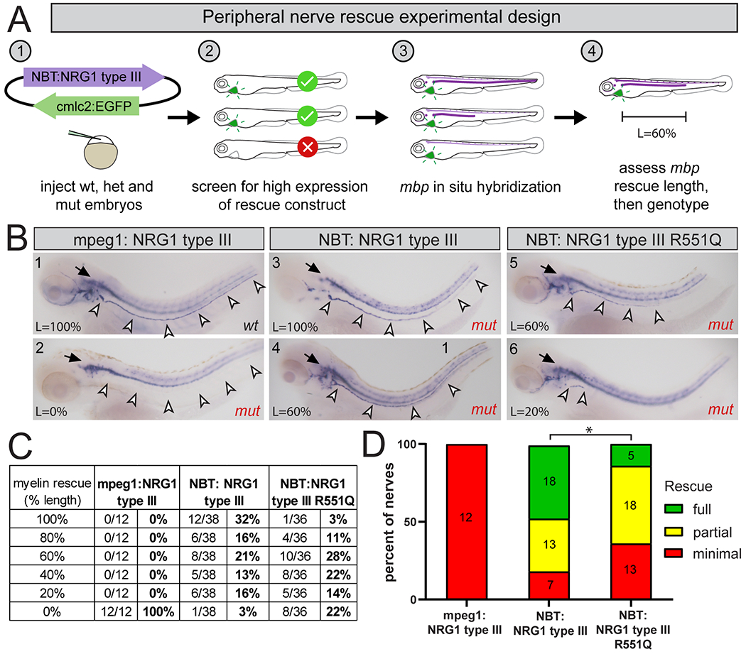
(A) Peripheral nerve rescue experimental design. A construct expressing human NRG1 type III is microinjected into embryos at the one-cell stage. This construct also expresses GFP in heart tissue (cmlc2:EGFP). After 4.5 days of development, larvae are visually screened for high expression of the rescue construct using the GFP heart signal, and myelin basic protein (mbp) mRNA is detected through in situ hybridization. Larvae are imaged and the length of mbp expression is analyzed, then larvae are genotyped individually. (B1) Control; expression of NRG1 type III in the macrophage lineage (mpeg1) does not affect mbp expression in wild type (wt) embryos. Normal CNS (black arrows) and PNS (white arrowheads) mbp expression extending the full length of the larva (L=100%). (B2) In nrg1 st153mutant embryos (mut), NRG1 expression in the macrophage lineage does not rescue PNS mbp expression (L=0%). (B3) NRG1 expression in neurons fully rescues PNS mbp expression in many injected mutants and (B4) partially rescues myelination in others. (B5) NRG1 type III R551Q expression in neurons only partially or (B6) minimally rescues PNS mbp expression. (C) Table showing the number of larvae in each rescue category out of total mutant larvae injected with the indicated construct. Percentages show the proportion of larvae in each rescue category relative to the total number of larvae in that condition. (D) Summary chart generated from the data in C by characterizing the 100% and 80% categories as “full rescue”, the 0% and 20% categories as “minimal”, and 40% and 60% as “partial”. Whereas both NRG1 type III constructs can rescue myelination, the patient variant is significantly less active than wildtype (Chi-square test, p=0.0277).
NRG1 expression vectors and synthetic mRNA encoding Tol2 transpose were injected into embryos from a cross of nrg1st153/+ heterozygotes, and larvae with strong GFP expression in the heart were fixed to detect mbp expression by in situ hybridization at 4.5 dpf. The extent of mbp expression was assessed, and then the genotypes of all larvae were determined. The ability of the expression constructs to rescue the mutants was assessed by the extent of mbp expression along the posterior lateral line nerve (Figure 2B). Whereas expressing human NRG1 type IIIa in the macrophage lineage did not rescue mbp expression along the lateral line nerve, expressing NRG1 type IIIa in neurons strongly rescued mbp expression in nrg1st153/st153 mutants: 32% of nerves in injected mutants were fully rescued, and only 3% of nerves showed no evidence of rescue (Figure 2C). The R551Q variant had significantly less rescuing activity than wildtype human NRG1 type IIIa in four independent experiments: only 3% of nerves in injected mutants appeared fully rescued, while 22% of nerves showed no evidence of rescue (Figure 2C,D; Chi-square test, p=0.0277). These results indicate that the R551Q substitution identified in family 1 is a partial loss-of-function variant that reduces the ability of NRG1 type IIIa to direct Schwann cell myelination in peripheral nerves.
Extensive previous studies have demonstrated that Nrg1 signals and their ErbB receptors control Schwann cell development and myelination (Britsch et al., 1998; Garratt et al., 2000; Lyons et al., 2005; Meyer & Birchmeier, 1995; Perlin et al., 2011; Raphael et al., 2011; Taveggia et al., 2005; Wolpowitz et al., 2000). The >30 isoforms encoded by the NRG1 gene share a central EGF-related domain that is flanked by variable N- and C-terminal regions generated via different promoters and alternative mRNA splicing (Falls, 2003). Nrg1 type III signals are prominently expressed in developing sensory and motor neurons in mouse and zebrafish (Honjo et al., 2008; Meyer et al., 1997), and mutational studies provide compelling evidence that these signals are key regulators of myelination in peripheral nerves (Perlin et al., 2011; Wolpowitz et al., 2000). Mice homozygous for a mutation that disrupts all Nrg1 isoforms die during mid-embryogenesis with defects in heart trabeculation and lacking Schwann cell precursors in peripheral nerves (Meyer & Birchmeier, 1995). Several lines of evidence indicate that Nrg1 type III isoforms are the key neuregulins that govern Schwann cell development and myelination. Mice homozygous for a targeted mutation that specifically disrupts Nrg1 type III specific sequences die at the time of birth with a severe reduction in Schwann cell numbers in peripheral nerves (Wolpowitz et al., 2000). Similarly, zebrafish nrg1 type III specific homozygous mutants lack Schwann cells in peripheral nerves (Perlin et al., 2011), because both Schwann cell proliferation and migration are disrupted in the absence of these essential signals. Nrg1 type III signals also control the radial sorting process, during which axons are paired with Schwann cells that will subsequently myelinate them, and it is thought that axons expressing the highest levels of Nrg1 type III are the first to be myelinated (Domènech-Estévez et al., 2016; Goebbels et al., 2010; Raphael et al., 2011; Taveggia et al., 2005). In addition, the level of Nrg1 type III expressed on an axon determines the thickness of the myelin produced by its associated Schwann cells (Michailov et al., 2004). The fact that Nrg1 type III is the rate-limiting signal controlling so many steps of Schwann cell development may explain the loss of function constraint in gnomAD (pLI = 1) and the lack of individuals homozygous for loss of function variants. Not only would individuals entirely lacking NRG1 signaling be expected to have early, lethal abnormalities, even a partial reduction of NRG1 activity might disrupt peripheral nerves and other organs that are finely regulated by the level of NRG1 signaling.
Our analysis has identified a recessive, partial loss-of-function variant in NRG1 that segregates with peripheral neuropathy in a consanguineous pedigree. The variant changes an incompletely conserved Arg residue encoded by one of the alternative 3’ exons of the reference sequence to Gln. We provide evidence that this R>Q change partially reduces the function of NRG1 type III signals using our functional tests in zebrafish. Expression of the reference human NRG1 type III-beta1a protein in neurons can rescue Schwann cell development in all but a few percent of zebrafish nrg1 type III mutants. In contrast, about ten times fewer zebrafish mutants were fully rescued when the R>Q variant form was expressed. It is important to emphasize that the R>Q variant does retain significant activity, which likely accounts for the relatively mild clinical phenotype of the patient when compared to the embryonic and perinatal lethality of mice homozygous for complete knockout (Meyer & Birchmeier, 1995) and type III specific (Wolpowitz et al., 2000) Nrg1 mutations, respectively.
There are several possible mechanisms by which an R>Q missense variant in the C-terminal region of NRG1 proteins could reduce function. One possibility is the R>Q substitution might impair trafficking of NRG1 signals through the secretory pathway or proper proteolytic processing (Liu et al., 1998). Consistent with this hypothesis, a previous study reported that the C-terminal region of Nrg1 signals determines the extent to which the proteins are retained in the cytoplasm of sensory neurons (Zhang et al., 2006). Another possibility is suggested by previous work showing that the C-terminal region of Nrg1 type III controls its subcellular localization on the membrane of pyramidal neurons (Exposito-Alonso et al., 2020); thus, the R>Q variant might mislocalize NRG1 signals within peripheral neurons. Finally, it has been reported that a C-terminal fragment of Nrg1 type III proteins is involved in “back signaling”, in which Nrg1—ErbB interactions can have transcriptional (Bao et al., 2003, 2004) and non-transcriptional effects (Canetta et al., 2011; Hancock et al., 2008) on the neurons expressing Nrg1; thus, the R>Q substitution might disrupt such a back signaling function in peripheral neurons.
Our analysis provides evidence that suggests partial loss of NRG1 function may cause peripheral neuropathy in humans. This finding complements prior studies that identified pathogenic variants in ErbB2 and ErbB3, the Schwann cell receptors for axonal NRG1 signals, in patients with peripheral neuropathy and arthrogryposis (Le et al., 2021; Narkis et al., 2007). Analysis of further cases is required to determine the extent to which different types of NRG1 variants may contribute to peripheral neuropathy in humans.
Supplementary Material
Supp. Table S2: Rare, coding homozygous variants identified in the proband. Here we present additional variants identified in the proband and show the segregation status of those screened in the family.
Supp. Table S3: Neurogenetic disease gene panel (version 5).
No causative pathogenic or likely pathogenic variants were identified within this list.
Supp. Figure S1: Generation of nrg1 type III loss of function allele (nrg1st153) in zebrafish.
(A) nrg1 gene structure in zebrafish. Alternative promoters (arrows) are used to transcribe unique N-terminal exons that distinguish types I, II, and III. Exons encoding Ig, EGF, linker, transmembrane (TM) and C-terminal domains are highly conserved (white boxes, 5’- or 3’-UTRs; exons to scale). The st153 lesion was created in the type III-specific N-terminal exon (red arrowhead). (B) nrg1 mRNA isoforms, illustrating that the st153 lesion affects only the nrg1 type III isoform. (C) st153 is a 7-bp deletion created just upstream of the PAM site of the targeted guide RNA (orange boxes). (D) nrg1 protein isoform diagrams showing the predicted consequence of the st153 lesion. st153 causes a frameshift and amino acid change at Proline 40 and a premature stop codon, truncating the protein at 68 amino acids, predicted to eliminate function of nrg1 type III. (E) Expression of mbp is not detectable in peripheral nerves of nrg1-st153 mutants at 4.5 dpf. Wild type embryos (nrg1+/+) show extensive mbp expression in the hindbrain and spinal cord (black arrowheads – CNS; purple stripe extending the length of the larva) and peripheral lateral line nerve (white arrowheads – PNS; more superficial purple stripe extending the length of the larva). In nrg1 type III mutant embryos (nrg1 st153/st153) mbp expression appears normal in the CNS (black arrows) but is absent in the PNS (white arrowheads).
Supp. Table S1: Nerve conduction studies (NCS) performed in the Proband.
Acknowledgements
We thank members of our labs for helpful discussion and critical comment on the manuscript and T. Reyes and C. Hill for fish care.
Funding
This work was supported by the National Institutes of Health [R35 NS111584 to W.S.T., 1F32NS095466 to D.E.L.]. This work was supported by the Australian National Health and Medical Research Council [APP1122952 to G.R.] and the Australian Genomics Health Alliance [NHMRC GNT1113531].
Footnotes
Conflicts of Interest Statement
The authors have no competing interests to declare.
Data Availability Statement
Data available on request from the authors.
References
- Bao J, Lin H, Ouyang Y, Lei D, Osman A, Kim T-W, Mei L, Dai P, Ohlemiller KK, & Ambron RT (2004). Activity-dependent transcription regulation of PSD-95 by neuregulin-1 and Eos. Nature Neuroscience, 7(11), 1250–1258. 10.1038/nn1342 [DOI] [PubMed] [Google Scholar]
- Bao J, Wolpowitz D, Role LW, & Talmage DA (2003). Back signaling by the Nrg-1 intracellular domain. The Journal of Cell Biology, 161(6), 1133–1141. 10.1083/jcb.200212085 [DOI] [PMC free article] [PubMed] [Google Scholar]
- Britsch S, Li L, Kirchhoff S, Theuring F, Brinkmann V, Birchmeier C, & Riethmacher D (1998). The ErbB2 and ErbB3 receptors and their ligand, neuregulin-1, are essential for development of the sympathetic nervous system. Genes & Development, 12(12), 1825–1836. 10.1101/gad.12.12.1825 [DOI] [PMC free article] [PubMed] [Google Scholar]
- Canetta SE, Luca E, Pertot E, Role LW, & Talmage DA (2011). Type III Nrg1 back signaling enhances functional TRPV1 along sensory axons contributing to basal and inflammatory thermal pain sensation. PloS One, 6(9), e25108. 10.1371/journal.pone.0025108 [DOI] [PMC free article] [PubMed] [Google Scholar]
- Chou C-F, & Ozaki M (2010). In silico analysis of neuregulin 1 evolution in vertebrates. Bioscience Reports, 30(4), 267–275. 10.1042/BSR20090097 [DOI] [PubMed] [Google Scholar]
- Derick LH, Liu SC, Chishti AH, & Palek J (1992). Protein immunolocalization in the spread erythrocyte membrane skeleton. European Journal of Cell Biology, 57(2), 317–320. [PubMed] [Google Scholar]
- Domènech-Estévez E, Baloui H, Meng X, Zhang Y, Deinhardt K, Dupree JL, Einheber S, Chrast R, & Salzer JL (2016). Akt Regulates Axon Wrapping and Myelin Sheath Thickness in the PNS. The Journal of Neuroscience: The Official Journal of the Society for Neuroscience, 36(16), 4506–4521. 10.1523/JNEUROSCI.3521-15.2016 [DOI] [PMC free article] [PubMed] [Google Scholar]
- Drew AP, Zhu D, Kidambi A, Ly C, Tey S, Brewer MH, Ahmad-Annuar A, Nicholson GA, & Kennerson ML (2015). Improved inherited peripheral neuropathy genetic diagnosis by whole-exome sequencing. Molecular Genetics & Genomic Medicine, 3(2), 143–154. 10.1002/mgg3.126 [DOI] [PMC free article] [PubMed] [Google Scholar]
- Exposito-Alonso D, Osório C, Bernard C, Pascual-García S, Del Pino I, Marín O, & Rico B (2020). Subcellular sorting of neuregulins controls the assembly of excitatory-inhibitory cortical circuits. ELife, 9, e57000. 10.7554/eLife.57000 [DOI] [PMC free article] [PubMed] [Google Scholar]
- Falls DL (2003). Neuregulins: Functions, forms, and signaling strategies. Experimental Cell Research, 284(1), 14–30. 10.1016/s0014-4827(02)00102-7 [DOI] [PubMed] [Google Scholar]
- Fledrich R, Stassart RM, Klink A, Rasch LM, Prukop T, Haag L, Czesnik D, Kungl T, Abdelaal TAM, Keric N, Stadelmann C, Brück W, Nave K-A, & Sereda MW (2014). Soluble neuregulin-1 modulates disease pathogenesis in rodent models of Charcot-Marie-Tooth disease 1A. Nat Med, 20(9), 1055–1061. 10.1038/nm.3664 [DOI] [PubMed] [Google Scholar]
- Garratt AN, Voiculescu O, Topilko P, Charnay P, & Birchmeier C (2000). A dual role of erbB2 in myelination and in expansion of the schwann cell precursor pool. The Journal of Cell Biology, 148(5), 1035–1046. 10.1083/jcb.148.5.1035 [DOI] [PMC free article] [PubMed] [Google Scholar]
- Glenn TD, & Talbot WS (2013). Signals regulating myelination in peripheral nerves and the Schwann cell response to injury. Curr Opin Neurobiol, 23(6), 1041–1048. 10.1016/j.conb.2013.06.010 [DOI] [PMC free article] [PubMed] [Google Scholar]
- Goebbels S, Oltrogge JH, Kemper R, Heilmann I, Bormuth I, Wolfer S, Wichert SP, Möbius W, Liu X, Lappe-Siefke C, Rossner MJ, Groszer M, Suter U, Frahm J, Boretius S, & Nave K-A (2010). Elevated phosphatidylinositol 3,4,5-trisphosphate in glia triggers cell-autonomous membrane wrapping and myelination. J Neurosci, 30(26), 8953–8964. 10.1523/JNEUROSCI.0219-10.2010 [DOI] [PMC free article] [PubMed] [Google Scholar]
- Hancock ML, Canetta SE, Role LW, & Talmage DA (2008). Presynaptic type III neuregulin1-ErbB signaling targets {alpha}7 nicotinic acetylcholine receptors to axons. The Journal of Cell Biology, 181(3), 511–521. 10.1083/jcb.200710037 [DOI] [PMC free article] [PubMed] [Google Scholar]
- Honjo Y, Kniss J, & Eisen JS (2008). Neuregulin-mediated ErbB3 signaling is required for formation of zebrafish dorsal root ganglion neurons. Development, 135(15), 2615–2625. 10.1242/dev.022178 [DOI] [PMC free article] [PubMed] [Google Scholar]
- Hu X, Hicks CW, He W, Wong P, Macklin WB, Trapp BD, & Yan R (2006). Bace1 modulates myelination in the central and peripheral nervous system. Nature Neuroscience, 9(12), 1520–1525. 10.1038/nn1797 [DOI] [PubMed] [Google Scholar]
- Husain-Chishti A, Levin A, & Branton D (1988). Abolition of actin-bundling by phosphorylation of human erythrocyte protein 4.9. Nature, 334(6184), 718–721. 10.1038/334718a0 [DOI] [PubMed] [Google Scholar]
- Khanna R, Chang SH, Andrabi S, Azam M, Kim A, Rivera A, Brugnara C, Low PS, Liu S-C, & Chishti AH (2002). Headpiece domain of dematin is required for the stability of the erythrocyte membrane. Proceedings of the National Academy of Sciences of the United States of America, 99(10), 6637–6642. 10.1073/pnas.052155999 [DOI] [PMC free article] [PubMed] [Google Scholar]
- La Marca R, Cerri F, Horiuchi K, Bachi A, Feltri ML, Wrabetz L, Blobel CP, Quattrini A, Salzer JL, & Taveggia C (2011). TACE (ADAM17) inhibits Schwann cell myelination. Nature Neuroscience, 14(7), 857–865. 10.1038/nn.2849 [DOI] [PMC free article] [PubMed] [Google Scholar]
- Lassuthova P, Rebelo AP, Ravenscroft G, Lamont PJ, Davis MR, Manganelli F, Feely SM, Bacon C, Brožková DŠ, Haberlova J, Mazanec R, Tao F, Saghira C, Abreu L, Courel S, Powell E, Buglo E, Bis DM, Baxter MF, … Zuchner S (2018). Mutations in ATP1A1 Cause Dominant Charcot-Marie-Tooth Type 2. American Journal of Human Genetics, 102(3), 505–514. 10.1016/j.ajhg.2018.01.023 [DOI] [PMC free article] [PubMed] [Google Scholar]
- Le T-L, Galmiche L, Levy J, Suwannarat P, Hellebrekers DM, Morarach K, Boismoreau F, Theunissen TE, Lefebvre M, Pelet A, Martinovic J, Gelot A, Guimiot F, Calleroz A, Gitiaux C, Hully M, Goulet O, Chardot C, Drunat S, … Bondurand N (2021). Dysregulation of the NRG1/ERBB pathway causes a developmental disorder with gastrointestinal dysmotility in humans. The Journal of Clinical Investigation, 131(6), 145837. 10.1172/JCI145837 [DOI] [PMC free article] [PubMed] [Google Scholar]
- Liu X, Hwang H, Cao L, Buckland M, Cunningham A, Chen J, Chien KR, Graham RM, & Zhou M (1998). Domain-specific gene disruption reveals critical regulation of neuregulin signaling by its cytoplasmic tail. Proceedings of the National Academy of Sciences of the United States of America, 95(22), 13024–13029. 10.1073/pnas.95.22.13024 [DOI] [PMC free article] [PubMed] [Google Scholar]
- Lu Y, Hanada T, Fujiwara Y, Nwankwo JO, Wieschhaus AJ, Hartwig J, Huang S, Han J, & Chishti AH (2016). Gene disruption of dematin causes precipitous loss of erythrocyte membrane stability and severe hemolytic anemia. Blood, 128(1), 93–103. 10.1182/blood-2016-01-692251 [DOI] [PMC free article] [PubMed] [Google Scholar]
- Lyons DA, Pogoda H-M, Voas MG, Woods IG, Diamond B, Nix R, Arana N, Jacobs J, & Talbot WS (2005). Erbb3 and erbb2 are essential for schwann cell migration and myelination in zebrafish. Current Biology: CB, 15(6), 513–524. 10.1016/j.cub.2005.02.030 [DOI] [PubMed] [Google Scholar]
- McMullan TW, Crolla JA, Gregory SG, Carter NP, Cooper RA, Howell GR, & Robinson DO (2002). A candidate gene for congenital bilateral isolated ptosis identified by molecular analysis of a de novo balanced translocation. Human Genetics, 110(3), 244–250. 10.1007/s00439-002-0679-5 [DOI] [PubMed] [Google Scholar]
- Meyer D, & Birchmeier C (1995). Multiple essential functions of neuregulin in development. Nature, 378(6555), 386–390. 10.1038/378386a0 [DOI] [PubMed] [Google Scholar]
- Meyer D, Yamaai T, Garratt A, Riethmacher-Sonnenberg E, Kane D, Theill LE, & Birchmeier C (1997). Isoform-specific expression and function of neuregulin. Development (Cambridge, England), 124(18), 3575–3586. [DOI] [PubMed] [Google Scholar]
- Michailov GV, Sereda MW, Brinkmann BG, Fischer TM, Haug B, Birchmeier C, Role L, Lai C, Schwab MH, & Nave K-A (2004). Axonal neuregulin-1 regulates myelin sheath thickness. Science, 304(5671), 700–703. 10.1126/science.1095862 [DOI] [PubMed] [Google Scholar]
- Morris JK, Lin W, Hauser C, Marchuk Y, Getman D, & Lee KF (1999). Rescue of the cardiac defect in ErbB2 mutant mice reveals essential roles of ErbB2 in peripheral nervous system development. Neuron, 23(2), 273–283. [DOI] [PubMed] [Google Scholar]
- Morrissey TK, Levi AD, Nuijens A, Sliwkowski MX, & Bunge RP (1995). Axon-induced mitogenesis of human Schwann cells involves heregulin and p185erbB2. Proc Natl Acad Sci U S A, 92(5), 1431–1435. [DOI] [PMC free article] [PubMed] [Google Scholar]
- Nakamura E, Hata K, Takahata Y, Kurosaka H, Abe M, Abe T, Kihara M, Komori T, Kobayashi S, Murakami T, Inubushi T, Yamashiro T, Yamamoto S, Akiyama H, Kawaguchi M, Sakata N, & Nishimura R (2021). Zfhx4 regulates endochondral ossification as the transcriptional platform of Osterix in mice. Communications Biology, 4(1), 1258. 10.1038/s42003-021-02793-9 [DOI] [PMC free article] [PubMed] [Google Scholar]
- Narkis G, Ofir R, Manor E, Landau D, Elbedour K, & Birk OS (2007). Lethal congenital contractural syndrome type 2 (LCCS2) is caused by a mutation in ERBB3 (Her3), a modulator of the phosphatidylinositol-3-kinase/Akt pathway. Am J Hum Genet, 81(3), 589–595. 10.1086/520770 [DOI] [PMC free article] [PubMed] [Google Scholar]
- Nave K-A, & Salzer JL (2006). Axonal regulation of myelination by neuregulin 1. Curr Opin Neurobiol, 16(5), 492–500. 10.1016/j.conb.2006.08.008 [DOI] [PubMed] [Google Scholar]
- Perlin JR, Lush ME, Stephens WZ, Piotrowski T, & Talbot WS (2011). Neuronal Neuregulin 1 type III directs Schwann cell migration. Development, 138(21), 4639–4648. 10.1242/dev.068072 [DOI] [PMC free article] [PubMed] [Google Scholar]
- Raphael AR, Lyons DA, & Talbot WS (2011). ErbB signaling has a role in radial sorting independent of Schwann cell number. Glia, 59(7), 1047–1055. 10.1002/glia.21175 [DOI] [PMC free article] [PubMed] [Google Scholar]
- Ravenscroft G, Clayton JS, Faiz F, Sivadorai P, Milnes D, Cincotta R, Moon P, Kamien B, Edwards M, Delatycki M, Lamont PJ, Chan SH, Colley A, Ma A, Collins F, Hennington L, Zhao T, McGillivray G, Ghedia S, … Davis MR (2021). Neurogenetic fetal akinesia and arthrogryposis: Genetics, expanding genotype-phenotypes and functional genomics. Journal of Medical Genetics, 58(9), 609–618. 10.1136/jmedgenet-2020-106901 [DOI] [PMC free article] [PubMed] [Google Scholar]
- Scott EM, Halees A, Itan Y, Spencer EG, He Y, Azab MA, Gabriel SB, Belkadi A, Boisson B, Abel L, Clark AG, Greater Middle East Variome Consortium, Alkuraya FS, Casanova J-L, & Gleeson JG (2016). Characterization of Greater Middle Eastern genetic variation for enhanced disease gene discovery. Nature Genetics, 48(9), 1071–1076. 10.1038/ng.3592 [DOI] [PMC free article] [PubMed] [Google Scholar]
- Senderek J, Lassuthova P, Kabzińska D, Abreu L, Baets J, Beetz C, Braathen GJ, Brenner D, Dalton J, Dankwa L, Deconinck T, De Jonghe P, Dräger B, Eggermann K, Ellis M, Fischer C, Stojkovic T, Herrmann DN, Horvath R, … Auer-Grumbach M (2020). The genetic landscape of axonal neuropathies in the middle-aged and elderly: Focus on MME. Neurology, 95(24), e3163–e3179. 10.1212/WNL.0000000000011132 [DOI] [PMC free article] [PubMed] [Google Scholar]
- Taveggia C, Zanazzi G, Petrylak A, Yano H, Rosenbluth J, Einheber S, Xu X, Esper RM, Loeb JA, Shrager P, Chao MV, Falls DL, Role L, & Salzer JL (2005). Neuregulin1 type III determines the ensheathment fate of axons. Neuron, 47(5), 681–694. 10.1016/j.neuron.2005.08.017 [DOI] [PMC free article] [PubMed] [Google Scholar]
- Willem M, Garratt AN, Novak B, Citron M, Kaufmann S, Rittger A, DeStrooper B, Saftig P, Birchmeier C, & Haass C (2006). Control of peripheral nerve myelination by the beta-secretase BACE1. Science (New York, N.Y.), 314(5799), 664–666. 10.1126/science.1132341 [DOI] [PubMed] [Google Scholar]
- Wolpowitz D, Mason TB, Dietrich P, Mendelsohn M, Talmage DA, & Role LW (2000). Cysteine-rich domain isoforms of the neuregulin-1 gene are required for maintenance of peripheral synapses. Neuron, 25(1), 79–91. 10.1016/s0896-6273(00)80873-9 [DOI] [PubMed] [Google Scholar]
- Zhang Z, Prentiss L, Heitzman D, Stahl RC, DiPino F, & Carey DJ (2006). Neuregulin isoforms in dorsal root ganglion neurons: Effects of the cytoplasmic domain on localization and membrane shedding of Nrg-1 type I. Journal of Neuroscience Research, 84(1), 1–12. 10.1002/jnr.20861 [DOI] [PubMed] [Google Scholar]
Associated Data
This section collects any data citations, data availability statements, or supplementary materials included in this article.
Supplementary Materials
Supp. Table S2: Rare, coding homozygous variants identified in the proband. Here we present additional variants identified in the proband and show the segregation status of those screened in the family.
Supp. Table S3: Neurogenetic disease gene panel (version 5).
No causative pathogenic or likely pathogenic variants were identified within this list.
Supp. Figure S1: Generation of nrg1 type III loss of function allele (nrg1st153) in zebrafish.
(A) nrg1 gene structure in zebrafish. Alternative promoters (arrows) are used to transcribe unique N-terminal exons that distinguish types I, II, and III. Exons encoding Ig, EGF, linker, transmembrane (TM) and C-terminal domains are highly conserved (white boxes, 5’- or 3’-UTRs; exons to scale). The st153 lesion was created in the type III-specific N-terminal exon (red arrowhead). (B) nrg1 mRNA isoforms, illustrating that the st153 lesion affects only the nrg1 type III isoform. (C) st153 is a 7-bp deletion created just upstream of the PAM site of the targeted guide RNA (orange boxes). (D) nrg1 protein isoform diagrams showing the predicted consequence of the st153 lesion. st153 causes a frameshift and amino acid change at Proline 40 and a premature stop codon, truncating the protein at 68 amino acids, predicted to eliminate function of nrg1 type III. (E) Expression of mbp is not detectable in peripheral nerves of nrg1-st153 mutants at 4.5 dpf. Wild type embryos (nrg1+/+) show extensive mbp expression in the hindbrain and spinal cord (black arrowheads – CNS; purple stripe extending the length of the larva) and peripheral lateral line nerve (white arrowheads – PNS; more superficial purple stripe extending the length of the larva). In nrg1 type III mutant embryos (nrg1 st153/st153) mbp expression appears normal in the CNS (black arrows) but is absent in the PNS (white arrowheads).
Supp. Table S1: Nerve conduction studies (NCS) performed in the Proband.
Data Availability Statement
Data available on request from the authors.