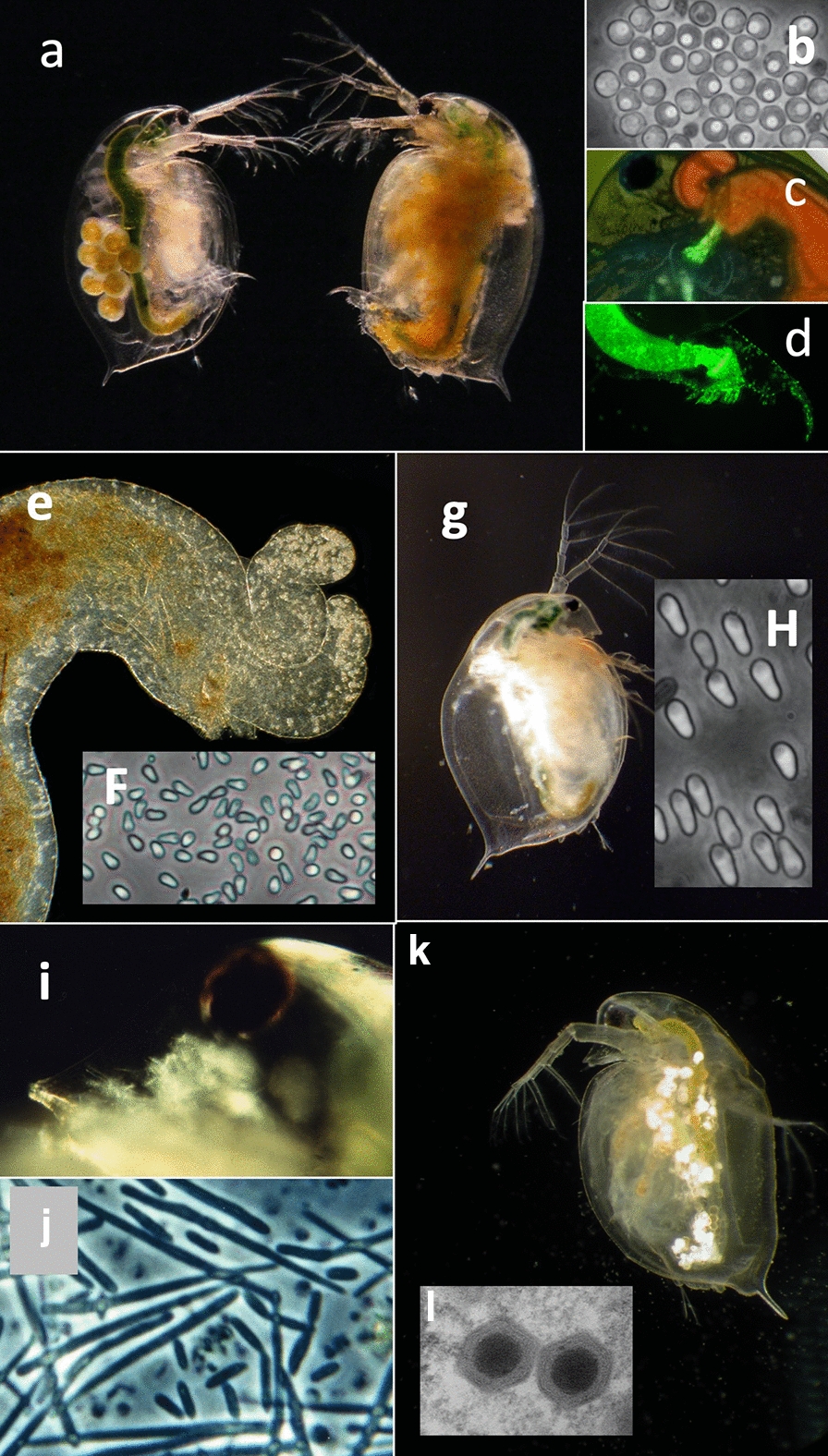
Fig. 7.

Examples of frequently studied parasites of Daphnia. a–d Bacterium Pasteuria ramosa colonizes the body cavity of the host. a Infected (right) and uninfected (left) D. magna. b Transmission stages (= spores) of P. ramosa. c, d Attachment of green fluorescent labelled P. ramosa spores to the oesophagus (c) and hindgut (d) of the host. Attachment of spores is required for the subsequent infection of the host [72, 131]. e Upper midgut of D. magna with spore clusters of the microsporidium Ordospora colligata in the appendices (upper right corner). The parasite colonized the gut epithelium of the host. f Spores of O. colligata. g D. magna infected with the microsporidian Hamiltosporidium tvaerminnensis. The parasite colonized the ovaries and fat body of the host. h Spores of H. tvaerminnensis. i Head of D. magna infected with Metschnikowia bicuspidata. The needles-like spores are visible through the transparent cuticle. j Spores of M. bicuspidata. k Daphnia pulex infected with the Daphnia Iridovirus (DIV-1), the causative agent of White Fat Cell Disease [68]. l Two DIV-1 particles. Picture taken by Jason Andras (a), David Duneau (c), Benjamin Hüssy (d), Patrick Mucklow (g) and Elena Toenshoff (l). All other pictures by Dieter Ebert
