Abstract
The aim of the present study was to compare the myoelectric activation and peak force (PF) between pullover (PO) and pulldown (PW) exercises in different shoulder joint positions during maximal isometric contractions (0o, 45o, 90o, 135o, and 180°). Fifteen young, healthy, resistance-trained men were recruited. The participants performed three maximal voluntary isometric contractions for each exercise at five shoulder joint positions. The myoelectric activation (iEMG) from pectoralis major (PM); latissimus dorsi (LD); posterior deltoid (PD), and PF were measured. For PF, there were significant main effects for exercise and joint positions (p < 0.001). For iEMG PM, there was significant a main effect for joint positions (p < 0.001). There was a significant interaction between exercises and joint positions (p < 0.001). For iEMG LD, there was a significant main effect for joint positions (p < 0.001). There was no significant interaction between exercises and joint positions. For iEMG PD, there was a significant main effect for joint positions (p < 0.001). There was no significant interaction between exercises and joint positions. For RPE, there were no significant differences between exercises and joint positions. The study concludes that specific shoulder joint positions affect PF production and iEMG during both exercises. RPE was not affected.
Keywords: Kinesiology, strength, muscle performance
INTRODUCTION
Among resistance training (RT) variables, exercise choice is one of the most important factors to develop muscle mass, strength, or muscular endurance (15). The exercise choice is based on movement specificity and it takes into account factors like the number of joints, range of motion, prime movers, level of balance, type of routine, frequency, periodization phase, etc (10). In this way, different exercises can be used to move specific joints, activate specific muscle groups, and produce different levels of force. Pullover (PO) and Pulldown (PW) are single-joint exercises(11) and present a similar shoulder joint movement during dynamic contractions (extension: concentric phase and flexion: eccentric phase), however, based on the mechanical characteristics of each exercise, such as the level of force per joint angle, these two exercises might stimuli the same muscles groups differently, including the latissimus dorsi (LD), posterior deltoid (PD), pectoralis major (PM), anterior deltoid (AD), and the long head of the triceps brachii (LTB) (7).
Several studies have reported greater myoelectric activation of the PM when compared to the LD for the PO exercise [with dumbbell (2, 3), straight bar (3, 12, 13), and W bar (1)] in dynamic contractions. On the other hand, no study was found comparing PM and LD for PW exercise. This comparison becomes important since both exercises present similar joint movements, but different emphases in relation to the range of motion. It is possible to assume that the myoelectric activation is angle-dependent and pennate muscles like PM and LD have an oblique disposition of their fibers in relation to the tendon (7). In this way, the myoelectric activation and the capacity to produce force can be affected by the disposition of the muscle fibers, as well as by the joint position throughout the movement cycle (12, 13). However, to the best of the authors’ knowledge, there is no study comparing force production and myoelectric activation between both exercises in similar mechanical conditions.
Therefore, the aim of the present study was to compare the myoelectric activation and peak force between PO and PW exercises in different shoulder joint positions during maximal isometric contractions. The main hypothesis considers that, due to the functional and morphological characteristics of different muscle groups around the shoulder joint, the interaction among these muscles will change based on the shoulder joint position and will produce a different peak force and maximal myoelectric activation (7, 13). The secondary hypothesis considers that the RPE will remain constant for both exercises and shoulder joint positions. The rationale of this study was to verify the force capacity and interaction among muscles between exercises without the influence of the direction of the external force or movement velocity. The understanding of specific shoulder joint changes might help in the adequate use of these exercises in rehabilitation programs and prescription of strength training.
METHODS
Participants
The sample size was justified by a pilot study that used peak force as a dependent variable [α=0.05 and power (1 − β) of 0.80] (6). Fifteen young, healthy, resistance-trained men were recruited (age: 27.3±5.7 years; total body mass: 81.1±8.3 kg; height: 175.3±6.1 cm; biacromial distance: 39.2±3.2, experience with resistance training: 3.6±2 years) and a minimum of 1 year of RT experience with both exercises [pullover (PO) and pulldown (PW)]. Participants had no previous injuries or surgeries on upper limbs and trunk and no history of injury with residual symptoms (pain, “giving-away” sensations) within the last year. The Institutional Review Board (IRB) approved this study (##2.871.351), the participants were informed of the risks and benefits of the study prior to any data collection, and then read and signed an institutionally approved informed consent document. This research was carried out fully in accordance to the ethical standards of the International Journal of Exercise Science (14).
Protocol
This study used a randomized and counterbalanced design. Participants attended one laboratory session and refrained from performing resistance training other than activities of daily living for at least 72 hours before testing. Before data collection, participants were asked to identify their preferred arm for writing, which was then considered their dominant arm. All participants were right arm dominant. In addition, anthropometric data were evaluated (total body mass, height, and biacromial distance).
All participants performed a specific dynamic warm-up (concentric and eccentric actions) of 15 repetitions at 10% of their body mass for the pulldown with a closed grip at 100% of the biacromial distance. The range of motion adopted was from 180o (initial position) to 0o (final position) of shoulder extension, in order to cover the entire range of movement that was used in the proposed exercises. A familiarization session was performed and each participant performed two voluntary isometric contractions at 50% of perceived exertion (RPE) for three seconds for both exercises (PO and PW) in all joint positions analyzed in the study (0o, 45o, 90o, 135o, and 180o).
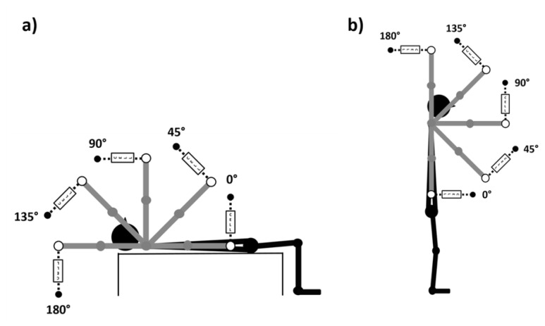
For PO exercise, all participants lay supine on a flat bench holding a short barbell connected to the cross-over equipment; and for PW exercise, all participants remained standing with the torso in the vertical position holding a short barbell connected to a cross over equipment. For both exercises and each shoulder joint position, the cross-over equipment was adjusted to maintain the cable perpendicular to the participant’s upper limb (Figure 1).
Figure 1.
Shoulder joint positions performed during both exercises: a) Pullover and b) Pulldown.
For both exercises, a grip of 100% of the bi-acromial distance was adopted (17) and marked with adhesive tape on the bar. The participants were instructed to maintain the pronated handgrip with elbows fully extended. A rest period of 5-min between conditions and 30-min between exercises (PO and PW) was used. At the end of each experimental condition, all participants were asked to report the rating of perceived exertion (RPE). All experimental conditions were randomized for all participants. All participants received verbal encouragement during all experimental conditions. All measures were performed at the same hour of the day (between 9 AM and 12 PM) by the same researcher.
Shoulder Joint Position
A fleximeter (model FL6010, Sanny, SP, Brazil) was used to control the shoulder joint position (0o, 45o, 90o, 135o, and 180°) in both exercises (PO and PW). The zero degrees was set when the upper limb was aligned with the greater trochanter. In order to determine the joint position, participants were instructed to flex their shoulders until they reached the angular position referring to 45o, 90o, 135o, and 180°. The fleximeter was positioned on the dominant upper limb (mid-arm) (Figure 1).
Maximal Voluntary Isometric Contraction (MVIC)
The MVIC was measured by a load cell acquisition system (EMG832C, EMG system, Sao Jose dos Campos, Brazil) with a sampling rate of 2KHz using a commercially designed software program (EMG system, Sao Jose dos Campos, Brazil). All MVIC data were synchronized with the sEMG data. In order to acquire MVIC, a load cell was fixed between the bar and the cross-over equipment. During all experimental conditions, the load cell was adjusted to remain perpendicular to the upper limbs of each participant. The digitized force data were low-pass filtered at 10 Hz using a fourth-order Butterworth filter with a zero lag. Then, the maximal value was defined as peak force (PF) for all exercises and shoulder joint positions.
Surface Electromyography (sEMG)
The participants’ skin was prepared before the placement of the sEMG electrodes. Hair at the site of electrode placement was shaved, and the skin was cleaned with alcohol. Bipolar passive disposable dual Ag/AgCl snap electrodes were used which were 1 cm in diameter for each circular conductive area with 2-cm center-to-center spacing. These were placed on the dominant upper limb for Pectoralis Major (PM): electrodes were positioned at 50% on the line between the muscular belly and the middle fibers (sternal-costal); Latissimus Dorsi (LD): the electrodes were positioned 2cm below the lower edge of the scapula on the midline between the spine and the side of the trunk, obliquely towards the first lumbar vertebra (L1), according to the Criswell protocol (5); and Posterior Deltoid (PD): the electrodes were positioned two fingers behind the angle of the acromion, following the alignment between the acromion and the little finger of the hand, according to the SENIAM protocol (9). The sEMG signals were recorded by an electromyography acquisition system (EMG832C, EMG system, Sao Jose dos Campos, Brazil) with a sampling rate of 2KHz using a commercially designed software program (EMG system, Brazil). EMG amplitude was amplified (bipolar differential amplifier, input impedance = 2 MΩ, common-mode rejection ratio >100 dB min (60 Hz), gain × 20, noise > 5 μV) and analog-to-digitally converted (12 bit). The ground electrode was placed on the bony prominence of the elbow (olecranon). All sEMG data were analyzed with a software program (EMG system, Sao Jose dos Campos, Brazil). The digitized sEMG data were band-pass filtered at 20–400 Hz using a fourth-order Butterworth filter with a zero lag. For myoelectric activation time-domain analysis, RMS (150ms moving window) was calculated during the MVIC. The area under the RMS sEMG curve was calculated, defining the integrated sEMG (iEMG). The reliability (ICC) of the EMG data between isometric contractions for each exercise and shoulder position ranged between 0.69 and 0.81
Rating of Perceived Exertion (RPE)
The session RPE was assessed with a CR-10 scale using the recommendations of Sweet et al., (16). Participants were asked to use an arbitrary unit (A.U.) on the scale to rate their overall effort for each RT exercise. A rating of 0 was associated with no effort and a rating of 10 was associated with maximal effort and the most stressful exercise ever performed. The RPE was requested after each experimental condition and all participants answered the following question based on the CR-10 scale: “How was your workout?”
Statistical Analysis
The normality and homogeneity of variances within the data were confirmed with the Shapiro-Wilk and Levene’s tests, respectively. Mean, standard deviation, and delta percentage (Δ%) were calculated. Interrater reliability was assessed for each condition and exercise. Test-retest reliability (ICC) was calculated and evaluated based on the following criteria: < 0.4 poor; 0.4 – < 0.75 satisfactory; ≥ 0.75 excellent. A two-way repeated-measures ANOVA (2×5) with exercises (PO and PW) and joint positions (0o, 45o, 90o, 135o, and 180°) were used for all dependent variables (iEMG, peak force, and RPE). The RPE was reported through a descriptive analysis using the mean and standard deviation. Cohen’s formula for effect size (d) were qualitatively interpreted using the following thresholds: <0.35 - trivial; 0.35–0.8 - small; 0.8–1.5 - moderate; >1.5 - large for recreationally trained (4). An alpha of 5% was used to determine statistical significance.
RESULTS
The results of the intraclass correlation coefficient (ICC) for peak force were excellent for all joint positions and exercises [0°: PO (0.97) and PW: (0.95); 45°: PO (0.97) and PW: (0.98); 90°: PO (0.91) and PW: (0.92); 135°: PO (0.95) and PW: (0.98); and 180°: PO (0.93) and PW: (0.94)].
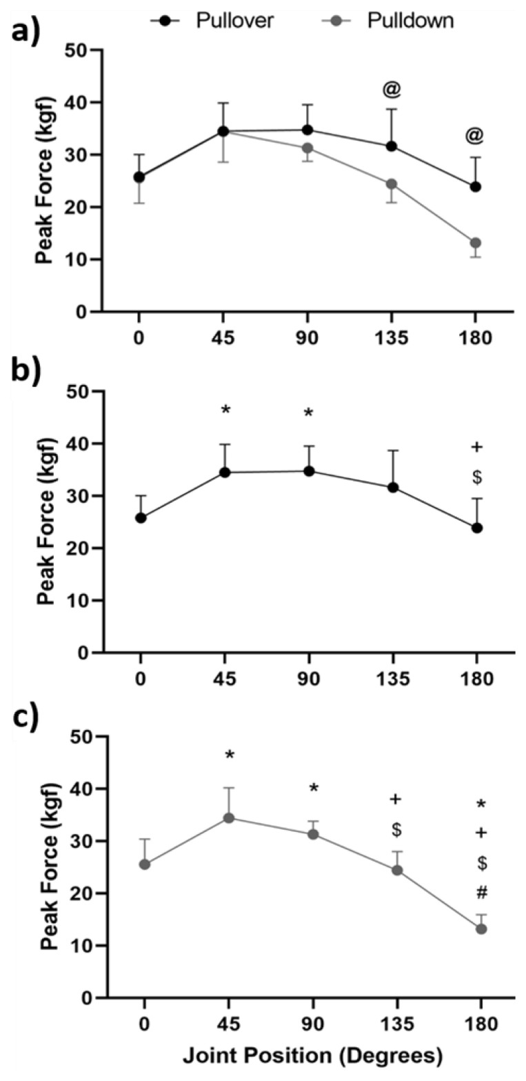
For Peak Force (PF), there were significant main effects for exercise (p < 0.001) and joint positions (p < 0.001). There was significant interaction between exercises and joint positions (p < 0.001). There were significant differences between PO and PW exercises only for 135° and 180°, the highest values were observed for PO exercise (p = 0.023, d = 1.28, Δ% = 22.8, and p = 0.001, d = 2.42, Δ% = 44.9, respectively) (Figure 2a). For PO exercise, there were significant differences between 45° vs 0° (p < 0.001, d = 6.57, Δ% = 25.3) and 90° vs 0° (p < 0.001, d = 10.61, Δ% = 25.8) and 45° vs 180° (p = 0.001, d = 1.92, Δ% = 30.7) and 90° vs 180° (p = 0.002, d = 1.21, Δ% = 31.2) (Figure 2b). For the PW exercise, there were observed significant differences between 45° vs 0° (p < 0.001, d = 1.65, Δ% = 25.7), 90° vs 0° (p = 0.001, d = 1.48; Δ% = 18.3), 45° vs 135° (p < 0.001, d = 2.06, Δ% = 40.8), 90° vs 135° (p = 0.001, d = 2.21, Δ% = 21.9), 45° vs 180° (p < 0.001, d = 4.66, Δ% = 61.7), and 90° vs 180° (p < 0.001; d = 6.88, Δ% = 57.9), 0° vs 180° (p < 0.001, d = 3.15, Δ% = 94.0), and 0° vs 180° (p < 0.001, d = 3.52, Δ% = 46.1) (Figure 2c).
Figure 2.
Mean and standard deviation of the peak force: a) comparison between exercises (Pullover vs. Pulldown) for each shoulder joint position; b) Pullover Exercise: comparison between shoulder joint positions (0°, 45°, 90°, 135°, and 180°), and c) Pulldown Exercise: comparison between shoulder joint positions (0°, 45°, 90°, 135°, and 180°). Legend: @Significant difference between exercises. *Significant difference between joint positions when compared to 0°; +Significant difference between joint positions when compared to 45°; $Significant difference between joint positions when compared to 90°, and #Significant difference between joint positions when compared to 135°, (p < 0.05).
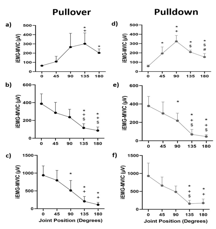
For iEMG PM, there was a significant main effect for joint positions (p < 0.001). There was a significant interaction between exercises and joint positions (p < 0.001). For iEMG LD, there was a significant main effect for joint positions (p < 0.001). There was not a significant interaction between exercises and joint positions (p = 0.070). For iEMG PD, there was a significant main effect for joint positions (p < 0.001). There was not a significant interaction between exercises and joint positions (p = 0.192). For PO exercise, there were observed significant differences for iEMG PM between 135° vs 0° (p = 0.001, d = 0.40, Δ% = 78.7), 180° vs 0° (p = 0.001, d = 1.42, Δ% = 68.4), and 135° vs 45° (p = 0.006, d = 0.24, Δ% = 48.4) (Figure 3a). For iEMG LD, there were significant differences between 135° vs 0° (p = 0.001, d = 3.17, Δ% = 70.1), 135° vs 45° (p = 0.031, d = 1.91, Δ% = 59.4), 135° vs 90° (p = 0.009, d = 1.69, Δ% = 51.0), 180° vs 0° (p < 0.001, d = 3.59, Δ% = 77.8), 180° vs 45° (p = 0.027, d = 2.28, Δ% = 69.9), and 180° vs 90°(p = 0.005, d = 2.17 Δ% = 63.6) (Figure 3b). For iEMG PD, there were significant differences between 0° vs 90° (p = 0.033, d = 0.15, Δ% = 45.4), 0° vs 135° (p < 0.001, d = 0.34, Δ% = 77.9), 0° vs 180° (p < 0.001, d = 0.42, Δ% = 88.0), 45° vs 135° (p = 0.002, d = 0.26 Δ% = 74.0), and 45° vs 180° (p = 0.001, d = 0.33, Δ% = 85.9) (Figure 3c).
Figure 3.
Mean and standard deviation of iEMG for both exercises (Pullover: graphs a-b-c and Pulldown: graphs d-e-f) at all shoulder joint angles. Pectoralis Major (a and d); Latissimus Dorsi (b and e); and Posterior Deltoid (c and f). Legend: @Significant difference between exercises. *Significant difference between joint positions when compared to 0°; +Significant difference between joint positions when compared to 45°; $Significant difference between joint positions when compared to 90°, and #Significant difference between joint positions when compared to 135°, (p < 0.05).
For PW exercise, there were observed significant differences for iEMG PM between 0° vs 45° (p = 0.007, d = 2.68, Δ% = 70.4), 0° vs 90° (p < 0.001, d = 5.77, Δ% = 82.3), 0° vs 135° (p < 0.001, d = 8.59 Δ% = 72.5), 0° vs 180° (p < 0.001, d = 3.43 Δ% = 63.1), 90° vs 45° (p = 0.003, d = 1.92, Δ% = 40.1), 90° vs 135° (p = 0.025, d = 2.41, Δ% = 35.5), 90° vs 180° (p = 0.001, d = 3.19, Δ% = 51.9), and 135° vs 180° (p = 0.020, d = 1.72, Δ% = 25.5) (Figure 3d). For iEMG LD, there were significant differences between 0° vs 90° (p = 0.002, d = 1.69 Δ% = 42.3), 0° vs 135° (p < 0.001, d = 3.71, Δ% = 81.7), 0° vs 180° (p < 0.001, d = 4.40, Δ% = 88.0), 45° vs 135° (p < 0.001, d = 2.38, Δ% = 76.7), 90° vs 135° (p = 0.003, d = 2.14, Δ% = 68.3), 45° vs 180° (p = 0.001, d = 2.82, Δ% = 84.7), and 90° vs 180° (p = 0.001, d = 2.87, Δ% = 79.1) (Figure 3e). For iEMG PD, there were significant differences between of 0° vs 135° (p = 0.001, d = 2.94 Δ% = 83.2), 45° vs 135° (p = 0.001, d = 2.77, Δ % = 76.3), 90° vs 135° (p = 0.005, d = 2.45, Δ% = 67.8), 0° vs 180° (p = 0.001, d = 2.84, Δ% = 81.3), and 45° vs 180° (p = 0.001, d = 2.60, Δ% = 73.7) (Figure 3f).
For RPE, there were no significant differences between exercises and joint positions: PO (0°: 9.6 ± 0.6, 45°: 9.7 ± 0.6, 90°: 9.8 ± 0.4, 135°: 9.7 ± 0.6, and 180: 9.5 ± 0.7) and PW (0°: 9.7 ± 0.6, 45°: 9.6±0.5, 90°: 9.8 ± 0.4, 135°: 9.6±0.6, and 180°: 9.7 ± 0.5) (p > 0.05).
DISCUSSION
The aim of the present study was to compare the myoelectric activation (PM, LD, and PD) and peak force between PO and PW exercises in different shoulder joint positions (0o, 45o, 90o, 135o, and 180°) during maximal isometric contractions. In order to achieve this goal, this study measured peak force and myoelectric activation in specific joint positions and adjusted the external force always perpendicular to the upper limb in order to understand both, the peak force and myoelectric activity pattern. The main findings of this study are 1. Peak force was similar between exercises, however, PW showed lower values between 135° and 180°; 2. Peak force was greater between 45° and 90° for both exercises; 3. PM presented similar maximal myoelectric activation between exercises, with higher values between 90° and 135°; 4. LD presented similar maximal myoelectric activation between exercises, with higher values between 0° and 45°; 5. LD and PD presented a similar pattern for both exercises; 6. The RPE was similar for both exercises and shoulder joint positions. The results of this study corroborated the main hypothesis because; peak force is angle-dependent and the functional and morphological characteristics of muscle groups around the shoulder joint were affected by the shoulder joint position.
Based on the authors’ knowledge no study compared the peak force between PO and PW exercises. Regarding peak force, both exercises presented a similar force-angle curve between 0° and 90° with higher values at 45° and 90°. However, differences in peak force were observed at angles 135° and 180° between exercises (PO>PD: 22.8% and 44.9%, respectively). Therefore, in general, the PO exercise presented high values of peak force in the entire range of motion when compared to the PD exercise. Possibly, the highest force values between 45° and 90° might be attributed to the combined participation of the PM and LD, and between 135° and 180°, the participation of the LD was substantially reduced. This discussion is only valid for the quasi-linear relationship between muscle activity and force production in isometric conditions. In addition, the lower values of force observed in the PD exercise at angles of 135° and 180° might be attributed to differences in the participants’ body posture during both exercises. During the PO exercise, the participants remained to lie on a bench, which possibly increased trunk stability throughout the entire range of motion; on the other hand, the PD exercise was performed while standing, which could negatively affect trunk stability and make cable traction more difficult, especially at 135° and 180° of shoulder extension.
Regarding the maximal myoelectric activation, both exercises presented a similar iEMG-angle curve, however, different patterns were observed between prime movers (PM vs. LD). The PM activation presented an ascendant-descendent pattern, with higher values between 90° and 135°; on the other hand, the LD and PD activation presented a decrescent pattern with higher values between 0° and 45°. The results of this study corroborated the main hypothesis that all muscle groups around the shoulder joint were affected by the joint position. Previous studies have shown that the myoelectric activation of the PM and LD varied according to the direction of the external load in relation to the shoulder joint position for shoulder extension exercises (Pullover and Ab Wheel Rollout) (12, 13). According to Hamil et al., (8) pennate muscles have fibers that advance diagonally in relation to the tendon, thus, the maximal myoelectric activation might be affected by the shoulder joint position. These differences might help in the correct choice of the exercise aiming to emphasize specific ranges of motion, for example, in the pull phase of the crawl style in swimming.
For RPE, similar scores were observed for both exercises and shoulder joint positions corroborating the secondary hypothesis. It is well known that RPE is affected by the level of neuromuscular fatigue after each RT dynamic exercise for recreationally-trained participants, thus, the type of contraction (isometric) and the level of contraction (maximal) might not affect the perception of effort, considering that both exercises reached similar levels of intensity.
This research may be of benefit to recreational athletes, bodybuilders, and rehabilitation programs. Isometric contractions are of great importance in increasing the strength at specific joint positions, which may aid in rehabilitation programs/testing or at certain phases of training of recreational athletes/bodybuilders when the aim is to increase strength in joint positions with lower mechanical advantage (sticking point) or even increase metabolic stress aiming at greater neuromuscular fatigue. The results of this research support the notion that both exercises produce similar force-angle curves. However, when the training goal is to maximally activate different prime movers, the use of specific ranges of motion could be prescribed (PM: between 90° to 135°; and LD/PD: between 0° and 90°). Additionally, the maximal isometric contractions did not affect the RPE for both exercises and joint positions.
We recognize that this study has some limitations. We did not measure all muscles around the shoulder joint complex which could help to understand some effects on peak force. We did not control for skinfold thickness of the sEMG detection area which is considered to be a low-pass filter, and there may have been some inherent differences in the musculotendinous tightness between participants. The position of the body might affect the peak force in both exercises (PO: lying down; PW: standing); however, the position of the body used was the closest to the practical application of each exerciser. Both exercises were evaluated with the load cell perpendicular to the position of the upper limb, which limits the extrapolation of results to other directions of the force. We also used a healthy, resistance-trained population, and our results, therefore, are not directly generalizable to dynamic contractions, populations, or athletes. Our results showed that specific shoulder joint positions affect maximal force production or myoelectric activation during both exercises (Pullover and Pulldown). The present findings suggest that the highest level of force is observed between 45° and 90° for both exercises; The pectoralis major and latissimus dorsi present similar iEMG-angle curves between exercises, however, specific iEMG patterns are observed between shoulder joint positions. RPE is not affected by exercise or shoulder joint positions.
ACKNOWLEDGEMENTS
The authors thank the participants for their participation.
REFERENCES
- 1.Borges E, Mezencio B, Pinho J, Soncin R, Barbosa J, Araujo F, Gianolla F, Amadio C, Serrao JC. Resistance training acute session: Pectoralis major, latissimus dorsi and triceps brachii electromyographic activity. J Phys Educ Sport. 2018;18(2):648–653. [Google Scholar]
- 2.Campos YAC, da Silva SF. Comparison of electromyographic activity during the bench press and barbell pullover exercises. Motriz. 2014;20(2):200–205. [Google Scholar]
- 3.Campos YAC, Rodrigues HL, da Silva SF, Marchetti PH. The use of barbell or dumbbell does not affect muscle activation during pullover exercise. Rev Bras Med Esporte. 2017;23(5):386–389. [Google Scholar]
- 4.Cohen J. Statistical power analysis for the behavioral sciences. New Jersey: Lawrence Erlbaum Associates; 1988. [Google Scholar]
- 5.Criswell E. Cram’s introduction to surface electromyography. Burlington, MA: Jones and Bartlett; 2011. [Google Scholar]
- 6.Eng J. Sample size estimation: How many individuals should be studied? Radiol. 2003;227(2):309–313. doi: 10.1148/radiol.2272012051. [DOI] [PubMed] [Google Scholar]
- 7.Floyd RT. Manual of structural kinesiology. 21th ed. New York, NY: McGraw Hill; 2021. [Google Scholar]
- 8.Hamill J, Knutzen KM, Derrick TR. Biomechanical basis of human movement. 5th ed. Philadelphia, PA: Wolters Kluwer; 2021. [Google Scholar]
- 9.Hermens HJ, Freriks B, Disselhorst-Klug C, Rau G. Development of recommendations for semg sensors and sensor placement procedures. J Electromyogr Kinesiol. 2000;10(5):361–374. doi: 10.1016/s1050-6411(00)00027-4. [DOI] [PubMed] [Google Scholar]
- 10.Marchetti PH. Applied science. 2nd ed. Dubuque, IA: Kendall Hunt; 2022. Strength training manual. [Google Scholar]
- 11.Marchetti PH, Calheiros R, Charro M. Biomecanica aplicada: Uma abordagem para o treinamento de forca. Sao Paulo: Phorte Editora; 2019. [Google Scholar]
- 12.Marchetti PH, Schoenfeld BJ, Silva JJ, Guiselini MA, Freitas FS, Pecoraro SL, Gomes WA, Lopes CR. Muscle activation pattern during isometric ab wheel rollout exercise in different shoulder angle-positions. Med Express. 2015;2(4):1–5. [Google Scholar]
- 13.Marchetti PH, Uchida MC. Effects of the pullover exercise on the pectoralis major and latissimus dorsi muscles as evaluated by emg. J Appl Biomech. 2011;27(4):380–384. doi: 10.1123/jab.27.4.380. [DOI] [PubMed] [Google Scholar]
- 14.Navalta JW, Stone WJ, Lyons TS. Ethical issues relating to scientific discovery in exercise science. Int J Exerc Sci. 2019;12(1):1–8. doi: 10.70252/EYCD6235. [DOI] [PMC free article] [PubMed] [Google Scholar]
- 15.Ratamess NA, Alvar BA, Evetoch TK, Houst TJ, Kibler WB, Kraemer WJ, Triplett NT. Progression models in resistance training for healthy adults. Med Sci Sports Exerc. 2009;41(3):687–708. doi: 10.1249/MSS.0b013e3181915670. [DOI] [PubMed] [Google Scholar]
- 16.Sweet TW, Foster C, McGuigan MR, Brice G. Quantitation of resistance training using the session rating of perceived exertion method. J Strength Cond Res. 2004;18(4):796–802. doi: 10.1519/14153.1. [DOI] [PubMed] [Google Scholar]
- 17.Wagner LL, Evans SA, Weir JP, Housh TJ, Johnson GO. The effect of grip width on bench press performance. J Appl Biomech. 1992;8(1):1–10. [Google Scholar]


