

Abstract
Ganodermataceae is one of the main families of macrofungi since species in the family are both ecologically and economically important. The double-walled basidiospores with ornamented endospore walls are the characteristic features of Ganodermataceae. It is a large and complex family; although many studies have focused on Ganodermataceae, the global diversity, geographic distribution, taxonomy and molecular phylogeny of Ganodermataceae still remained incompletely understood. In this work, taxonomic and phylogenetic studies on worldwide species of Ganodermataceae were carried out by morphological examination and molecular phylogenetic analyses inferred from six gene loci including the internal transcribed spacer regions (ITS), the large subunit of nuclear ribosomal RNA gene (nLSU), the second largest subunit of RNA polymerase II gene (rpb2), the translation elongation factor 1-α gene (tef1), the small subunit mitochondrial rRNA gene (mtSSU) and the small subunit nuclear ribosomal RNA gene (nSSU). A total of 1 382 sequences were used in the phylogenetic analyses, of which 817 were newly generated, including 132 sequences of ITS, 139 sequences of nLSU, 83 sequences of rpb2, 124 sequences of tef1, 150 sequences of mtSSU and 189 sequences of nSSU. The combined six-gene dataset included sequences from 391 specimens representing 146 taxa from Ganodermataceae. Based on morphological and phylogenetic analyses, 14 genera were confirmed in Ganodermataceae: Amauroderma, Amaurodermellus, Cristataspora, Foraminispora, Furtadoella, Ganoderma, Haddowia, Humphreya, Magoderna, Neoganoderma, Sanguinoderma, Sinoganoderma, Tomophagus and Trachydermella. Among these genera, Neoganoderma gen. nov. is proposed for Ganoderma neurosporum; Sinoganoderma gen. nov. is proposed for Ganoderma shandongense; Furtadoella gen. nov. is proposed to include taxa previously belonging to Furtadoa since Furtadoa is a homonym of a plant genus in the Araceae; Trachydermella gen. nov. is proposed to include Trachyderma tsunodae since Trachyderma is a homonym of a lichen genus in the Pannariaceae. Twenty-three new species, viz., Ganoderma acaciicola, G. acontextum, G. alpinum, G. bubalinomarginatum, G. castaneum, G. chuxiongense, G. cocoicola, G. fallax, G. guangxiense, G. puerense, G. subangustisporum, G. subellipsoideum, G. subflexipes, G. sublobatum, G. tongshanense, G. yunlingense, Haddowia macropora, Sanguinoderma guangdongense, Sa. infundibulare, Sa. longistipitum, Sa. melanocarpum, Sa. microsporum and Sa. tricolor are described. In addition, another 33 known species are also described in detail for comparison. Scanning electron micrographs of basidiospores of 10 genera in Ganodermataceae are provided. A key to the accepted genera of Ganodermataceae and keys to the accepted species of Ganoderma, Haddowia, Humphreya, Magoderna, Sanguinoderma and Tomophagus are also provided. In total, 278 species are accepted as members of Ganodermataceae including 59 species distributed in China.
Taxonomic novelties: New genera: Furtadoella B.K. Cui & Y.F. Sun, Neoganoderma B.K. Cui & Y.F. Sun, Sinoganoderma B.K. Cui, J.H. Xing & Y.F. Sun and Trachydermella B.K. Cui & Y.F. Sun; New species: Ganoderma acaciicola B.K. Cui, J.H. Xing & Y.F. Sun, G. acontextum B.K. Cui, J.H. Xing & Vlasák, G. alpinum B.K. Cui, J.H. Xing & Y.F. Sun, G. bubalinomarginatum B.K. Cui, J.H. Xing & Y.F. Sun, G. castaneum B.K. Cui, J.H. Xing & Y.F. Sun, G. chuxiongense B.K. Cui, J.H. Xing & Y.F. Sun, G. cocoicola B.K. Cui, J.H. Xing & Y.F. Sun, G. fallax B.K. Cui, J.H. Xing & Vlasák, G. guangxiense B.K. Cui, J.H. Xing & Y.F. Sun, G. puerense B.K. Cui, J.H. Xing & Y.F. Sun, G. subangustisporum B.K. Cui, J.H. Xing & Y.F. Sun, G. subellipsoideum B.K. Cui, J.H. Xing & Y.F. Sun, G. subflexipes B.K. Cui, J.H. Xing & Y.F. Sun, G. sublobatum B.K. Cui, J.H. Xing & Y.F. Sun, G. tongshanense B.K. Cui, J.H. Xing & Y.F. Sun, G. yunlingense B.K. Cui, J.H. Xing & Y.F. Sun, Haddowia macropora B.K. Cui, Vlasák & Y.F. Sun, Sanguinoderma guangdongense B.K. Cui & Y.F. Sun, Sa. infundibulare B.K. Cui & Y.F. Sun, Sa. longistipitum B.K. Cui & Y.F. Sun, Sa. melanocarpum B.K. Cui & Y.F. Sun, Sa. microsporum B.K. Cui & Y.F. Sun and Sa. tricolor B.K. Cui & Y.F. Sun; New combinations: Furtadoella biseptata (Costa-Rezende et al.) B.K. Cui & Y.F. Sun, Fu. brasiliensis (Singer) B.K. Cui & Y.F. Sun, Fu. corneri (Gulaid & Ryvarden) B.K. Cui & Y.F. Sun, Neoganoderma neurosporum (J.S. Furtado) B.K. Cui & Y.F. Sun, Sinoganoderma shandongense (J.D. Zhao & L.W. Xu) B.K. Cui, J.H. Xing & Y.F. Sun and Trachydermella tsunodae (Yasuda ex Lloyd) B.K. Cui & Y.F. Sun.
Citation: Sun Y-F, Xing J-H, He X-L, Wu D-M, Song C-G, Liu S, Vlasák J, Gates G, Gibertoni TB, Cui B-K (2022). Species diversity, systematic revision and molecular phylogeny of Ganodermataceae (Polyporales, Basidiomycota) with an emphasis on Chinese collections. Studies in Mycology 101: 287–415. doi: 10.3114/sim.2022.101.05.
Keywords: Ganoderma, macro fungi, medicinal mushrooms, new taxa, phylogeny, ultrastructure, white-rot fungi
INTRODUCTION
Ganodermataceae as an important family in the Polyporales has been researched for many decades due to its high medicinal and ecological values. As traditional medicine, Ganoderma lingzhi, G. sinense and Amauroderma rugosum have been used for anti-cancer treatment, for lowering blood pressure and for improving immunity (Dai et al. 2009, Cao et al. 2012, Chan et al. 2013, Zhou et al. 2015, Zhang et al. 2019). Tree pathogens such as G. boninense can cause a basal stem rot on oil palm trees (Pilotti 2005), and G. philippii can cause a red root rot on Acacia mangium (Glen et al. 2009). Besides, Ganoderma lucidum and A. rugosum have been used biotechnologically in the production of biofuel and degradation of environmental pollutants (Jong et al. 2017, Wang et al. 2021).
The systematics of the Ganodermataceae have been carried out for about 100 years. Donk (1948) introduced Ganodermataceae as a family based on its unique double-walled basidiospores with obvious ornamentation on the endospore walls, which only included Ganoderma with a laccate pileal surface and truncated basidiospores and Amauroderma with globose to ellipsoid basidiospores without a truncated apex. Murrill (1905) introduced a genus Tomophagus that included pale-coloured basidiomata with pale and soft context and truncated basidiospores. In the past decades, specimens of Ganodermataceae have been collected from all over the world (Steyaert 1972, Moncalvo & Ryvarden 1997, Ryvarden 2004b, Hapuarachchi et al. 2019b) except for the polar region. More genera have been established by evidence of morphological characters and/or molecular data. Imazeki (1952) presented Trachyderma as a genus with a fleshy succulent context and truncated basidiospores with spinules. Steyaert (1972) examined several known species in Ganoderma, then established Haddowia based on non-truncated basidiospores with longitudinal ridges partly connected with short transverse walls on the endospore walls; Humphreya with reticular or erratic irregularly ridged double walls on truncated basidiospores; and Magoderna based on anticlinal hyphae in pileipellis and ellipsoid to ovoid basidiospores without truncated apex. Costa-Rezende et al. (2017) established Foraminispora according to the hollow columnar endospore ornamentation, which persist to the exospore wall sometimes forming holes on the basidiospores, and Furtadoa based on a monomitic hyphal system in context with clamped and simple-septate generative hyphae. Sun et al. (2020) studied one group of Amauroderma with a unique pore surface that changes to blood red when bruised, and established Sanguinoderma. Costa-Rezende et al. (2020b) established Amaurodermellus with a dull pileal surface and ovoid basidiospores, and Cristataspora with double-walled basidiospores which have endosporic ornamentation as vertical or transverse ridges.
However, the uncertainty about the family level of Ganodermataceae should not be ignored. Binder et al. (2013) carried out the phylogenetic and phylogenomic analyses of Polyporales based on 356 single-copy genes from 10 genomes of this order, which showed that Ganoderma was involved in the ‘core polyporoid clade’. Justo et al. (2017) suggested Ganodermataceae as a synonym of Polyporaceae based on ITS, nLSU and rpb1 sequences. The conclusions based on phylogenetic evidence have certain credibility, while in this study, Ganodermataceae is still regarded as an independent family based on its remarkable morphological features to clarify the intergeneric and interspecific relationships. For the time being, we prefer to treat Ganodermataceae as an independent family different from Polyporaceae according to previous specialised studies on the Ganoderma group (Moncalvo & Ryvarden 1997, Robledo et al. 2015, Zhou et al. 2015, Hapuarachchi et al. 2018a, b, 2019a, b, Xing et al. 2018, Costa-Rezende et al. 2020a, b, Sun et al. 2020, Luangharn et al. 2021). The scientific status of Ganodermataceae within the Polyporales should be considered in morphology, phylogeny, and even whole genome sequences.
Ongoing taxonomic studies of Ganodermataceae from Asia, Africa, Europe, Neotropics and North America have been conducted for a long time with many new species and combinations continually being reported (Otieno 1968, Steyaert 1972, Moncalvo & Ryvarden 1997, Ryvarden 2004a, b, Gibertoni et al. 2008, Cao et al. 2012, Le et al. 2012, Coetzee et al. 2015, Gomes-Silva et al. 2015, Hapuarachchi et al. 2019b, Sun et al. 2020, Costa-Rezende et al. 2020b). China has a complex and diverse natural environment resulting in high species richness, and a total of 130 species of Ganodermataceae have been reported (Zhao & Zhang 2000, Dai 2012, Cao & Yuan 2012, Wang & Wu 2014, Li et al. 2015, Zhou et al. 2015, Hapuarachchi et al. 2018b, Xing et al. 2018, Ye et al. 2019, Sun et al. 2020). The great variability in the macroscopic characters of the basidiomata and the relatively uniform macro- and micro-morphology of most species in Ganodermataceae have resulted in many confusions in taxonomy. As of 10 March 2022, there were 642 records of Ganodermataceae recorded in Index Fungorum (http://www.indexfungorum.org/), and 698 records in MycoBank (http://www.mycobank.org/). Nearly half of these records have been identified as synonyms, especially in Ganoderma and Amauroderma and it is necessary to assess the validity of these records.
With the rapid development of molecular techniques in recent years, DNA sequence data have been widely used in the taxonomic studies of Ganodermataceae. Moncalvo (1995) used ITS sequences and the D2 region of nLSU sequences to construct the relationships among species in Ganoderma, and concluded that the combined data is useful for intrageneric segregation while the D2 region is suitable for intergeneric or higher ranks segregation. Subsequently, ITS and nLSU sequences were often used to identify species (Cao et al. 2012, Le et al. 2012, de Lima Júnior et al. 2014, Gomes-Silva et al. 2015, Li et al. 2015). It is worth mentioning that Fryssouli et al. (2020) carried out a phylogenetic study of Ganoderma based only on 3 970 ITS sequences obtained from the GenBank/ENA/DDBJ database which evaluated the accuracy of sequences and showed that Ganoderma can be divided into five main lineages. However, for the complex groups in Ganoderma or for the higher rank classification of Ganodermataceae, most researchers use multi-gene datasets to construct phylogenetic trees (Zhou et al. 2015, Costa-Rezende et al. 2017, 2020b, Justo et al. 2017, Cabarroi-Hernández et al. 2019, Hapuarachchi et al. 2019b, Luangharn et al. 2020, Sun et al. 2020). At present, eight genes have been applied to the phylogenetic analyses in Ganodermataceae, viz., the internal transcribed spacer regions (ITS), the large subunit of nuclear ribosomal RNA gene (nLSU), the largest subunit of RNA polymerase II gene (rpb1), the second largest subunit of RNA polymerase II gene (rpb2), the translation elongation factor 1-α gene (tef1), the β-tubulin gene (tub), the small subunit mitochondrial rRNA gene (mtSSU) and the small subunit nuclear ribosomal RNA gene (nSSU). According to the records in GenBank (https://www.ncbi.nlm.nih.gov/) as of 21 April 2021, 150 801 items were found by searching ‘Ganodermataceae’ directly, but only about 65 000 items among them were identified as species of Ganodermataceae. The number of sequences is considerable, but repetitive sequences of the same species or specimens, inaccurate identification and low quality of sequences make it necessary to select only the reliable molecular data for phylogenetic analyses.
In this study, the specimens collected from all over the world were studied by macromorphological and microscopic examinations together with ultrastructural observations and phylogenetic analyses based on six gene loci (ITS, nLSU, rpb2, tef1, mtSSU and nSSU). A total of 146 species in Ganodermataceae with available DNA sequences were involved in the phylogenetic analyses. Based on morphological characters and phylogenetic evidence, 14 genera were confirmed within Ganodermataceae, Furtadoella gen. nov., Neoganoderma gen. nov., Sinoganoderma gen. nov. and Trachydermella gen. nov. were proposed as new genera; 278 species were confirmed in Ganodermataceae including 23 new species which are listed in Table 2. The ultrastructural features observed under SEM of basidiospores of 10 genera in Ganodermataceae were described and photographed. In total, 56 species and nine genera are described and illustrated here. A key to accepted genera of Ganodermataceae and keys to accepted species of Ganoderma, Haddowia, Humphreya, Magoderna, Sanguinoderma, Tomophagus are also provided.
Table 1.
Taxa information and GenBank accession numbers of the sequences used in this study. Species in bold are new species or new combinations.
a Newly generated sequences for this study.
MATERIALS AND METHODS
Morphological studies
The studied specimens are deposited at the fungaria of the Institute of Microbiology, Beijing Forestry University (BJFC, Beijing, China), the Institute of Applied Ecology, Chinese Academy of Sciences (IFP, Shenyang, China), the private fungarium of J. Vlasák of Czech Republic (JV) and the Universidade Federal de Pernambuco, Brazil (URM). Macro-morphological descriptions of the new taxa (or selected taxa) were based on field notes and fungarium specimens. Special colour terms followed Petersen (1996). Micro-morphological data were obtained from dried specimens and observed under a compound microscope following Cui et al. (2019) and Sun et al. (2020). Sections were studied at a magnification up to 1 000× using Nikon E80i microscope and phase contrast illumination (Nikon, Tokyo, Japan). Line drawings were made with the aid of a drawing tube. Ultrastructure of basidiospores was observed with Scanning Electron Microscopy (SEM) using a Field Emission Scanning Electron Microscope (FESEM) Hitachi SU-8010 (Hitachi, Ltd, Tokyo, Japan) at Beijing Forestry University, China (BJFU). Microscopic features, measurements and drawings were made from slide preparations stained with Cotton Blue and Melzer’s reagent. Spores were measured from sections cut from the tubes. To represent the variation in the size of the basidiospores, 5 % of measurements were excluded from each end of the range, and are given in parentheses. The following abbreviations are used: IKI = Melzer’s reagent, IKI – = neither amyloid nor dextrinoid, KOH = 5 % potassium hydroxide, CB = Cotton Blue, CB + = cyanophilous, L = mean spore length (arithmetic average of all spores), W = mean spore width (arithmetic average of all spores), Q = variation in the L/W ratios between the specimens studied, n (a/b) = number of spores: (a) measured from given number, (b) of specimens.
DNA extraction, amplification and sequencing
A cetyl trimethylammonium bromide (CTAB) rapid plant genome extraction kit-DN14 (Aidlab Biotechnologies Co., Ltd, Beijing, China) and a FH plant DNA kit II (Demeter Biotech Co., Ltd., Beijing, China) were used to extract total genomic DNA from dried specimens and to perform the polymerase chain reaction (PCR) according to the manufacturer’s instructions with some modifications (Xing et al. 2018, Sun et al. 2020). The ITS regions were amplified with primer pairs ITS5 and ITS4 (White et al. 1990). The nLSU regions were amplified with primer pairs LR0R and LR7, and the primer LR5 was used sometimes as an alternative to LR7 (Vilgalys & Hester 1990). The rpb2 regions were amplified with primer pairs fRPB2-5F and fRPB2-7CR (Liu et al. 1999). The tef1 regions were amplified with primer pairs EF1-983F and EF1-1567R (Rehner & Buckley 2005). The mtSSU regions were amplified with primer pairs MS1 and MS2 (White et al. 1990). The nSSU regions were amplified with primer pairs PNS1 and NS41 (White et al. 1990).
The PCR cycling schedule for ITS, tef1 and mtSSU included an initial denaturation at 95 °C for 3 min, followed by 35 cycles at 94 °C for 40 s, 54 °C for ITS and mtSSU, 55 °C for tef1 for 45 s, 72 °C for 1 min, and a final extension at 72 °C for 10 min. The PCR cycling schedule for nLSU and nSSU included an initial denaturation at 94 °C for 1 min, followed by 35 cycles at 94 °C for 30 s, 50 °C for nLSU and 53 °C for nSSU for 1 min, 72 °C for 1.5 min, and a final extension at 72 °C for 10 min. The PCR cycling schedule for rpb2 included an initial denaturation at 94 °C for 2 min, followed by 10 cycles at 94 °C for 40 s, 60 °C for 40 s and 72 °C for 2 min, then followed by 37 cycles at 94 °C for 45 s, 55 °C for 1.5 min and 72 °C for 2 min, and a final extension of 72 °C for 10 min. The PCR products were purified and sequenced at the Beijing Genomics Institute (BGI), China, with the same primers. All sequences analysed in this study were deposited at GenBank and are listed in Table 1.
Phylogenetic analyses
The sequences generated in this study and retrieved from GenBank were combined with ITS, nLSU, rpb2, tef1, mtSSU and nSSU. Perenniporia subtephropora was selected as the outgroup (Xing 2019). Phylogenetic analyses used in this study followed the approach of Song & Cui (2017) and Shen et al. (2019). All sequences of ITS, nLSU, rpb2, tef1, mtSSU and nSSU were respectively aligned in MAFFT v. 7 (Katoh & Standley 2013, https://mafft.cbrc.jp/alignment/server/) and manually adjusted in BioEdit v. 7.0.9. (Hall 1999). Alignments were spliced in Mesquite v. 3.2. (Maddison & Maddison 2017). The partition homogeneity test (PHT) (Farris et al. 1994) of the six-gene dataset was tested by PAUP v. 4.0b10 (Swofford 2002) under 1 000 homogeneity replicates. The best-fit evolutionary model was selected by hierarchical likelihood ratio tests (hLRT) and Akaike information criterion (AIC) in MrModeltest v. 2.3 (Nylander 2004) after scoring 24 models of evolution by PAUP v. 4.0b10.
The Maximum Likelihood (ML) and Bayesian Inference (BI) analyses were performed based on the combined dataset. Each gene of ITS, nLSU, rpb2, tef1, mtSSU and nSSU was used to perform ML analyses respectively. The ML analyses were performed in RAxML-HPC v. 8.2.3 (Stamatakis 2014) and involved 1 000 ML searches under the GTRGAMMA model, and only the Maximum Likelihood best tree from all searches was provided. In addition, 1 000 rapid bootstrap replicates were run with the GTRCAT model to assess ML bootstrap values of the nodes. Bayesian Inference was calculated using MrBayes v. 3.1.2 (Ronquist & Huelsenbeck 2003) with four Markov chains, starting trees for 80 M generations until the split deviation frequency < 0.01, and trees were sampled every 100 generations. The first 25 % of the sampled trees were discarded as burn-in and the remaining ones were used to reconstruct a majority rule consensus and calculate Bayesian Posterior Probabilities (BPP) of the clades.
All trees were viewed in FigTree v. 1.4.2 (http://tree.bio.ed.ac.uk/software/figtree/). The ML bootstrap support values ≥ 50 % and Bayesian Posterior Probabilities ≥ 0.95 were presented on topologies from ML analyses respectively. The final alignments and the retrieved topologies were deposited in TreeBASE (http://www.treebase.org), under accession ID: 27788 (http://purl.org/phylo/treebase/phylows/study/TB2:S27788).
RESULTS
Molecular phylogeny
In this study, 1 382 sequences derived from six gene loci (ITS, nLSU, rpb2, tef1, mtSSU and nSSU) were used to reconstruct phylogenetic trees of Ganodermataceae, including 374 sequences of ITS, 242 sequences of nLSU, 173 sequences of rpb2, 242 sequences of tef1, 158 sequences of mtSSU and 193 sequences of nSSU. The combined six-gene dataset (ITS + nLSU + rpb2 + tef1 + mtSSU + nSSU) included sequences from 391 specimens representing 146 taxa from Ganodermataceae and Perenniporia subtephropora as the outgroup. The partition homogeneity test indicated all the six different genes displayed a congruent phylogenetic signal (P value = 1.00). The best-fit evolutionary models selected by MrModeltest v. 2.3 for each region of the six genes were GTR + I + G (ITS1), K80 (5.8S), HKY + I + G (ITS2), GTR + I + G (nLSU), K80 + I + G (rpb2 introns), K80 + G (rpb2 1st codon), GTR + I + G (rpb2 2nd codon), GTR + I + G (tef1 introns), HKY + I + G (tef1 1st codon), SYM + I + G (tef1 2nd codon), SYM + I + G (tef1 3rd codon), GTR + I + G (mtSSU) and GTR + I + G (nSSU). These models were applied in Bayesian analyses for the combined dataset.
The combined six-gene dataset has an aligned length of 5 172 total characters including gaps, of which 3 780 are constant, 197 are variable and parsimony-uninformative, and 1 195 are parsimony-informative. The average standard deviation of split frequencies in the Bayesian analyses reached 0.008329. The calculated values based on the combined six-gene dataset are shown in Fig. 1. Thirteen clades were obtained in the phylogenetic analyses of Ganodermataceae: Amauroderma clade (100 % ML, 1.00 BPP), Amaurodermellus clade (100 % ML, 1.00 BPP), Cristataspora clade (100 % ML, 1.00 BPP), Foraminispora clade (99 % ML, 1.00 BPP), Furtadoella gen. nov. clade (100 % ML, 1.00 BPP), Ganoderma clade (58 % ML), Haddowia clade (85 % ML, 0.99 BPP), Magoderna clade (100 % ML, 1.00 BPP), Neoganoderma gen. nov. clade (100 % ML, 1.00 BPP), Sanguinoderma clade (88 % ML, 0.98 BPP), Sinoganoderma gen. nov. clade (100 % ML, 1.00 BPP), Tomophagus clade (100 % ML, 1.00 BPP) and Trachydermella gen. nov. clade (100 % ML, 1.00 BPP)
Fig. 1.


Maximum Likelihood analyses of Ganodermataceae based on dataset of ITS + nLSU + rpb2 + tef1 + mtSSU + nSSU. Maximum Likelihood bootstrap values higher than 50 % and Bayesian posterior probabilities values more than 0.95 are shown. New species are in bold. Ganoderma clade is divided by laccate or dull pileal surface.
The Ganoderma clade is composed of 95 taxa including 16 new species. All taxa in this clade were divided into two groups according to laccate or dull pileal surface, and 10 subclades are separated by this feature: subclade I-laccate/dull (84 % ML, 1.00 BPP), subclade II-laccate (100 % ML, 1.00 BPP), subclade III-laccate (100 % ML, 1.00 BPP), subclade IV-dull (100 % ML, 1.00 BPP), subclade V-laccate/dull (93 % ML, 1.00 BPP), subclade VI-dull (98 % ML, 1.00 BPP), subclade VII-laccate/dull (75 % ML, 0.99 BPP), subclade VIII, subclade IX (100 % ML, 1.00 BPP) and subclade X (99 % ML, 1.00 BPP), these subclades were shown in Fig. 1.
The phylogenetic topologies of Ganodermataceae based on ITS, nLSU, rpb2, tef1, mtSSU and nSSU sequences respectively with ML bootstrap support values ≥ 50 % are shown in Figs 2, 3, 4, 5, 6, 7. Besides, including Perenniporia subtephropora as outgroup, there were 146 taxa included in the ITS dataset, 107 taxa included in the nLSU dataset, 87 taxa included in the rpb2 dataset, 102 taxa included in the tef1 dataset, 70 taxa included in the mtSSU dataset, and 81 taxa included in the nSSU dataset.
Fig. 2.


Maximum Likelihood analyses of Ganodermataceae based on dataset of ITS. Maximum Likelihood bootstrap values higher than 50 % are shown. New species are in bold.
Fig. 3.


Maximum Likelihood analyses of Ganodermataceae based on dataset of nLSU. Maximum Likelihood bootstrap values higher than 50 % are shown. New species are in bold.
Fig. 4.


Maximum Likelihood analyses of Ganodermataceae based on dataset of rpb2. Maximum Likelihood bootstrap values higher than 50 % are shown. New species are in bold.
Fig. 5.


Maximum Likelihood analyses of Ganodermataceae based on dataset of tef1. Maximum Likelihood bootstrap values higher than 50 % are shown. New species are in bold.
Fig. 6.


Maximum Likelihood analyses of Ganodermataceae based on dataset of mtSSU. Maximum Likelihood bootstrap values higher than 50 % are shown. New species are in bold.
Fig. 7.


Maximum Likelihood ML analyses of Ganodermataceae based on dataset of nSSU. Maximum Likelihood bootstrap values higher than 50 % are shown. New species are in bold.
Taxonomy
Ganodermataceae Donk, Bull. Bot. Gdns Buitenz. 17: 474. 1948. Fig. 8. MycoBank MB 80782.
Fig. 8.

Scanning Electron Micrograph (SEM) of basidiospores of 10 genera in Ganodermataceae. A. Amauroderma schomburgkii (JV 1908/9). B. Foraminispora rugosa (JV 1608/889-ND). C. Furtadoella brasiliensis (JV 1909/75). D. Ganoderma lucidum (Cui 14405). E. Haddowia macropora (JV 1908/46). F. Magoderna subresinosum (Cui 18280). G. Sanguinoderma rude (MEL 2150776). H. Sinoganoderma shandongense (Dai 20244). I. Tomophagus cattienensis (Dai 18487). J. Trachydermella tsunodae (Dai 3221c). Scale bars = 2 μm.
Type genus: Ganoderma P. Karst.
Description: Basidiomata annual to perennial, sessile to stipitate, pileate, fleshy to woody hard. Pilei variable in shape and colour, with or without laccate surface. Hyphal system dimitic to trimitic, rarely monomitic in context; generative hyphae mostly bearing clamp connections, rarely simple-septate. Basidiospores subglobose to ovoid or reniform, truncated or not, double-walled and slightly to distinctly thick-walled with varied ornamentation.
Notes: In this study, 12 genera of Ganodermataceae: Amauroderma, Amaurodermellus, Cristataspora, Foraminispora, Furtadoella, Ganoderma, Haddowia, Humphreya, Magoderna, Sanguinoderma, Tomophagus, Trachydermella and two new genera: Neoganoderma and Sinoganoderma were confirmed based on morphological and molecular studies. Humphreya was not included in the phylogenetic analyses since there are no available specimens to obtain sequences, but it is treated as an independent genus within Ganodermataceae based on its unique basidiospore ornamentation.
Key to accepted genera of Ganodermataceae
1a. Colour of fresh pore surface becoming blood red when bruised ....................................................................................
Sanguinoderma
1b. Colour of fresh pore surface darkening or unchanged when bruised .................................................................................................... 2
2a. Basidiospores non-truncated ................................................................................................................................................................. 3
2b. Basidiospores truncated ........................................................................................................................................................................ 8
3a. Hyphal system monomitic in context, generative hyphae clamped to simple-septate ........................................................... Furtadoella
3b. Hyphal system di-trimitic in context, generative hyphae clamped .......................................................................................................... 4
4a. Exospore wall incomplete and smooth, endospore wall with two longitudinal crests and transverse membranes .................. Haddowia
4b. Exospore wall complete and uneven to foveolate or verrucose, endospore wall with obvious spinules ................................................ 5
5a. Endospore wall with hollow spinules which persist until exospore wall forming holes ...................................................... Foraminispora
5b. Endospore wall with solid spinules, exospore wall verrucose ................................................................................................................ 6
6a. Basidiospores globose to oblong ..................................................................................................................................... Amauroderma
6b. Basidiospores ellipsoid to ovoid ............................................................................................................................................................. 7
7a. Basidiomata woody hard, with short stipe or sessile .............................................................................................................. Magoderna
7b. Basidiomata corky, with long stipe .............................................................................................................................. Amaurodermellus
8a. Basidiomata soft to fleshy when fresh ................................................................................................................................................... 9
8b. Basidiomata soft corky to woody hard when fresh .............................................................................................................................. 10
9a. Hyphal system dimitic, generative hyphae branched .......................................................................................................... Tomophagus
9b. Hyphal system trimitic, generative hyphae unbranched .................................................................................................. Trachydermella
10a. Endospore wall with spiny ornamentation ......................................................................................................................................... 11
10b. Endospore wall with ridged ornamentation ........................................................................................................................................ 12
11a. Pore dissepiments thin, context cream; exospore wall uneven to foveolate ................................................................ Sinoganoderma
11b. Pore dissepiments thick, context pale white to dark brown; exospore wall verrucose to vermicular ................................... Ganoderma
12a. Basidiomata sessile to subsessile; basidiospores inconspicuously truncated .............................................................. Neoganoderma
12b. Basidiomata stipitate; basidiospores conspicuously truncated .......................................................................................................... 13
13a. Context white; endospore wall with vertical or transverse ridges ..................................................................................... Cristataspora
13b. Context honey; endospore wall with reticular ridges ............................................................................................................ Humphreya
Amauroderma Murrill, Bull. Torrey Bot. Club 32: 366. 1905. MycoBank MB 17052.
Type species: Amauroderma schomburgkii (Mont. & Berk.) Torrend.
For a detailed description of Amauroderma, see Costa-Rezende et al. (2016) and Sun et al. (2020).
Notes: The Amauroderma clade is composed of species from the Neotropics. According to Costa-Rezende et al. (2020a), 24 species of Amauroderma have been recorded from the Neotropics, 16 species with available DNA sequences were included in the current phylogenetic analyses. Besides these species, this genus contains 40 taxa which have been recorded from Africa, Southeast Asia and Central America, and the sequences of these taxa are not available. Until now, 58 species (Table 2) can be recognised in Amauroderma based on previous literature (Furtado 1967b, Steyaert 1972, Corner 1983, Henao-M 1997, Moncalvo & Ryvarden 1997, Gulaid & Ryvarden 1998, Aime et al. 2003, Ryvarden 2004b, Gomes-Silva et al. 2015, Ryvarden 2020).
Table 2.
The list of confirmed species in Ganodermataceae. Species in bold occur in China.
| Genus | Species | Type locality | Sequences | References |
|---|---|---|---|---|
| Amauroderma (58) | A. africanum | Liberia | – | Ryvarden (2004b) |
| A. albocontextum | Cameroon | – | Ryvarden (2020) | |
| A. albostipitatum | Brazil | – | Gomes-Silva et al. (2015) | |
| A. andinum | Venezuela | – | Ryvarden (2004b) | |
| A. argenteofulvum | Zimbabwe | – | Moncalvo & Ryvarden (1997) | |
| A. aurantiacum | Brazil | √ | Gibertoni et al. (2008) | |
| A. boleticeum | Venezuela | – | Ryvarden (2004a) | |
| A. buloloi | Papua New Guinea | – | Moncalvo & Ryvarden (1997) | |
| A. calcigenum | Brazil | √ | Ryvarden (2004a) | |
| A. calcitum | Brazil | √T | Costa-Rezende et al. (2016) | |
| A. camerarium | Brazil | √ | Ryvarden (2004a) | |
| A. coltricioides | Guyana | – | Aime et al. (2003) | |
| A. congregatum | Malaysia | – | Corner (1983) | |
| A. conicum | Madagascar | – | Moncalvo & Ryvarden (1997) | |
| A. conjunctum | Africa | – | Moncalvo & Ryvarden (1997) | |
| A. deviatum | Ecuador | – | Ryvarden (2004a) | |
| A. ealaense | Zaire | – | Moncalvo & Ryvarden (1997) | |
| A. elegantissimum | Venezuela | √ | Ryvarden (2004a) | |
| A. exile | Brazil | √ | Ryvarden (2004a) | |
| A. faculum | Colombia | – | Henao-M (1997) | |
| A. flabellatum | Guyana | – | Aime et al. (2007) | |
| A. floriformum | Brazil | √T | Gomes-Silva et al. (2015) | |
| A. fuscatum | Uganda | – | Moncalvo & Ryvarden (1997) | |
| A. fuscoporia | Zimbabwe | – | Moncalvo & Ryvarden (1997) | |
| A. grandisporum | Burundi | – | Gulaid & Ryvarden (1998) | |
| A. insulare | Pacific: New Caledonia | – | Moncalvo & Ryvarden (1997) | |
| A. intermedium | Brazil | √ | Ryvarden (2004a) | |
| A. kwiluense | Zaire | – | Ryvarden (1974) | |
| A. laccatostipitatum | Brazil | √ | Gomes-Silva et al. (2015) | |
| A. leptopus | New Guinea | – | Furtado (1967b) | |
| A. leucosporum | Singapore | – | Corner (1983) | |
| A. malesianum | Malaysia | – | Corner (1983) | |
| A. minuta | Zimbabwe | – | Ryvarden (2018) | |
| A. nigrum | Cameroon | – | Moncalvo & Ryvarden (1997) | |
| A. oblongisporum | Angola | – | Campacci & Gugliotta (2009) | |
| A. omphalodes | Brazil | √ | Ryvarden (2004a) | |
| A. parasiticum | Singapore | – | Corner (1983) | |
| A. partitum | Brazil | √ | Gomes-Silva et al. (2010) | |
| A. picipes | Brazil | – | Gomes-Silva & Gibertoni (2012) | |
| A. praetervisum | Brazil | √ | Ryvarden (2004a) | |
| A. preussii | Cameroon | – | Steyaert (1972) | |
| A. pseudoboletus | Paraguay | √ | Ryvarden (2004a) | |
| A. pudens | India | – | Moncalvo & Ryvarden (1997) | |
| A. renidens | Brazil | – | Furtado (1967b), Ryvarden (2004a) | |
| A. robledoi | Brazil | √ | Costa-Rezende et al. (2020a) | |
| A. ryvardenii | Zambia | – | Ryvarden (2020) | |
| A. salisburiense | Zimbabwe | – | Moncalvo & Ryvarden (1997) | |
| A. schomburgkii | Guyana | √ | Ryvarden (2004a) | |
| A. secedens | Malaysia: Pahang | – | Corner (1983) | |
| A. sericatum | Nigeria | – | Moncalvo & Ryvarden (1997) | |
| A. sessile | Brazil | – | Gomes-Silva et al. (2015) | |
| A. solomonense | Solomon Islands | – | Corner (1983) | |
| A. subrugosum | Samoa Islands | – | Moncalvo & Ryvarden (1997) | |
| A. subsessile | Brazil | √ | Gomes-Silva et al. (2015) | |
| A. tapetellum | Colombia | – | Henao-M (1997) | |
| A. trichodermatum | Brazil | – | Robledo et al. (2015) | |
| A. unilaterum | Brazil | – | Ryvarden (2004a) | |
| A. velutina | Cameroon | – | Ryvarden (2020) | |
| Amaurodermellus (1) | Amaurodermellus ovisporum | Brazil | √ | Gomes-Silva et al. (2015), Costa-Rezende et al. (2020b) |
| Cristataspora (2) | C. coffeata | St. Vincent | √ | Costa-Rezende et al. (2020b) |
| C. flavipora | Jamaica | √ | Costa-Rezende et al. (2020b) | |
| Foraminispora (5) | Fo. austrosinensis | China: Hainan | √ | Zhao et al. (1984), Sun et al. (2020) |
| Fo. concentrica | China: Sichuan | √T | Song et al. (2016), Sun et al. (2020) | |
| Fo. rugosa | Brazil | √ | Costa-Rezende et al. (2017) | |
| Fo. yinggelingensis | China: Hainan | √T | Sun et al. (2020) | |
| Fo. yunnanensis | China: Yunnan | √ | Zhao & Zhang (1986b), Sun et al. (2020) | |
| Furtadoella (3) | Fu. biseptata comb. nov. | Brazil | √T | Costa-Rezende et al. (2017) |
| Fu. brasiliensis comb. nov. | Brazil | √ | Costa-Rezende et al. (2017) | |
| Fu. corneri comb. nov. | Brazil | – | Costa-Rezende et al. (2017) | |
| Ganoderma (181) | G. acaciicola sp. nov. | Australia | √T | This study |
| G. acontextum sp. nov. | Guatemala | √T | This study | |
| G. adspersum | Slovenia: Vinkovce | √ | Steyaert (1972), this study | |
| G. aetii | Indonesia: Kalimantan | – | Zmitrovich (2018) | |
| G. ahmadii | Pakistan: Sialkot | – | Steyaert (1972) | |
| G. alluaudii | Kenya: Nairobi | – | Ryvarden (1983) | |
| G. alpinum sp. nov. | China: Yunnan | √T | This study | |
| G. amazonense | Brazil: Para State | – | Furtado (1967a) | |
| G. angustisporum | China: Fujian | √T | Xing et al. (2018) | |
| G. applanatum | Europe | √ | Patouillard (1887) | |
| G. aridicola | South Africa: Durban | √T | Xing et al. (2016) | |
| G. aureolum | Angola: Tchivinguiro | – | Moncalvo & Ryvarden (1997) | |
| G. australe | Pacific island | √ | Ryvarden (2004a) | |
| G. austroafricanum | South Africa: Gauteng | √ | Crous et al. (2014) | |
| G. barretoi | Brazil: Madeira | – | Moncalvo & Ryvarden (1997) | |
| G. baudonii | Central African Republic | – | Moncalvo & Ryvarden (1997) | |
| G. bilobum | – | – | – | |
| G. boninense | Japan: Bonin Islands | √ | Ryvarden (1983) | |
| G. brownii | USA: California | √ | Steyaert (1972) | |
| G. bruggemanii | Indonesia: Java | – | Steyaert (1972) | |
| G. bubalinomarginatum sp. nov. | China: Guangxi | √T | This study | |
| G. calidophilum | China: Hainan | √ | Cao (2013), this study | |
| G. capense | South Africa | – | Teng (1963) | |
| G. carnosum | France: Pyrenees | √ | Moncalvo & Ryvarden (1997) | |
| G. carocalcareum | Cameroon | √T | Douanla-Meli & Langer (2009) | |
| G. castaneum sp. nov. | China: Hainan | √T | This study | |
| G. casuarinicola | China: Guangdong | √T | Xing et al. (2018) | |
| G. cervinum | Papua New Guinea | – | Moncalvo & Ryvarden (1997) | |
| G. chalceum | Sierra Leone: Kenema | √ | Steyaert (1967) | |
| G. chocoense | Ecuador: Esmeraldas | √T | Crous et al. (2018) | |
| G. chonoides | Zaire: Shaba | – | Moncalvo & Ryvarden (1997) | |
| G. chuxiongense sp. nov. | China: Yunnan | √T | This study | |
| G. cinnamomea | Cameroon | – | Ryvarden (2020) | |
| G. citriporum | Venezuela: Yutaje | – | Ryvarden (2004a) | |
| G. cocoicola sp. nov. | Australia | √T | This study | |
| G. concinnum | Colombia: Choco State | √ | Ryvarden (2000) | |
| G. corrugatum | Zaire: Kasai | – | Steyaert (1961) | |
| G. cupreum | Guinea | √ | Moncalvo & Ryvarden (1997), this study | |
| G. curranii | Philippines: Luzon | – | Murrill (1908a) | |
| G. curtisii | USA: South Carolina | √ | Murrill (1908b) | |
| G. dejongii | Indonesia: Java | – | Steyaert (1972) | |
| G. destructans | South Africa: Gauteng | √T | Coetzee et al. (2015) | |
| G. dianzhongense | China: Yunnan | √T | He et al. (2021) | |
| G. dimidiatum | Japan | – | Papp (2016) | |
| G. donkii | Indonesia: West Java | – | Steyaert (1972) | |
| G. dorsale | Brazil | – | Moncalvo & Ryvarden (1997) | |
| G. dubio-cochlear | Madagascar | – | Moncalvo & Ryvarden (1997) | |
| G. dunense | South Africa: Western Cape | √T | Tchotet Tchoumi et al. (2018) | |
| G. dussii | Guadeloupe | – | Moncalvo & Ryvarden (1997) | |
| G. ecuadorense | Ecuador: Orellana | √T | Crous et al. (2016) | |
| G. eickeri | South Africa | √T | Tchotet Tchoumi et al. (2019) | |
| G. elegantum | Ecuador: Yasuni National Park | – | Ryvarden (2004a) | |
| G. ellipsoideum | China: Hainan | √T | Hapuarachchi et al. (2018b) | |
| G. endochrum | Uganda: Entebbe | – | Moncalvo & Ryvarden (1997) | |
| G. enigmaticum | South Africa: Gauteng | √T | Coetzee et al. (2015) | |
| G. esculentum | China: Yunnan | √T | He et al. (2021) | |
| G. fallax sp. nov. | USA | √T | This study | |
| G. fassii | Congo: Ubangi | – | Steyaert (1961) | |
| G. fassioides | Congo: Yangambi | – | Steyaert (1961) | |
| G. fici | Tunisia: Gafsa | – | Moncalvo & Ryvarden (1997) | |
| G. flexipes | Vietnam: Tonkin | √ | Steyaert (1972) | |
| G. fuscum | Zaire: Shaba | – | Moncalvo & Ryvarden (1997) | |
| G. gabonensis | Gabon | – | Decock & Ryvarden (2020) | |
| G. ghesquierei | Zaire: Lukoleka | – | Moncalvo & Ryvarden (1997) | |
| G. gibbosum | Indonesia: Java | √ | Moncalvo & Ryvarden (1997), this study | |
| G. gilletii | Zaire: Moanda | – | Moncalvo & Ryvarden (1997) | |
| G. guangxiense sp. nov. | China: Guangxi | √T | This study | |
| G. guianensis | French Guiana | – | Ryvarden (2004a) | |
| G. hildebrandii | Comores Islands | – | Moncalvo & Ryvarden (1997) | |
| G. hinnuleum | Zaire: Yangambi | – | Moncalvo & Ryvarden (1997) | |
| G. hochiminhense | Vietnam | √T | Luangharn et al. (2021) | |
| G. hoehnelianum | Indonesia: Java | √ | Luangharn et al. (2021) | |
| G. hoploides | Congo: Virunga National Park | – | Steyaert (1961) | |
| G. impolitum | Malaysia: Pahang | – | Moncalvo & Ryvarden (1997) | |
| G. insulare | Seychelles | – | Ryvarden (2020) | |
| G. knysnamense | South Africa | √T | Tchotet Tchoumi et al. (2019) | |
| G. kosteri | The Netherlands: Gouda | – | Steyaert (1972) | |
| G. lamaoense | Philippines: Lamao | – | Steyaert (1972) | |
| G. leucocontextum | China: Tibet | √T | Li et al. (2015) | |
| G. leucocreas | Zaire: Loango | – | Moncalvo & Ryvarden (1997) | |
| G. leytense | Philippines: Leyte | – | Steyaert (1972) | |
| G. lingua | Indonesia: Java | – | Moncalvo & Ryvarden (1997) | |
| G. lingzhi | China: Hubei | √T | Cao et al. (2012) | |
| G. lobatoideum | Guyana | – | Steyaert (1980) | |
| G. lobatum | USA: North Carolina | √ | Steyaert (1980) | |
| G. lobenense | Cameroon | – | Kinge & Mih (2014) | |
| G. longistipitatum | Venezuela | – | Ryvarden (2000) | |
| G. lucidum | England: London | √ | Steyaert (1972) | |
| G. luteicinctum | Singapore | – | Foroutan & Vaidya (2007) | |
| G. magniporum | China: Guangxi | √ | This study | |
| G. mangiferae | Tahiti | – | Moncalvo & Ryvarden (1997) | |
| G. manoutchehrii | Iran: Ramsar | – | Steyaert (1972) | |
| G. martinicense | Martinique | √T | Welti & Courtecuisse (2010) | |
| G. mbrekobenum | Ghana | √T | Crous et al. (2016) | |
| G. megalosporum | Kenya: Nairobi | – | Moncalvo & Ryvarden (1997) | |
| G. melanophaeum | Zaire: Shaba | – | Moncalvo & Ryvarden (1997) | |
| G. mexicanum | Mexico | √ | Torres-Torres & Guzmán-Dávalos (2012) | |
| G. miniatocinctum | Malaysia: Banting | – | Steyaert (1967) | |
| G. mirabile | Malaysia: Pahang | √ | Steyaert (1972) | |
| G. mizoramense | India: Mizoram | √T | Crous et al. (2017a) | |
| G. multicornum | Venezuela | – | Ryvarden (2000) | |
| G. multipileum | China: Taiwan | √ | Wang et al. (2009) | |
| G. multiplicatum | French Guiana | √ | Steyaert (1980), Ryvarden (2000) | |
| G. mutabile | China: Yunnan | √T | Cao & Yuan (2012) | |
| G. myanmarense | Myanmar | √T | Luangharn et al. (2021) | |
| G. namutambalaense | Uganda | – | Moncalvo & Ryvarden (1997) | |
| G. nasalaense | Laos | √T | Hapuarachchi et al. (2019b) | |
| G. neogibbosum | Martinica insula | – | Welti & Courtecuisse (2010) | |
| G. neojaponicum | Japan: Tokyo | √T | This study | |
| G. nitidum | Honduras: Puerto Sierra | √ | Moncalvo & Ryvarden (1997), this study | |
| G. ochrolaccatum | Philippines: Manila | – | Moncalvo & Ryvarden (1997) | |
| G. oerstedii | USA: Puerto Rico | √ | Moncalvo & Ryvarden (1997) | |
| G. orbiforme | Guinea | √ | Ryvarden (2000) | |
| G. oregonense | USA: Oregon | √ | Murrill (1908b) | |
| G. ostracodes | Vietnam: Tonkin | – | Moncalvo & Ryvarden (1997) | |
| G. parvigibbosum | Martinique | – | Welti & Courtecuisse (2010) | |
| G. parvulum | Nicaragua | √ | Ryvarden (2004a), this study | |
| G. petchii | Sri Lanka: Hakgala | – | Steyaert (1972) | |
| G. pfeifferi | Germany | √ | Foroutan & Vaidya (2007) | |
| G. philippii | Myanmar: Mergui | √ | Steyaert (1972) | |
| G. piceum | Malaysia | – | Ryvarden (2015) | |
| G. platense | Argentina | √ | Moncalvo & Ryvarden (1997), this study | |
| G. podocarpense | Ecuador | √T | Crous et al. (2017b) | |
| G. polychromum | USA: California | √ | Moncalvo & Ryvarden (1997), this study | |
| G. puerense sp. nov. | China: Yunnan | √T | This study | |
| G. puglisii | Italy: Potenza | – | Steyaert (1972) | |
| G. pulchella | – | – | Bresadola (1912) | |
| G. pygmoideum | Brazil | – | Moncalvo & Ryvarden (1997) | |
| G. ramosissimum | China: Yunnan | √ | Zhao (1989a), this study | |
| G. ravenelii | USA: South Carolina | √ | Steyaert (1980) | |
| G. resinaceum | France: Blois | √ | Ryvarden (2000), Ryvarden (2004a) | |
| G. reticulatosporum | Zimbabwe: Harare | – | Moncalvo & Ryvarden (1997) | |
| G. rhacodes | – | – | Patouillard (1914) | |
| G. rothwellii | Zimbabwe | – | Steyaert (1980) | |
| G. rufoalbum | Venezuela | – | Moncalvo & Ryvarden (1997) | |
| G. ryvardenii | Cameroon | √T | Kinge & Mih (2011) | |
| G. sanduense | China: Guizhou | √T | Hapuarachchi et al. (2019b) | |
| G. sarasinii | New Caledonia: Yate | – | Steyaert (1961) | |
| G. sculpturatum | Madagascar | – | Moncalvo & Ryvarden (1997) | |
| G. septatum | Zaire: Kivu | – | Moncalvo & Ryvarden (1997) | |
| G. sessile | USA: New York | √ | Steyaert (1972), this study | |
| G. sessiliforme | Mexico | √ | Torres-Torres & Guzmán-Dávalos (2012) | |
| G. shanxiense | China: Shanxi | √T | Liu et al. (2019) | |
| G. sichuanense | China: Sichuan | √T | Zhao et al. (1983) | |
| G. silveirae | Brazil: Madeire | – | Moncalvo & Ryvarden (1997) | |
| G. sinense | China: Hainan | √ | Zhao et al. (1979) | |
| G. soyeri | Zaire: Shaba | – | Steyaert (1961) | |
| G. sp. | Vietnam | √ | Le et al. (2018) | |
| G. steyaertianum | Indonesia: Tirtaganga | √ | Smith & Sivasithamparam (2003) | |
| G. stipitatum | Nicaragua | √ | Murrill (1908b) | |
| G. subangustisporum sp. nov. | China: Yunnan | √T | This study | |
| G. subellipsoideum sp. nov. | Malaysia | √T | This study | |
| G. subflexipes sp. nov. | China: Guangdong | √T | This study | |
| G. sublobatum sp. nov. | Australia | √T | This study | |
| G. sublucidum | Zaire: Eala | – | Moncalvo & Ryvarden (1997) | |
| G. substipitata | – | – | Bresadola (1915) | |
| G. subumbraculum | Japan | – | Moncalvo & Ryvarden (1997) | |
| G. testaceum | Brazil | – | Moncalvo & Ryvarden (1997) | |
| G. thailandicum | Thailand | √T | Luangharn et al. (2019) | |
| G. tongshanense sp. nov. | China: Hubei | √T | This study | |
| G. tornatum | Mariana Island | √ | Moncalvo & Ryvarden (1997), this study | |
| G. torosum | Thailand: Nakhawn Strithamarat | – | Moncalvo & Ryvarden (1997) | |
| G. trengganuense | Malaysia: Trengganu | – | Foroutan & Vaidya (2007) | |
| G. tropicum | Indonesia: Java | √ | Steyaert (1972) | |
| G. trulla | Indonesia: Java | – | Moncalvo & Ryvarden (1997) | |
| G. trulliforme | Indonesia: Java | – | Moncalvo & Ryvarden (1997) | |
| G. tsugae | USA: New York | √ | Murrill (1902) | |
| G. tuberculosum | Belize | √ | Murrill (1908b) | |
| G. turbinatum | Uganda: Kabale | – | Ipulet & Ryvarden (2005) | |
| G. umbrinum | Indonesia: Java | – | Moncalvo & Ryvarden (1997) | |
| G. valesiacum | Switzerland: Valais | – | Moncalvo & Ryvarden (1997) | |
| G. vanheurnii | Indonesia: Java | – | Steyaert (1972) | |
| G. vanmeelii | Zaire: Shaba | – | Steyaert (1961) | |
| G. vivianimercedianum | Mexico | – | Torres-Torres (2008) | |
| G. weberianum | Samoa Islands | √ | Steyaert (1972) | |
| G. weixiense | China: Yunnan | √T | Ye et al. (2019) | |
| G. wiiroense | Ghana | √T | Crous et al. (2015) | |
| G. williamsianum | Philippines: Luzon | √ | Murrill (1907) | |
| G. xylonoides | Zaire: Bongabo | – | Steyaert (1961) | |
| G. yunlingense sp. nov. | China: Yunnan | √T | This study | |
| G. zonatum | USA: Florida | √ | Murrill (1902) | |
| Haddowia (2) | Ha. longipes | French Guyana | √ | Steyaert (1972) |
| Ha. macropora sp. nov. | French Guyana | √T | This study | |
| Humphreya (3) | Hu. eminii | Tanzania | – | Moncalvo & Ryvarden (1997) |
| Hu. endertii | Indonesia | – | Steyaert (1972) | |
| Hu. lloydii | – | – | Steyaert (1972) | |
| Magoderna (2) | M. infundibuliforme | Uganda | – | Steyaert (1972) |
| M. subresinosum | Philippines: Luzon | √ | Steyaert (1972) | |
| Neoganoderma gen. nov. (1) | N. neurosporum comb. nov. | Panama | √ | Furtado (1967a), Ryvarden (2004a) |
| Sanguinoderma (16) | Sa. bataanense | Philippines: Luzon | √ | Murrill (1908a), Sun et al. (2020) |
| Sa. elmerianum | Philippines: Luzon | √ | Murrill (1907), Sun et al. (2020) | |
| Sa. flavovirens | Zambia | √T | Sun et al. (2020) | |
| Sa. guangdongense sp. nov. | China: Guangdong | √T | This study | |
| Sa. infundibulare sp. nov. | China: Guangdong | √T | This study | |
| Sa. laceratum | China: Yunnan | √T | Sun et al. (2020) | |
| Sa. longistipitum sp. nov. | China: Yunnan | √T | This study | |
| Sa. melanocarpum sp. nov. | Malaysia | √T | This study | |
| Sa. microporum | China: Hainan | √T | Sun et al. (2020) | |
| Sa. microsporum sp. nov. | Thailand | √T | This study | |
| Sa. perplexum | Malaysia | √ | Corner (1983), Sun et al. (2020) | |
| Sa. reniforme | Zambia | √T | Sun et al. (2020) | |
| Sa. rude | Australia: Tasmania | √ | Sun et al. (2020) | |
| Sa. rugosum | Indonesia: Java | √ | Sun et al. (2020) | |
| Sa. sinuosum | Australia: Queensland | √T | Sun et al. (2020) | |
| Sa. tricolor sp. nov. | Malaysia | √T | This study | |
| Sinoganoderma gen. nov. (1) | Si. shandongense comb. nov. | China: Shandong | √ | This study |
| Tomophagus (2) | To. cattienensis | Vietnam | √T | Le et al. (2012) |
| To. colossus | Costa Rica | √ | Le et al. (2012) | |
| Trachydermella (1) | Tr. tsunodae comb. nov. | Japan | √ | Imazeki (1952) |
T Sequences from type specimens.
Amaurodermellus Costa-Rezende et al., Mycol. Prog. 19: 727. 2020. MycoBank MB 833561.
Type species: Amaurodermellus ovisporum (Gomes-Silva et al.) Costa-Rezende et al.
For a detailed description of Amaurodermellus, see Costa-Rezende et al. (2020b).
Notes: Amaurodermellus was established by Costa-Rezende et al. (2020b) with type species, Amaurodermellus ovisporum. It can be distinguished from the other genera in Ganodermataceae by ovoid basidiospores with inconspicuous spinules on the endospore wall. Several species in Ganoderma also have ovoid basidiospores such as G. sichuanense, but Amaurodermellus ovisporum has a dark dull pileal surface and non-truncated basidiospores which is similar to Amauroderma. In this study, the taxonomic status of Amaurodermellus was further confirmed by multi-gene based phylogenetic analyses (Fig. 1).
Cristataspora Robledo & Costa-Rezende, Mycol. Prog. 19: 733. 2020. MycoBank MB 833558.
Type species: Cristataspora coffeata (Berk.) Robledo et al.
For a detailed description of Cristataspora, see Costa-Rezende et al. (2020b).
Notes: Cristataspora coffeata as the only species in Cristataspora was previously placed in Humphreya due to the reticulate or disjointed crests on the endospore wall (Steyaert 1972). Costa-Rezende et al. (2020b) examined the specimens of C. coffeata collected from neotropical areas, and the vertical or transverse ridges on the endospore wall observed under SEM rendered it distinct from Humphreya. Therefore, Cristataspora was established as a new genus based on its different basidiospores and independent clade in the phylogenetic analysis (Fig. 1).
Foraminispora Robledo et al., Persoonia 39: 258. 2017. MycoBank MB 819015.
Type species: Foraminispora rugosa (Berk.) Costa-Rezende et al.
For a detailed description of Foraminispora, see Costa-Rezende et al. (2017) and Sun et al. (2020).
Notes: Foraminispora is characterised by the unique ultrastructure of its basidiospores, which shows an uneven exospore wall with holes caused by hollow and columnar spinules on the endospore wall. In this study, Foraminispora is recognised as an independent clade including five species with high support (Fig. 1). Species of this genus were reported from East Asia and Neotropics, and the descriptions of these species can be found in Costa-Rezende et al. (2017) and Sun et al. (2020).
Furtadoella B.K. Cui & Y.F. Sun, gen. nov. MycoBank MB 840977.
Diagnosis: Differs from other genera by its soft basidiomata, white context, monomitic hyphal system in context, with both clamped and simple-septate generative hyphae.
Etymology: furtadoella (Lat.), refers to the Dr João Salvador Furtado who contributed significantly to the taxonomy of Ganodermataceae.
Type species: Furtadoella biseptata (Costa-Rezende et al.) B.K. Cui & Y.F. Sun.
Description: Basidiomata annual, stipitate, soft to corky. Pilei solitary, orbicular to flabelliform or infundibuliform. Pileal surface yellowish brown to greyish brown, dull, glabrous to tomentose, obviously concentrically zonate. Pore surface white to straw colour; pores circular to angular; dissepiments thin to thick, entire to lacerate. Context white to pale brown, with dark resinous lines, soft corky. Hyphal system dimitic in trama and monomitic in context; context composed of clamped to simple-septate generative hyphae, thin- to slightly thick-walled; tubes composed of clamped generative hyphae and arboriform skeletal hyphae. Basidiospores subglobose to ellipsoid, colourless, double-walled with verrucose to reticulate exospore wall, IKI–.
Notes: Furtadoa is an illegitimate name as it is a homonym of one genus in Araceae and was renamed as Furtadoella in this study. Furtadoella was described from the Neotropics comprising three species in Costa-Rezende et al. (2017). In this study one specimen collected in French Guiana supported the views of Costa-Rezende et al. (2017) and Sun et al. (2020) in the morphological and phylogenetic analyses. Under SEM, the ornamentation of basidiospores in Furtadoella (Fig. 8C) was obviously shown to have a verrucose to reticulate exospore wall which is similar with the ultrastructural features of Amauroderma and Trachydermella. However, Furtadoella can be distinguished from other genera in Ganodermataceae by its soft basidiomata, a monomitic hyphal structure in context and non-truncated basidiospores.
Furtadoella biseptata (Costa-Rezende et al.) B.K. Cui & Y.F. Sun, comb. nov. MycoBank MB 843287.
Basionym: Furtadoa biseptata Costa-Rezende et al., Persoonia 39: 265. 2017.
Notes: Furtadoa biseptata was described as a new species by its simple septate generative hyphae in the context. However, due to the illegality of Furtadoa, this species was transferred to Furtadoella as a new combination in this study. The description of Fu. biseptata can be found in Costa-Rezende et al. (2017).
Furtadoella brasiliensis (Singer) B.K. Cui & Y.F. Sun, comb. nov. MycoBank MB 843289.
Basionym: Scutiger brasiliensis Singer, Beih. Nova Hedwigia 77: 22. 1983.
Notes: Costa-Rezende et al. (2017) transferred Scutiger brasiliensis to Furtadoa based on its similar morphological characters. But the name Furatadoa is illegitimate, and therefore S. brasiliensis is placed in Furtadoella. The description of Fu. brasiliensis can be found in Coelho et al. (2007).
Furtadoella corneri (Gulaid & Ryvarden) B.K. Cui & Y.F. Sun, comb. nov. MycoBank MB 843290.
Basionym: Amauroderma corneri Gulaid & Ryvarden, Mycol. Helv. 10: 28. 1998.
Notes: This species was firstly described from Brazil, and it was recombined to Furtadoa by its monomitic hyphal system in context (Costa-Rezende et al. 2017). In this study, Amauroderma corneri was treated as a new combination in Furtadoella due to the illegality of Furtadoa. The detailed description of Fu. corneri can be found in Gulaid & Ryvarden (1998).
Ganoderma P. Karst., Revue Mycol., Toulouse 3: 17. 1881. MycoBank MB 17639.
Type species: Ganoderma lucidum (Curtis) P. Karst.
Description: Basidiomata annual to perennial, sessile or subsessile to stipitate. Pilei solitary or imbricate, of variable shape. Pileal surface pale brown, reddish brown to almost black, dull to laccate, with variable ornamentation. Context homogeneous or heterogeneous. Tubes stratified or not. Hyphal system trimitic, generative hyphae usually with clamp connections. Basidiospores subglobose to ellipsoid or ovoid, truncated, double-walled with thick walls, exospore wall semi-reticulate, endospore wall smooth or with conspicuous spinules.
Notes: In this study, 95 species of Ganoderma were included to construct the phylogenetic tree, and they formed an independent clade with proper support (Fig. 1). Based on morphological characters and phylogenetic evidence, 16 new species are described and illustrated. In addition, 28 known species are also described, and a key to confirmed species of Ganoderma in China is provided.
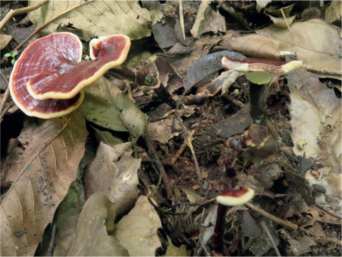
Ganoderma acaciicola B.K. Cui, J.H. Xing & Y.F. Sun, sp. nov. MycoBank MB 839670. Figs 9, 10.
Fig. 9.

Basidiomata of Ganoderma acaciicola.
Fig. 10.

Microscopic structures of Ganoderma acaciicola (drawn from Cui 16815). A. Basidiospores. B. Apical cells from cuticle. C. Basidia and basidioles. D. Hyphae from trama. E. Hyphae from context. Scale bars = 10 μm.
Diagnosis: Differs from other species in the genus by its sessile and concrescent basidiomata with reddish brown and laccate pileal surface, homogeneous context, non-stratified tubes, cream to buff pore surface unchanging when bruised, broadly ellipsoid to ovoid basidiospores with truncated apex.
Etymology: acaciicola (Lat.), refers to this species growing on Acacia.
Typus: Australia, Queensland, Cairns, on stump of Acacia, 18 May 2018, Cui 16815 (holotype BJFC030114).
Additional materials examined: Australia, Queensland, Cairns, on stump of Acacia, 18 May 2018, Cui 16813 (BJFC030112), Cui 16814 (BJFC030113); on root of living Acacia, 18 May 2018, Cui 16817 (BJFC030116).
Description: Basidiomata annual, sessile or subsessile and broadly attached, usually concrescent, hard corky to woody hard. Pilei sub-circular to flabelliform, up to 16.5 cm diam and 3 cm thick. Pileal surface rusty orange brown to reddish brown, laccate, glabrous, pileal margin distinct, cream buff; margin obtuse, entire, irregularly wavy. Pore surface cream to buff when fresh, unchanging when bruised, pale straw yellow when dry; pores circular to angular, 4–6 per mm; dissepiments moderately thick, entire. Context cinnamon brown to dark brown, homogeneous, with black melanoid lines, hard corky, up to 2 cm thick. Tubes yellowish brown to greyish brown, non-stratified, up to 1 cm long. Hyphal system trimitic; generative hyphae with clamp connections; all hyphae IKI –, CB +; tissues darkening in KOH. Generative hyphae in context colourless, thin-walled, 2–4 μm diam; skeletal hyphae in context dark brown, thick-walled with a wide to narrow lumen or sub-solid, arboriform and flexuous, 2–6 μm diam; binding hyphae in context colourless, thick-walled, branched and flexuous, up to 1 μm diam. Generative hyphae in tubes colourless, thin-walled, 2–3 μm diam; skeletal hyphae in tubes pale yellowish brown, thick-walled with a wide to narrow lumen or sub-solid, arboriform and flexuous, 2–4 μm diam; binding hyphae in tubes colourless, thick-walled, branched and flexuous, up to 1 μm diam. Pileipellis composed of clamped generative hyphae, thick-walled to sub-solid, apical cells clavate, inflated and flexuous, pale yellow to golden yellow, about 25–38 × 6–10 μm, forming a regular palisade. Cystidia and cystidioles absent. Basidia barrel-shaped, colourless, thin-walled, 15–20 × 9–12 μm; basidioles clavate, colourless, thin-walled, 15–20 × 6–10 μm. Basidiospores broadly ellipsoid to ovoid, truncated, yellowish brown, IKI –, CB +, double-walled with distinctly thick walls, exospore wall smooth, endospore wall with dense spinules, (9–)9.2–11.3(–11.6) × (5–)5.6–7 μm, L = 10.12 μm, W = 6.29 μm, Q = 1.61 (n = 60/2, with the turgid vesicular appendix excluded); (10.5–)10.7–11.8(–12.1) × (5.5–)5.8–7 μm, L = 11.12 μm, W = 6.31 μm, Q = 1.72–1.81 (n = 60/2, with the turgid vesicular appendix included).
Notes: Ganoderma acaciicola was collected from Australia on Acacia. It can be characterised by concrescent basidiomata without a stipe, rusty orange brown to reddish brown and laccate pileal surface, pore surface unchanging when bruised. In the phylogenetic analyses, G. acaciicola is closely related to G. mizoramense which was described from Mizoram, India; however, its stipitate basidiomata with irregular pileal surface, ellipsoid basidiospores in larger size (10–12.5 × 6–9 μm, Crous et al. 2017a) differentiate it from G. acaciicola.
Ganoderma acontextum B.K. Cui, J.H. Xing & Vlasák, sp. nov. MycoBank MB 805754. Figs 11, 12.
Fig. 11.

Basidiomata of Ganoderma acontextum.
Fig. 12.

Microscopic structures of Ganoderma acontextum (drawn from JV 0611/21G). A. Basidiospores. B. Basidia and basidioles. C. Hyphae from trama. D. Hyphae from context. Scale bars = 10 μm.
Diagnosis: Differs from other species in the genus by its ungulate pilei with non-laccate pileal surface, heterogeneous and thin context, non-stratified tubes, almond-shaped basidiospores without spinules on the endospore wall.
Etymology: acontextum (Lat.), refers to the basidiomata having extremely thin context.
Typus: Guatemala, San Mateo, on angiosperm tree, 22 Nov. 2006 (holotype JV 0611/21G).
Additional materials examined: USA, Virginia, Woodbridge, Mason Neck State Park, on Quercus, 11 Aug. 2012, JV 1208/11J, JV 1407/64 (JV).
Diagnosis: Basidiomata perennial, sessile, woody hard. Pilei solitary, ungulate to columnar at maturity, up to 6 cm diam and 4 cm thick. Pileal surface reddish brown to dark brown, dull, glabrous, with dense concentric furrows; margin obtuse, entire, wavy. Pore surface white when fresh, turning darker when bruised, clay-buff to dark brown when dry; pores circular, 4–6 per mm; dissepiments thick, entire. Context dark brown, heterogeneous, composed of a strikingly light brown to ochre zone under pileal crust, followed by black melanoid lines, and dark brown context above the tubes, corky, thin, up to 3 mm thick altogether. Tubes dark brown, non-stratified, up to 4 cm long. Hyphal system trimitic; generative hyphae with clamp connections; skeletal hyphae occasionally with simple septa; all hyphae IKI –, CB +; tissues darkening in KOH. Generative hyphae in context colourless, thin-walled, 2–3 μm diam; skeletal hyphae in context yellow to brown, thick-walled with a wide to narrow lumen or sub-solid, arboriform and flexuous, 2–5 μm diam; binding hyphae in context colourless, thick-walled, branched and flexuous, 1.2–2.5 μm diam. Generative hyphae in tubes colourless, thin-walled, 2.2–2.8 μm diam; skeletal hyphae in tubes pale brown to brown, thick-walled with a wide to narrow lumen or sub-solid, arboriform and flexuous, 2.3–4 μm diam; binding hyphae in tubes colourless, thick-walled, branched and flexuous, 1–2.5 μm diam. Cystidia and cystidioles absent. Basidia broadly clavate, colourless, thin-walled, 20–30 × 7–11 μm; basidioles in shape like the basidia, colourless, thin-walled, 14–25 × 6–10 μm. Basidiospores almond-shaped, not obviously truncated, golden-brown, IKI –, CB +, double-walled with slightly thick walls, exospore wall smooth, endospore wall without spinules, (9–)9.8–10.2(–11) × (4–)5.5–6.3(–6.5) μm, L = 9.89 μm, W = 5.73 μm, Q = 1.73 (n = 30/1, with the turgid vesicular appendix excluded).
Notes: Ganoderma acontextum was collected from Central America and the USA belonging to the non-laccate group. It has distinctive features such as ungulate pilei with densely concentrically furrowed pileal surface, heterogeneous and thin context, non-stratified tubes, almond-shaped basidiospores without an obviously truncated apex, and no spinules on the endospore wall.
Ganoderma alpinum B.K. Cui, J.H. Xing & Y.F. Sun, sp. nov. MycoBank MB 839671. Figs 13, 14.
Fig. 13.

Basidiomata of Ganoderma alpinum.
Fig. 14.

Microscopic structures of Ganoderma alpinum (drawn from Cui 17467). A. Basidiospores. B. Apical cells from cuticle. C. Basidia and basidioles. D. Hyphae from trama. E. Hyphae from context. Scale bars = 10 μm.
Diagnosis: Differs from other species in the genus by its perennial and sessile basidiomata, pale brown to greyish brown pileal surface with concentric furrows and radial wrinkles, cracked margin, homogeneous context and non-stratified tubes.
Etymology: alpinum (Lat.), refers to this species being collected from an alpine area.
Typus: China, Yunnan, Shangri-La, Daxueshan, on stump of Populus, 12 Aug. 2019, Cui 17467 (holotype BJFC034326).
Additional materials examined: China, Sichuan, Yajiang County, on stump of Pinus, 8 Aug. 2019, Cui 17325 (BJFC034183); Xizang, Chayu County, on stump of Cupressus, 10 Sep. 2020, Cui 18402 (BJFC035263).
Description: Basidiomata perennial, sessile, woody hard. Pilei flabelliform to shell-shaped, applanate, up to 15 cm diam and 4 cm thick. Pileal surface pale brown to greyish brown, dull, glabrous, with concentric furrows and radial wrinkles; margin subacute to obtuse, entire, slightly wavy, cracked when dry. Pore surface white to cream when fresh, turning darker when bruised, clay buff to dark brown when dry; pores circular, 5–7 per mm; dissepiments slightly thick, entire. Context cinnamon brown to dark brown, homogeneous, with black melanoid lines, hard corky and fibrous, up to 2 cm thick. Tubes yellowish brown to dark brown, non-stratified, up to 2 cm long. Hyphal system trimitic; generative hyphae with clamp connections; all hyphae IKI –, CB +; tissues darkening in KOH. Generative hyphae in context colourless, thin-walled, 3–4 μm diam; skeletal hyphae in context pale yellowish brown, thick-walled with a wide to narrow lumen or sub-solid, arboriform and flexuous, 3–6 μm diam; binding hyphae in context colourless, thick-walled, branched and flexuous, up to 2 μm diam. Generative hyphae in tubes colourless, thin-walled, 2–4 μm diam; skeletal hyphae in tubes pale brown, thick-walled with a wide to narrow lumen or sub-solid, arboriform and flexuous, 3–5 μm diam; binding hyphae in tubes colourless, thick-walled, branched and flexuous, up to 2 μm diam. Pileipellis composed of clamped generative hyphae, thick-walled, apical cells clavate, slightly inflated and flexuous, yellowish brown, about 20–30 × 5–8 μm, forming a regular palisade. Cystidia and cystidioles absent. Basidia barrel-shaped, colourless, thin-walled, 20–30 × 11–16 μm; basidioles clavate, colourless, thin-walled, 12–18 × 6–10 μm. Basidiospores broadly ellipsoid to ovoid, truncated, yellowish brown, IKI –, CB +, double-walled with distinctly thick walls, exospore wall smooth, endospore wall with dense spinules, (6.2–)6.3–7.6(–7.8) × (4–)4.1–5.4(–5.5) μm, L = 6.97 μm, W = 4.83 μm, Q = 1.43–1.45 (n = 60/2, with the turgid vesicular appendix excluded); (7.7–)7.8–9.2(–9.4) × (4–)4.1–5.4(–5.8) μm, L = 8.37 μm, W = 4.85 μm, Q = 1.71–1.74 (n = 60/2, with the turgid vesicular appendix included).
Notes: Ganoderma alpinum was collected from high altitude areas of southwestern China. It is hard to distinguish G. alpinum from G. applanatum on morphology, however, G. alpinum can be separated from G. applanatum by phylogenetic analyses and ecological distribution.
Ganoderma bubalinomarginatum B.K. Cui, J.H. Xing & Y.F. Sun, sp. nov. MycoBank MB 839672. Figs 15, 16.
Fig. 15.

Basidiomata of Ganoderma bubalinomarginatum.
Fig. 16.

Microscopic structures of Ganoderma bubalinomarginatum (drawn from Dai 20075). A. Basidiospores. B. Apical cells from cuticle. C. Basidia and basidioles. D. Hyphae from trama. E. Hyphae from context. Scale bars = 10 μm.
Diagnosis: Differs from other species in the genus by its pale-coloured basidiomata without stipe, laccate pileal surface with buff margin, homogeneous context and non-stratified tubes.
Etymology: bubalinomarginatum (Lat.), refers to the pilei with buff margin.
Typus: China, Guangxi, Nanning, Guangxi Academy of Forestry, on stump of Castanopsis, 4 Jul. 2019, Dai 20075 (holotype BJFC031749).
Additional material examined: China, Guangxi, Nanning, Guangxi Academy of Forestry, on living tree of Phoebe, 4 Jul. 2019, Dai 20074 (BJFC031748).
Description: Basidiomata annual, sessile and broadly attached, usually concrescent, hard corky. Pilei solitary, flabelliform to shell-shaped, up to 7.5 cm diam and 6 mm thick. Pileal surface reddish brown at the base, yellowish brown at the centre, buff at the margin, laccate, glabrous, with wide concentric furrows and slightly radial wrinkles; margin obtuse, entire, wavy when dry. Pore surface white to greyish white when fresh, turning darker when bruised, pale wood brown to greyish brown when dry; pores circular to angular, 5–6 per mm; dissepiments moderately thick, entire. Context straw yellow, homogeneous, without black melanoid lines, hard corky, up to 4 mm thick. Tubes pale brown, non-stratified, up to 3 mm long. Hyphal system trimitic; generative hyphae with clamp connections; all hyphae IKI –, CB +; tissues darkening in KOH. Generative hyphae in context colourless, thin-walled, 2–4 μm diam; skeletal hyphae in context pale yellow, thick-walled with a wide to narrow lumen or sub-solid, arboriform and flexuous, 2–5 μm diam; binding hyphae in context colourless, thick-walled, rarely branched and flexuous, up to 2 μm diam. Generative hyphae in tubes colourless, thin-walled, 2–3 μm diam; skeletal hyphae in tubes pale yellow, thick-walled with a wide to narrow lumen or sub-solid, arboriform and flexuous, 2–4 μm diam; binding hyphae in tubes colourless, thick-walled, rarely branched and flexuous, up to 1.5 μm diam. Pileipellis composed of clamped generative hyphae, thick-walled to sub-solid, apical cells clavate, inflated and flexuous, pale golden yellow, about 28–42 × 9–11 μm, forming a regular palisade. Cystidia and cystidioles absent. Basidia broadly clavate, colourless, thin-walled, 15–22 × 7–11 μm; basidioles in shape like the basidia, colourless, thin-walled, 12–20 × 5–9 μm. Basidiospores broadly ellipsoid to ovoid, truncated, pale yellowish brown, IKI –, CB +, with oily drop, double-walled with slightly thick walls, exospore wall smooth, endospore wall with dense spinules, (6.2–)6.5–7.4(–7.6) × (4.2–) 4.5–5.3(–5.8) μm, L = 6.91 μm, W = 4.87 μm, Q = 1.41–1.43 (n = 60/2, with the turgid vesicular appendix excluded); (7–)7.2–8.3(–8.8) × (4.3–)4.5–5.6(–5.8) μm, L = 7.82 μm, W = 4.96 μm, Q = 1.57–1.59 (n = 60/2, with the turgid vesicular appendix included).
Notes: Ganoderma bubalinomarginatum has pale-coloured basidiomata without a stipe which differentiates it from the species described from Guangxi Autonomous Region: G. daiqingshanense, G. guinanense and G. magniporum. Ganoderma bubalinomarginatum is like G. sessile, described from America: it shares connate and sessile basidiomata, reddish brown to yellowish brown pileal surface, absence of black melanoid lines, white pore surface when fresh, homogeneous context and non-stratified tubes. However, G. sessile differs by the white, thin and acute pileal margin, larger pores (3–5 per mm), and larger basidiospores (9–11 × 6–8 μm, Murrill 1902).
Ganoderma castaneum B.K. Cui, J.H. Xing & Y.F. Sun, sp. nov. MycoBank MB 839673. Figs 17, 18.
Fig. 17.

Basidiomata of Ganoderma castaneum.
Fig. 18.

Microscopic structures of Ganoderma castaneum (drawn from Cui 17283). A. Basidiospores. B. Apical cells from cuticle. C. Basidia and basidioles. D. Hyphae from trama. E. Hyphae from context. Scale bars = 10 μm.
Diagnosis: Differs from other species in the genus by its chestnut brown and laccate pileal surface with wide concentric ridges, heterogeneous context, and broadly ellipsoid basidiospores with smooth endospore wall.
Etymology: castaneum (Lat.), refers to the reddish brown pileal surface like a chestnut.
Typus: China, Hainan, Ledong County, Jianfengling Nature Reserve, on stump of angiosperm tree, 3 Jul. 2019, Cui 17283 (holotype BJFC034139).
Additional materials examined: China, Hainan, Ledong County, Jianfengling Nature Reserve, on stump of angiosperm tree, 17 Jun. 2014, Dai 13710 (BJFC017447); on fallen branch of angiosperm tree, 19 Jun. 2016, Cui 13893 (BJFC028759); Wuzhishan, Wuzhishan Forest Park, on stump of Acacia, 11 Jun. 2016, Dai 16500 (BJFC022616), Dai 16501 (BJFC022617).
Description: Basidiomata annual, sessile, broadly attached, hard corky to woody hard. Pilei solitary, flabelliform, applanate, up to 8.5 cm diam and 2 cm thick. Pileal surface reddish brown like chestnut, laccate, glabrous, with wide concentric ridges and slightly radial wrinkles; margin obtuse, entire, wavy. Pore surface white to cream when fresh, turning darker when bruised, buff to pale straw yellow when dry; pores circular, 4–6 per mm; dissepiments moderately thick, entire. Context heterogeneous, the upper layer pale straw yellow, the lower layer cinnamon brown to dark brown, with black melanoid lines, hard corky, up to 1.6 cm thick. Tubes pale greyish brown, non-stratified, up to 5 mm long. Hyphal system trimitic; generative hyphae with clamp connections; all hyphae IKI –, CB +; tissues darkening in KOH. Generative hyphae in context colourless, thin-walled, 2–4 μm diam; skeletal hyphae in context pale yellowish brown, thick-walled with a wide to narrow lumen or sub-solid, arboriform and flexuous, 3–6 μm diam; binding hyphae in context colourless, thick-walled, branched and flexuous, up to 2 μm diam. Generative hyphae in tubes colourless, thin-walled, 2–4 μm diam; skeletal hyphae in tubes pale yellowish brown, thick-walled with a wide to narrow lumen or sub-solid, arboriform and flexuous, 2–6 μm diam; binding hyphae in tubes colourless, thick-walled, branched and flexuous, up to 1 μm diam. Pileipellis composed of clamped generative hyphae, slightly thick-walled, apical cells clavate, flexuous, pale yellow, about 25–40 × 3–5 μm, forming a regular palisade. Cystidia and cystidioles absent. Basidia barrel-shaped, colourless, thin-walled, 14–21 × 7–12 μm; basidioles broadly clavate, colourless, thin-walled, 9–12 × 5–10 μm. Basidiospores broadly ellipsoid, not obviously truncated, golden yellow, IKI –, slightly CB +, double-walled with distinctly thick walls, exospore and endospore walls smooth, (6.2–)6.7–8.3(–8.5) × (4.2–)4.8–6(–6.3) μm, L = 7.42 μm, W = 5.43 μm, Q = 1.37 (n = 60/1, with the turgid vesicular appendix excluded).
Notes: Ganoderma castaneum was collected from a tropical rainforest of Hainan Province. When compared with the species in the checklist of Ganoderma reported from Hainan Island by Hapuarachchi et al. (2018b), G. castaneum has distinguished features such as chestnut-coloured and laccate pileal surface with wide concentric ridges, heterogeneous context, broadly ellipsoid and basidiospores not obviously truncated with smooth endospore walls.
Ganoderma chuxiongense B.K. Cui, J.H. Xing & Y.F. Sun, sp. nov. MycoBank MB 840397. Figs 19, 20.
Fig. 19.
Basidiomata of Ganoderma chuxiongense.
Fig. 20.

Microscopic structures of Ganoderma chuxiongense (drawn from Cui 17262). A. Basidiospores. B. Apical cells from cuticle. C. Basidia and basidioles. D. Hyphae from trama. E. Hyphae from context. Scale bars = 10 μm.
Diagnosis: Differs from other species in the genus by its thin basidiomata, dimidiate and lobate pileus with reddish brown and laccate pileal surface, pale light-yellow pore surface.
Etymology: chuxiongense (Lat.), refers to the holotype of this species being found in Chuxiong City of Yunnan Province.
Typus: China, Yunnan, Chuxiong, Xishan Park, on stump of angiosperm tree, 25 Aug. 2018, Cui 17262 (holotype BJFC034120).
Description: Basidiomata annual, laterally stipitate, corky. Pilei solitary, flabelliform, dimidiate and lobate, up to 6 cm diam and 4 mm thick. Pileal surface reddish brown when fresh becoming dark-red when dry, laccate, glabrous, with concentric bands and slightly radial rugose; margin pale yellow, acute to obtuse, entire. Pore surface pale light-yellow when fresh, turning darker when bruised, buff to pale straw yellow when dry; pores circular, 4–6 per mm; dissepiments slightly thick, entire. Context dark wood brown, not obviously stratified, without black melanoid lines, soft corky, up to 2 mm thick. Tubes pale to dark wood brown, non-stratified, up to 2 mm long. Stipe concolorous with pileal surface, cylindrical and solid, up to 7 cm long and 6 mm diam. Hyphal system trimitic; generative hyphae with clamp connections; all hyphae IKI –, CB +; tissues darkening in KOH. Generative hyphae in context colourless, thin-walled, 2–3 μm diam; skeletal hyphae in context pale yellow, thick-walled with a wide to narrow lumen or sub-solid, arboriform and flexuous, 2–4 μm diam; binding hyphae in context colourless, thick-walled, branched and flexuous, up to 2 μm diam. Generative hyphae in tubes colourless, thin-walled, 2–3 μm diam; skeletal hyphae in tubes pale yellow, thick-walled with a wide to narrow lumen or sub-solid, arboriform and flexuous, 2–4 μm diam; binding hyphae in tubes colourless, thick-walled, branched and flexuous, up to 1.5 μm diam. Pileipellis composed of clamped generative hyphae, slightly thick-walled, apical cells clavate, slightly inflated and flexuous, golden yellow, about 23–30 × 6–11 μm, forming a regular palisade. Cystidia and cystidioles absent. Basidia barrel-shaped, colourless, thin-walled, 15–20 × 10–12 μm; basidioles broadly clavate, colourless, thin-walled, 13–18 × 7–11 μm. Basidiospores broadly ellipsoid to ovoid, truncated, pale yellowish brown, IKI –, CB +, double-walled with distinctly thick walls, exospore wall smooth, endospore wall with dense spinules, (7.9–)8–9(–9.2) × (6–)6.2–7(–7.1) μm, L = 8.45 μm, W = 6.58 μm, Q = 1.28 (n = 60/1, with the turgid vesicular appendix excluded); (9.5–)10–11.3(–11.5) × (6.2–)6.5–7.3 μm, L = 10.35 μm, W = 6.88 μm, Q = 1.5 (n = 60/1, with the turgid vesicular appendix included).
Notes: Ganoderma chuxiongense is characterised by its stipitate basidiomata with lobate pilei, reddish brown pileal surface and pale-yellow pore surface when fresh. Ganoderma kunmingense described from Yunnan Province is like G. chuxiongense in the thin basidiomata with laccate and reddish brown pileal surface, margin entire or incised, but it has no concentric bands on the pileal surface, a cream pore surface, and broadly ellipsoid to subglobose basidiospores without obvious spinules on the endospore wall (Zhao 1989a).
Ganoderma cocoicola B.K. Cui, J.H. Xing & Y.F. Sun, sp. nov. MycoBank MB 839674. Figs 21, 22.
Fig. 21.

Basidiomata of Ganoderma cocoicola.
Fig. 22.

Microscopic structures of Ganoderma cocoicola (drawn from Cui 16791). A. Basidiospores. B. Apical cells from cuticle. C. Basidia and basidioles. D. Hyphae from trama. E. Hyphae from context. Scale bars = 10 μm.
Diagnosis: Differs from other species in the genus by its small and hard basidiomata without stipe, dark and laccate pileal surface, homogeneous context, non-stratified tubes, oblong-ellipsoid and truncated basidiospores.
Etymology: cocoicola (Lat.), refers to this species growing on Cocos.
Typus: Australia, Queensland, Cairns, Cairns Botanical Garden, on stump of Cocos, 17 May 2018, Cui 16791 (holotype BJFC030090).
Additional material examined: Australia, Queensland, Cairns, Cairns Botanical Garden, on stump of Cocos, 17 May 2018, Cui 16792 (BJFC030091).
Description: Basidiomata annual to perennial, sessile or sometimes sub-stipitate, hard corky to woody hard. Pilei solitary, flabelliform to ungulate, up to 3.5 cm diam and 1.5 cm thick. Pileal surface dark-red to near black, laccate, glabrous, with concentric furrows and slightly radial wrinkles; margin obtuse, entire. Pore surface white when fresh, turning darker when bruised, straw yellow to pale brown when dry; pores circular to angular, 4–6 per mm; dissepiments distinctly thick, entire. Context dark brown to cinnamon brown, homogeneous, without black melanoid lines, hard corky, up to 3 mm thick. Tubes dark grey to greyish brown, non-stratified, up to 1.2 cm long. Hyphal system trimitic; generative hyphae with clamp connections; all hyphae IKI –, CB +; tissues darkening in KOH. Generative hyphae in context colourless, thin-walled, 2–3 μm diam; skeletal hyphae in context pale yellowish brown, thick-walled with a wide to narrow lumen or sub-solid, frequently arboriform and flexuous, 2.5–5 μm diam; binding hyphae in context colourless, thick-walled, branched and flexuous, up to 2 μm diam. Generative hyphae in tubes colourless, thin-walled, 2–3 μm diam; skeletal hyphae in tubes pale yellowish brown, thick-walled with a wide to narrow lumen or sub-solid, frequently arboriform and flexuous, 3–5 μm diam; binding hyphae in tubes colourless, thick-walled, branched and flexuous, up to 1.5 μm diam. Pileipellis composed of clamped generative hyphae, thick-walled to sub-solid, apical cells clavate, flexuous, yellowish brown, about 27–35 × 5–7 μm, forming a regular palisade. Cystidia and cystidioles absent. Basidia broadly clavate, colourless, thin-walled, 16–22 × 7–10 μm; basidioles in shape like the basidia, colourless, thin-walled, 13–20 × 7–10 μm. Basidiospores oblong-ellipsoid, truncated, pale yellowish brown, IKI –, CB +, double-walled with moderately thick walls, exospore wall smooth, endospore wall with dense spinules, 9–9.8(–10) × 4.2–5.4(–5.6) μm, L = 9.42 μm, W = 4.77 μm, Q = 1.91–2.04 (n = 60/2, with the turgid vesicular appendix excluded); (9.6–)9.7–10.7(–10.8) × (4.2–)4.4–5.4(–5.5) μm, L = 10.15 μm, W = 4.83 μm, Q = 2.08–2.12 (n = 60/2, with the turgid vesicular appendix included).
Notes: Ganoderma cocoicola was collected from Australia on Cocos. It is characterised by small and hard basidiomata without a stipe, blackish brown and laccate pileal surface, homogeneous context, non-stratified tubes, oblong-ellipsoid and truncated basidiospores. In the phylogenetic analyses, G. cocoicola clustered with G. zonatum, G. ryvardenii, G. boninense, and G. hochiminhense with high support (Fig. 1); all these species grow on palm trees (Patouillard 1889, Murrill 1902, Kinge & Mih 2011, Zhou et al. 2015), except G. hochiminhense which grows on Areca (Luangharn et al. 2021).
Ganoderma fallax B.K. Cui, J.H. Xing & Vlasák, sp. nov. MycoBank MB 839677. Figs 23, 24.
Fig. 23.

Basidiomata of Ganoderma fallax.
Fig. 24.

Microscopic structures of Ganoderma fallax (drawn from JV 1009/27). A. Basidiospores. B. Basidia and basidioles. C. Hyphae from trama. D. Hyphae from context. Scale bars = 10 μm.
Diagnosis: Differs from other species in the genus by its non-laccate pileal surface with faintly concentric furrows, stratified tubes, and basidiospores not obviously truncated with dense spinules on the endospore wall.
Etymology: fallax (Lat.), refers to this species being easily confused morphologically with other non-laccate species.
Typus: USA, Pennsylvania (holotype JV 1009/27).
Materials examined: USA, Arizona, JV 1209/60J (JV); New Jersey, JV 0109/B1-J (JV); Pennsylvania, JV 0709/39 (JV); Tennessee, JV 0509/93K, JV 1410/14J (JV).
Description: Basidiomata perennial, sessile, hard corky to woody hard. Pilei solitary, flabelliform to ungulate or shell-shaped, up to 11 cm diam and 3 cm thick. Pileal surface yellowish brown to dark brown, dull, glabrous, with faint concentric furrows; margin acute, entire, slightly wavy. Pore surface white to pale brown when fresh, turning darker when bruised, straw yellow when dry; pores circular, 4–6 per mm; dissepiments slightly thick, entire. Context yellowish brown to dark brown, homogeneous, with black melanoid lines, corky, up to 7 mm thick. Tubes concolorous with context, stratified, up to 1.2 cm long. Hyphal system trimitic; generative hyphae with clamp connections; all hyphae IKI –, CB +; tissues darkening in KOH. Generative hyphae in context colourless, thin-walled, 2.5–5.5 μm diam; skeletal hyphae in context pale yellowish brown, thick-walled with narrow lumen or sub-solid, arboriform and flexuous, 3–6 μm diam; binding hyphae in context colourless, thick-walled, branched and flexuous, 2–3 μm diam. Generative hyphae in tubes colourless, thin-walled, 2–3.5 μm diam; skeletal hyphae in tubes pale brown, thick-walled with narrow lumen or sub-solid, arboriform and flexuous, 2–5 μm diam; binding hyphae in tubes colourless, thick-walled, branched and flexuous, 2–2.5 μm diam. Cystidia and cystidioles absent. Basidia barrel-shaped to clavate, colourless, thin-walled, 25–30 × 14–21 μm; basidioles in shape like the basidia, colourless, thin-walled, 16–17 × 9–18 μm. Basidiospores ovoid, not obviously truncated, pale yellowish brown, IKI –, CB +, double-walled with distinctly thick walls, exospore wall smooth, endospore wall with dense spinules, (8.5–)9–10(–10.5) × 6–7.5 μm, L = 9.25 μm, W = 6.56 μm, Q = 1.37 (n = 30/1, with the turgid vesicular appendix included).
Notes: Ganoderma fallax may be confused with the non-laccate species G. adspersum, which has large basidiomata with a yellowish brown pileal surface, homogeneous context and stratified tubes, but G. adspersum has larger and more obviously truncated basidiospores, and lacks concentric ornamentation on the pileal surface. In the phylogenetic analyses, G. fallax formed an independent lineage then clustered with G. adspersum (Fig. 1).
Ganoderma guangxiense B.K. Cui, J.H. Xing & Y.F. Sun, sp. nov. MycoBank MB 839678. Figs 25, 26.
Fig. 25.

Basidiomata of Ganoderma guangxiense.
Fig. 26.

Microscopic structures of Ganoderma guangxiense (drawn from Cui 14453). A. Basidiospores. B. Apical cells from cuticle. C. Basidia and basidioles. D. Hyphae from trama. E. Hyphae from context. Scale bars = 10 μm.
Diagnosis: Differs from other species in the genus by its sessile basidiomata, greyish brown to near black pileal surface with obvious concentric furrows, cracked when dry, homogeneous context, non-stratified tubes, ellipsoid to ovoid and truncated basidiospores.
Etymology: guangxiense (Lat.), refers to the holotype of this species located at Guangxi.
Typus: China, Guangxi, Tianlin County, Cenwanglaoshan Nature Reserve, on stump of angiosperm tree, 8 Jul. 2017, Cui 14453 (holotype BJFC029321).
Additional materials examined: China, Guangxi, Tianlin County, Cenwanglaoshan Nature Reserve, on stump of angiosperm tree, 8 Jul. 2017, Cui 14454 (BJFC029322), Cui 14455 (BJFC029323); Jinxiu County, Dayaoshan Nature Reserve, on fallen trunk of angiosperm tree, 15 Jul. 2017, Cui 14500 (BJFC029369), Cui 14508 (BJFC029377).
Description: Basidiomata annual, sessile and broadly attached, hard corky to woody hard. Pilei solitary, flabelliform to shell-shaped, up to 11 cm diam and 4.3 cm thick. Pileal surface greyish brown to near black when fresh, dull, glabrous, with obvious concentric furrows, cracked when dry; margin obtuse, entire. Pore surface cream when fresh, turning darker when bruised, dark straw yellow to pale brown when dry; pores circular, 5–7 per mm; dissepiments moderately thick, entire. Context dark brown to cinnamon brown, homogeneous, with black melanoid lines, hard corky, up to 3 cm thick. Tubes slightly paler than context, non-stratified, up to 2 cm long. Hyphal system trimitic; generative hyphae with clamp connections; all hyphae IKI –, CB +; tissues darkening in KOH. Generative hyphae in context colourless, thin-walled, 2–3 μm diam; skeletal hyphae in context reddish brown, thick-walled with a wide to narrow lumen or sub-solid, arboriform and flexuous, 2–6 μm diam; binding hyphae in context colourless, thick-walled, branched and flexuous, up to 2 μm diam. Generative hyphae in tubes colourless, thin-walled, 2–3 μm diam; skeletal hyphae in tubes reddish brown, thick-walled with a wide to narrow lumen or sub-solid, arboriform and flexuous, 2–4 μm diam; binding hyphae in tubes colourless, thick-walled, branched and flexuous, up to 2 μm diam. Pileipellis composed of clamped generative hyphae, thick-walled to sub-solid, apical cells clavate, inflated and flexuous, golden yellow, about 28–37 × 5–8 μm, forming a regular palisade. Cystidia and cystidioles absent. Basidia barrel-shaped, colourless, thin-walled, 15–20 × 7–11 μm; basidioles broadly clavate, colourless, thin-walled, 12–15 × 7–9 μm. Basidiospores ellipsoid to ovoid, truncated, yellowish brown, IKI –, CB +, double-walled with moderately thick walls, exospore wall smooth, endospore wall with dense spinules, (7.5–)7.8–8.8(–8.9) × (4.9–)5–6(–6.5) μm, L = 8.23 μm, W = 5.49 μm, Q = 1.47–1.53 (n = 60/2, with the turgid vesicular appendix excluded); 8.7–9.8(–10) × (4.9–)5–6(–6.1) μm, L = 9.23 μm, W = 5.52 μm, Q = 1.67–1.68 (n = 60/2, with the turgid vesicular appendix included).
Notes: Morphologically, Ganoderma guangxiense is very similar to G. australe and they are not easy to separate. However, phylogenetically, G. guangxiense and G. australe are divided into two independent lineages with good support (Fig. 1). Ganoderma daiqingshanense also has sessile and hard basidiomata with blackish pilei, but it differs from G. guangxiense by having a heterogeneous context, larger pores (4–5 per mm) and irregular palisade of pileipellis structure (Zhao 1989a).
Ganoderma puerense B.K. Cui, J.H. Xing & Y.F. Sun, sp. nov. MycoBank MB 839679. Figs 27, 28.
Fig. 27.

Basidiomata of Ganoderma puerense.
Fig. 28.

Microscopic structures of Ganoderma puerense (drawn from Dai 20427). A. Basidiospores. B. Apical cells from cuticle. C. Basidia and basidioles. D. Hyphae from trama. E. Hyphae from context. Scale bars = 10 μm.
Diagnosis: Differs from other species in the genus by its woody hard and thin basidiomata, yellowish brown pileal surface with dense concentric black bands or furrows, and a margin lacerated like petals.
Etymology: puerense (Lat.), refers to this species being collected from Puer City of Yunnan Province.
Typus: China, Yunnan, Puer, Puer Forest Park, on living tree of Cinnamomum, 17 Aug. 2019, Dai 20427 (holotype BJFC032095).
Description: Basidiomata annual, sessile or with short stipe, usually growing together, imbricate, woody hard. Pilei solitary, sub-orbicular to flabelliform, applanate, up to 7.5 cm diam and 8 mm thick. Pileal surface yellowish brown to dark brown, dull, glabrous, with dense concentric black bands or furrows and slightly radial wrinkles; margin acute to obtuse, lacerated like petals. Pore surface white when fresh, turning darker when bruised, buff to pale brown when dry; pores circular to angular, 5–7 per mm; dissepiments moderately thick, entire. Context straw yellow to dark brown, homogeneous, with black melanoid lines, hard corky, up to 3 mm thick. Tubes dark brown, non-stratified, up to 5 mm long. Stipe concolorous with pileal surface, cylindrical and solid, up to 1.5 cm long and 3 mm diam. Hyphal system trimitic; generative hyphae with clamp connections; all hyphae IKI –, CB +; tissues darkening in KOH. Generative hyphae in context colourless, thin-walled, 2–3 μm diam; skeletal hyphae in context pale yellowish brown, thick-walled with a wide to narrow lumen or sub-solid, frequently arboriform and flexuous, 2–5 μm diam; binding hyphae in context colourless, thick-walled, rarely branched and flexuous, 1–2 μm diam. Generative hyphae in tubes colourless, thin-walled, 2–3 μm diam; skeletal hyphae in tubes pale yellowish brown, thick-walled with a wide to narrow lumen or sub-solid, arboriform and flexuous, 2–4 μm diam; binding hyphae in tubes colourless, thick-walled, rarely branched and flexuous, 1–2 μm diam. Pileipellis composed of clamped generative hyphae, thick-walled to sub-solid, apical cells clavate, faintly inflated and flexuous, yellowish brown, about 25–40 × 5–7 μm, forming a regular palisade. Cystidia and cystidioles absent. Basidia barrel-shaped, colourless, thin-walled, 12–20 × 7–10 μm; basidioles clavate to fusiform, colourless, thin-walled, 11–18 × 7–10 μm. Basidiospores ellipsoid to ovoid, truncated, yellowish brown, IKI –, CB +, double-walled with slightly thick walls, exospore wall smooth, endospore wall with dense spinules, (7–) 7.2–8.5(–8.8) × (4.8–)5–6(–6.4) μm, L = 7.83 μm, W = 5.31 μm, Q = 1.47 (n = 60/1, with the turgid vesicular appendix excluded); (8.5–)8.8–9.5(–9.8) × (4.8–)5–6.2(–6.8) μm, L = 9.1 μm, W = 5.52 μm, Q = 1.65 (n = 60/1, with the turgid vesicular appendix included).
Notes: Ganoderma bicharacteristicum also has a dull pileal surface, homogeneous context and was described from Yunnan Province, but G. bicharacteristicum can be distinguished from G. puerense by its stipitate basidiomata with black pileal surface, wood brown pore surface and subglobose basidiospores (6.3–9 × 5.6–8.7 μm, Zhang 1994).
Ganoderma subangustisporum B.K. Cui, J.H. Xing & Y.F. Sun, sp. nov. MycoBank MB 839680. Figs 29, 30.
Fig. 29.

Basidiomata of Ganoderma subangustisporum.
Fig. 30.

Microscopic structures of Ganoderma subangustisporum (drawn from Cui 18592). A. Basidiospores. B. Apical cells from cuticle. C. Basidia and basidioles. D. Hyphae from trama. E. Hyphae from context. Scale bars = 10 μm.
Diagnosis: Differs from other species in the genus by its dark-red to near black pileal surface with concentric bands and radial rugosities made by dark and continuous spots, margin acute and thin, and ellipsoid basidiospores with truncated apex.
Etymology: subangustisporum (Lat.), refers to this species being closely related to Ganoderma angustisporum.
Typus: China, Yunnan, Pingbian County, Daweishan National Nature Reserve, on stump of angiosperm tree, 3 Aug. 2019, Cui 18592 (holotype BJFC035453).
Materials examined: China, Yunnan, Pingbian County, Daweishan National Nature Reserve, on stump of angiosperm tree, 3 Aug. 2019, Cui 18593 (BJFC035454), Cui 18594 (BJFC035455), Cui 18595 (BJFC035456), Cui 18596 (BJFC035457), Cui 18597 (BJFC035458).
Description: Basidiomata annual, sessile or with short and lateral stipe, hard corky. Pilei solitary, sub-circular to flabelliform, up to 8 cm diam and 1.6 cm thick. Pileal surface dark reddish brown, laccate, glabrous, with concentric bands and radial rugose made by dark and continuous spots; margin acute, thin, entire. Pore surface white when fresh, turning darker when bruised, buff to pale straw yellow when dry; pores circular to angular, 4–6 per mm; dissepiments slightly thick, entire. Context cinnamon brown, homogeneous, with pale melanoid lines, hard corky, up to 9 mm thick. Tubes pale brown, non-stratified, up to 9 mm long. Stipe concolourous with pileal surface, cylindrical and solid, up to 1 cm long and 1.5 cm diam. Hyphal system trimitic; generative hyphae with clamp connections; all hyphae IKI –, CB +; tissues darkening in KOH. Generative hyphae in context colourless, thin-walled, 3–4 μm diam; skeletal hyphae in context yellowish brown, thick-walled with a wide to narrow lumen or sub-solid, arboriform and flexuous, 3–5 μm diam; binding hyphae in context colourless, thick-walled, branched and flexuous, up to 2 μm diam. Generative hyphae in tubes colourless, thin-walled, 3–4 μm diam; skeletal hyphae in tubes yellowish brown, thick-walled with a wide to narrow lumen or sub-solid, arboriform and flexuous, 2–5 μm diam; binding hyphae in tubes colourless, thick-walled, branched and flexuous, up to 2 μm diam. Pileipellis composed of clamped generative hyphae, thick-walled to sub-solid, apical cells clavate, inflated and flexuous, dark yellowish brown to reddish brown, about 32–50 × 7–12 μm, forming a regular palisade. Cystidia and cystidioles absent. Basidia barrel-shaped, colourless, thin-walled, 13–19 × 10–13 μm; basidioles broadly clavate, colourless, thin-walled, 13–18 × 6–12 μm. Basidiospores broadly ellipsoid to ellipsoid, truncated, yellowish brown, IKI –, CB +, double-walled with distinctly thick walls, exospore wall smooth, endospore wall with dense spinules, 10–11.8(–12.1) × (5–)5.1–7(–7.3) μm, L = 10.88 μm, W = 6.13 μm, Q = 1.72–1.84 (n = 60/2, with the turgid vesicular appendix excluded); (10.3–)10.7–12.8(–13.1) × (5–)5.2–7(–7.3) μm, L = 11.72 μm, W = 6.02 μm, Q = 1.93–1.97 (n = 60/2, with the turgid vesicular appendix included).
Notes: Ganoderma subangustisporum clusters with G. angustisporum with high support in the phylogenetic analyses (Fig. 1). Morphologically, G. angustisporum differs from G. subangustisporum by having large basidiomata with lacerated margin, darker pileal surface with concentric furrows, and narrower basidiospores (9–11.3 × 4–5.2 μm, Xing et al. 2018).
Ganoderma subellipsoideum B.K. Cui, J.H. Xing & Y.F. Sun, sp. nov. MycoBank MB 839681. Figs 31, 32.
Fig. 31.

Basidiomata of Ganoderma subellipsoideum.
Fig. 32.

Microscopic structures of Ganoderma subellipsoideum (drawn from Cui 18325). A. Basidiospores. B. Apical cells from cuticle. C. Basidia and basidioles. D. Hyphae from trama. E. Hyphae from context. Scale bars = 10 μm.
Diagnosis: Differs from other species in the genus by its subsessile and hard basidiomata, dark yellowish brown to blackish pileal surface with dense concentric furrows, small pores and stratified tubes, and ellipsoid basidiospores with truncated apex.
Etymology: subellipsoideum (Lat.), refers to this species being closely related to Ganoderma ellipsoideum.
Typus: Malaysia, Kuala Lumpur, Ecological Forest Park, on fallen trunk of angiosperm tree, 8 Dec. 2019, Cui 18325 (holotype BJFC035184).
Additional materials examined: Malaysia, Selangor, Kota Damansara, National Forest Reserve, on fallen trunk of angiosperm tree, 6 Dec. 2019, Cui 18241 (BJFC035100); Kuala Lumpur, Ecological Forest Park, on fallen trunk of angiosperm tree, 8 Dec. 2019, Cui 18327(BJFC035186).
Description: Basidiomata annual, sessile or with short and lateral stipe, woody hard. Pilei solitary, sub-circular to flabelliform, applanate, up to 9.5 cm diam and 2 cm thick. Pileal surface dark yellowish brown to near black when fresh becoming yellowish brown or greyish brown when dry, slightly laccate when fresh becoming dull when dry, glabrous, with dense concentric furrows; margin obtuse, entire. Pore surface white when fresh, turning darker when bruised, cream to buff when dry; pores circular, 6–8 per mm; dissepiments moderately thick, entire. Context cinnamon brown, homogeneous, with black melanoid lines, hard corky, up to 1 cm thick. Tubes dark brown, stratified by a layer of context, up to 9 mm long. Stipe concolourous with pileal surface, cylindrical and solid, up to 1.2 cm long and 1.3 cm diam. Hyphal system trimitic; generative hyphae with clamp connections; all hyphae IKI –, CB +; tissues darkening in KOH. Generative hyphae in context colourless, thin-walled, 2–4 μm diam; skeletal hyphae in context pale brown, thick-walled with a wide to narrow lumen or sub-solid, arboriform and flexuous, 2–5 μm diam; binding hyphae in context colourless, thick-walled, branched and flexuous, up to 1.5 μm diam. Generative hyphae in tubes colourless, thin-walled, 2–3 μm diam; skeletal hyphae in tubes yellowish brown, thick-walled with a wide to narrow lumen or sub-solid, arboriform and flexuous, 2–4 μm diam; binding hyphae in tubes colourless, thick-walled, branched and flexuous, up to 1 μm diam. Pileipellis composed of clamped generative hyphae, thick-walled, apical cells clavate, flexuous, pale yellowish brown, about 30–40 × 3–5 μm, forming a regular palisade. Cystidia and cystidioles absent. Basidia barrel-shaped, colourless, thin-walled, 12–17 × 9–11 μm; basidioles broadly clavate, colourless, thin-walled, 11–15 × 6–10 μm. Basidiospores ellipsoid, truncated, yellowish brown, IKI –, CB +, double-walled with moderately thick walls, exospore wall smooth, endospore wall with dense spinules, 6–6.9(–7) × 4–5.3(–5.5) μm, L = 6.46 μm, W = 4.62 μm, Q = 1.36–1.44 (n = 60/2, with the turgid vesicular appendix excluded); (8–)8.2–9.6(–10) × (4–)4.2–5.2(–6) μm, L = 8.89 μm, W = 4.69 μm, Q = 1.89–1.9 (n = 60/2, with the turgid vesicular appendix included).
Notes: In the phylogenetic analyses, G. subellipsoideum is closely related to G. ellipsoideum. Ganoderma ellipsoideum was described from Hainan Province and it has a yellowish brown pileal surface with alternating brownish orange to yellowish brown zones, heterogeneous context and non-stratified tubes, and ellipsoid basidiospores of smaller size (6.1–7.3 × 3.7–4.6 μm, Hapuarachchi et al. 2018b) which make it different from G. subellipsoideum.
Ganoderma subflexipes B.K. Cui, J.H. Xing & Y.F. Sun, sp. nov. MycoBank MB 839675. Figs 33, 34.
Fig. 33.

Basidiomata of Ganoderma subflexipes.
Fig. 34.

Microscopic structures of Ganoderma subflexipes (drawn from Cui 17257). A. Basidiospores. B. Apical cells from cuticle. C. Basidia and basidioles. D. Hyphae from trama. E. Hyphae from context. Scale bars = 10 μm.
Diagnosis: Differs from other species in the genus by its small and dorso-laterally stipitate basidiomata, dark yellow to orange-brown and laccate pileal surface, wavy margin, and obviously truncated basidiospores.
Etymology: subflexipes (Lat.), refers to this species being closely related to Ganoderma flexipes.
Typus: China, Guangdong, Renhua County, Danxiashan Nature Reserve, on stump of angiosperm tree, 4 Jun. 2019, Cui 17257 (holotype BJFC034115).
Additional materials examined: China, Guangdong, Renhua County, Danxiashan Nature Reserve, on root of angiosperm tree, 4 Jun. 2019, Cui 17247 (BJFC034105); on stump of angiosperm tree, 4 Jun. 2019, Cui 17258 (BJFC034116); Jiangxi, Shangrao, Daomaoshan Park, on the ground of Cyclobalanopsis jenseniana, 30 Aug. 2021, Dai 23665 (BJFC038237).
Description: Basidiomata annual, dorso-laterally stipitate, hard corky. Pilei solitary, flabelliform to shell-shaped, up to 3 cm diam and 1 cm thick. Pileal surface dark yellow to orange-brown, laccate, glabrous, with concentric bands and slightly radial rugose; margin obtuse, entire, wavy when fresh, incurved when dry. Pore surface buff to straw yellow when dry; pores circular to angular, 5–7 per mm; dissepiments slightly thick to moderately thick, entire. Context yellowish brown, homogeneous, without black melanoid lines, soft corky, up to 1.6 mm thick. Tubes buff, non-stratified, up to 7 mm long. Stipe dark-red, cylindrical and solid, sometimes budding without pilei, up to 14cm long and 6 mm diam. Hyphal system trimitic; generative hyphae with clamp connections; all hyphae IKI –, CB +; tissues darkening in KOH. Generative hyphae in context colourless, thin-walled, 2–3 μm diam; skeletal hyphae in context pale yellow, thick-walled with a wide to narrow lumen or sub-solid, arboriform and flexuous, 2–5 μm diam; binding hyphae in context colourless, thick-walled, branched and flexuous, up to 2 μm diam. Generative hyphae in tubes colourless, thin-walled, 2–3 μm diam; skeletal hyphae in tubes pale yellow, thick-walled with a wide to narrow lumen or sub-solid, arboriform and flexuous, 2–4 μm diam; binding hyphae in tubes colourless, thick-walled, branched and flexuous, up to 2 μm diam. Pileipellis composed of clamped generative hyphae, thick-walled to sub-solid, apical cells clavate, slightly inflated and flexuous, golden yellow, about 27–35 × 4–8 μm, forming a regular palisade. Cystidia and cystidioles absent. Basidia barrel-shaped, colourless, thin-walled, 15–22 × 9–11 μm; basidioles broadly clavate, colourless, thin-walled, 14–18 × 7–10 μm. Basidiospores ellipsoid to ovoid, truncated, yellowish brown, IKI –, CB +, double-walled with distinctly thick walls, exospore wall smooth, endospore wall with dense spinules, (6.3–)6.7–9.8(–10.1) × (4–)4.2–6.5(–7) μm, L = 8.2 μm, W = 5.37 μm, Q = 1.53–1.65 (n = 60/2, with the turgid vesicular appendix excluded); (7.6–)7.8–11(–11.5) × (4–)4.2–6.6(–6.8) μm, L = 9.25 μm, W = 5.46 μm, Q = 1.64–1.74 (n = 60/2, with the turgid vesicular appendix included).
Notes: Ganoderma subflexipes is distinguished by its small and dorso-laterally stipitate basidiomata, dark yellow to orange-brown pileal surface, wavy margin, and obviously truncated basidiospores. Ganoderma flexipes is sister to G. subflexipes in phylogenetic tree (Fig. 1), that species also has small and dorso-laterally stipitate basidiomata with a laccate pileal surface, but it differs from G. subflexipes by its reddish brown pileal surface, round and smooth margin, and basidiospores not obviously truncated (Steyaert 1972, Ryvarden 1983, Wang & Wu 2014).
Ganoderma sublobatum B.K. Cui, J.H. Xing & Y.F. Sun, sp. nov. MycoBank MB 839682. Figs 35, 36.
Fig. 35.

Basidiomata of Ganoderma sublobatum.
Fig. 36.

Microscopic structures of Ganoderma sublobatum (drawn from Cui 16804). A. Basidiospores. B. Basidia and basidioles. C. Hyphae from trama. D. Hyphae from context. Scale bars = 10 μm.
Diagnosis: Differs from other species in the genus by its annual basidiomata, non-laccate pileal surface with shallow concentric furrows and radial rugose, homogeneous context, non-stratified tubes and ellipsoid basidiospores with truncated apex.
Etymology: sublobatum (Lat.), refers to this species being closely related to Ganoderma lobatum.
Typus: Australia, Queensland, Cairns, Mount Whitfield Conservation Park, on stump of angiosperm tree, 18 May 2018, Cui 16804 (holotype BJFC030103).
Additional materials examined: Australia, Queensland, Cairns, Mount Whitfield Conservation Park, on stump of angiosperm tree, 18 May 2018, Cui 16805 (BJFC030104), Cui 16806 (BJFC030105).
Description: Basidiomata annual, sessile or subsessile, hard corky to woody hard. Pilei solitary, flabelliform to reniform or shell-shaped, applanate, up to 8 cm diam and 7 cm thick. Pileal surface pale brown to dark brown, dull, glabrous, with shallowly concentric furrows and radial rugose; margin obtuse, entire. Pore surface white to pale brown when fresh, turning darker when bruised, pale grey when dry; pores circular, 5–6 per mm; dissepiments moderately thick, entire. Context dark brown, homogeneous, with black melanoid lines, corky, up to 3 mm thick. Tubes dark brown, non-stratified, up to 7 mm long. Hyphal system trimitic; generative hyphae with clamp connections; all hyphae IKI –, CB +; tissues darkening in KOH. Generative hyphae in context colourless, thin-walled, 2.5–5 μm diam; skeletal hyphae in context pale brown, thick-walled with narrow lumen or sub-solid, arboriform and flexuous, 3.5–6.5 μm diam; binding hyphae in context colourless, thick-walled, branched and flexuous, 2–3 μm diam. Generative hyphae in tubes colourless, thin-walled, 2–3.5 μm diam; skeletal hyphae in tubes pale brown, thick-walled with narrow lumen or sub-solid, arboriform and flexuous, 2–4.5 μm diam; binding hyphae in tubes colourless, thick-walled, branched and flexuous, 1.2–2.5 μm diam. Pileipellis composed of clamped generative hyphae, thin to slightly thick-walled, apical cells clavate, faintly constricted with obvious branch, dark brown, about 30–40 × 3–6 μm, forming an untidy palisade. Cystidia and cystidioles absent. Basidia barrel-shaped, colourless, thin-walled, 15–25 × 10–17 μm; basidioles clavate, colourless, thin-walled, 16–22 × 7–14 μm. Basidiospores ellipsoid, truncated, pale yellowish brown, IKI –, CB +, double-walled with distinctly thick walls, exospore wall smooth, endospore wall with dense spinules, (7.5–)9–11(–11.5) × (6–)6.5–8(–8.5) μm, L = 9.67 μm, W = 7.3 μm, Q = 1.32 (n = 30/1, with the turgid vesicular appendix excluded); (10.5–)11–12.5(–13) × 6.5–8.5(–9) μm, L = 11.41 μm, W = 7.25 μm, Q = 1.57 (n = 30/1, with the turgid vesicular appendix included).
Notes: Ganoderma sublobatum was collected from Queensland in Australia, it is closely related to G. lobatum. However, G. lobatum has perennial basidiomata with darker pilei, white pore surface, and smaller basidiospores (7–8.6 × 5–5.5 μm) which are different from G. sublobatum.
Ganoderma tongshanense B.K. Cui, J.H. Xing & Y.F. Sun, sp. nov. MycoBank MB 839683. Figs 37, 38.
Fig. 37.
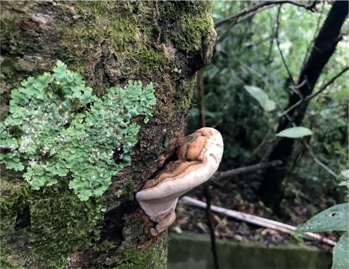
Basidiomata of Ganoderma tongshanense.
Fig. 38.

Microscopic structures of Ganoderma tongshanense (drawn from Cui 17168). A. Basidiospores. B. Apical cells from cuticle. C. Basidia and basidioles. D. Hyphae from trama. E. Hyphae from context. Scale bars = 10 μm.
Diagnosis: Differs from other species in the genus by its pale yellowish brown pileal surface, large pores, homogeneous context, non-stratified tubes, and ellipsoid and truncated basidiospores.
Etymology: tongshanense (Lat.), refers to the holotype of this species being found at Tongshan County of Hubei Province.
Typus: China, Hubei, Tongshan County, Jiugongshan National Park, on fallen trunk of angiosperm tree, 20 Oct. 2018, Cui 17168 (holotype BJFC030468).
Description: Basidiomata annual, sessile and broadly attached, hard corky. Pilei solitary, flabelliform, up to 7 cm diam and 2.5 cm thick. Pileal surface clay buff to pale yellowish brown, dull, glabrous, with concentric furrows and slightly radial wrinkles; margin obtuse, entire. Pore surface greyish white when fresh, turning darker when bruised, pale brown when dry; pores circular, shallow, 3–4 per mm; dissepiments moderately thick, entire. Context dark brown, homogeneous, without black melanoid lines, hard corky, up to 1.6 cm thick. Tubes slightly paler than context, non-stratified, stuffed with white mycelium, up to 9 mm long. Hyphal system trimitic; generative hyphae with clamp connections; all hyphae IKI –, CB +; tissues darkening in KOH. Generative hyphae in context colourless, thin-walled, 2–4 μm diam; skeletal hyphae in context dark brown, thick-walled with a wide to narrow lumen or sub-solid, arboriform and flexuous, 2–5 μm diam; binding hyphae in context colourless, thick-walled, branched and flexuous, up to 2 μm diam. Generative hyphae in tubes colourless, thin-walled, 2–4 μm diam; skeletal hyphae in tubes dark brown, thick-walled with a wide to narrow lumen or sub-solid, arboriform and flexuous, 2–5 μm diam; binding hyphae in tubes colourless, thick-walled, branched and flexuous, up to 2 μm diam. Pileipellis composed of clamped generative hyphae, thick-walled to sub-solid, apical cells clavate, inflated and flexuous, pale yellowish brown, about 30–45 × 6–8 μm, forming a regular palisade. Cystidia and cystidioles absent. Basidia clavate, colourless, thin-walled, 10–13 × 3–5 μm; basidioles in shape like the basidia, colourless, thin-walled, 8–11 × 4–6 μm. Basidiospores ellipsoid, truncated, pale yellowish brown, IKI –, CB +, double-walled with distinctly thick walls, exospore wall smooth, endospore wall with dense spinules, (8–)8.2–9.8(–10) × (5–)5.2–6.2(–6.5) μm, L = 8.91 μm, W = 5.62 μm, Q = 1.59 (n = 60/1, with the turgid vesicular appendix excluded); (9.2–)9.4–10.5(–11) × (5–)5.2–6.1(–6.3) μm, L = 9.81 μm, W = 5.65 μm, Q = 1.74 (n = 60/1, with the turgid vesicular appendix included).
Notes: Morphologically, G. tongshanense may be confused with G. australe which is widely distributed in South China. They share similar macro-morphological characters, but the latter has perennial and larger basidiomata and stratified tubes (Patouillard 1889, Ryvarden & Johansen 1980, Corner 1983). In the phylogenetic analyses, G. tongshanense and G. australe are distinct from each other (Fig. 1).
Ganoderma yunlingense B.K. Cui, J.H. Xing & Y.F. Sun, sp. nov. MycoBank MB 839684. Figs 39, 40.
Fig. 39.

Basidiomata of Ganoderma yunlingense.
Fig. 40.

Microscopic structures of Ganoderma yunlingense (drawn from Cui 16288). A. Basidiospores. B. Basidia and basidioles. C. Hyphae from trama. D. Hyphae from context. Scale bars = 10 μm.
Diagnosis: Differs from other species in the genus by its non-laccate pileal surface with concentrically irregular ridges, extremely thin context, stratified tubes, and ovoid to almond-shaped basidiospores without spinules on the endospore wall.
Etymology: yunlingense (Lat.), refers to this species being collected from the Yunling Mountains of Yunnan Province.
Typus: China, Yunnan, Lanping County, Luoguqing, on fallen trunk of Quercus, 9 Sep. 2017, Cui 16288 (holotype BJFC029587).
Additional materials examined: China, Yunnan, Lijiang, Yulongxueshan, on stump of Quercus, 16 Sep. 2018, Cui 17043 (BJFC030342), Cui 17060 (BJFC030359); Lanping County, Luoguqing, on fallen trunk of Quercus semecarpifolia, 18 Sep. 2018, Cui 17161 (BJFC030461).
Description: Basidiomata perennial, sessile, hard corky to woody hard. Pilei solitary, flabelliform to shell-shaped, occasionally dimidiate, up to 11.5 cm diam and 10 cm thick. Pileal surface greyish brown to grey, dull, glabrous, with concentrically irregular ridges; margin acute, entire, wavy and sometimes lacerated as petals. Pore surface white when fresh, turning darker when bruised, straw yellow when dry; pores circular to angular, 4–6 per mm; dissepiments moderately thick, entire. Context extremely thin, without black melanoid lines. Tubes brown to dark brown, distinctly stratified, up to 5 cm long. Hyphal system trimitic; generative hyphae with clamp connections; all hyphae IKI –, CB +; tissues darkening in KOH. Generative hyphae in context colourless, thin-walled, 2–3.5 μm diam; skeletal hyphae in context yellowish brown, thick-walled with narrow lumen or sub-solid, arboriform and flexuous, 3–5.5 μm diam; binding hyphae in context colourless, thick-walled, branched and flexuous, 1.5–3 μm diam. Generative hyphae in tubes colourless, thin-walled, 2–3 μm diam; skeletal hyphae in tubes yellowish brown, thick-walled with narrow lumen or sub-solid, arboriform and flexuous, 2.5–5 μm diam; binding hyphae in tubes colourless, thick-walled, branched and flexuous, 1.5–3 μm diam. Pileipellis composed of clamped generative hyphae, thick-walled, apical cells clavate, flexuous, golden yellow to pale brown, about 25–40 × 4–7 μm, forming a regular palisade. Cystidia and cystidioles absent. Basidia barrel-shaped, colourless, thin-walled, 15–20 × 8–13 μm; basidioles clavate, colourless, thin-walled, 13–17 × 6–10 μm. Basidiospores ovoid to almond-shaped, not obviously truncated, pale yellowish brown, IKI –, CB +, double-walled with slightly thick walls, exospore wall smooth, endospore wall without spinules, (7.5–)8–10(–10.5) × (4.3–)4.8–5.5(–6) μm, L = 8.93 μm, W = 5.16 μm, Q = 1.70–1.73 (n = 60/2, with the turgid vesicular appendix excluded).
Notes: Ganoderma yunlingense is characterised by its non-laccate pileal surface with concentrically irregular ridges, extremely thin context, stratified and long tubes, basidiospores not obviously truncated, without spinules on the endospore wall. It may be confused with G. acontextum because of the dull pileal surface, thin context and basidiospores not obviously truncated without any ornamentation; however, G. acontextum was collected from the United States of America and has reddish brown pileal surface with dense concentric furrows, and non-stratified tubes.
Notes on accepted species of Ganoderma recorded from China
Ganoderma ahmadii Steyaert, Persoonia 7: 91. 1972. MycoBank MB 314303.
For a detailed description of Ganoderma ahmadii, see Steyaert (1972) and Zhao (1989b).
Notes: Ganoderma ahmadii was described from Pakistan by Steyaert (1972). Zhao (1989b) examined an authentic specimen collected from Pakistan, but the description regarding the reddish brown pileal surface and homogeneous context is different to the original description. Wang (2005) studied one specimen of G. ahmadii collected from Sichuan Province in China and the ITS1-ITS2 sequences are similar to the records in GenBank. So, G. ahmadii was confirmed in Sichuan Province in China based on morphological and phylogenetic analyses.
Ganoderma angustisporum J.H. Xing et al., MycoKeys 34: 98. 2018. MycoBank MB 823320.
For a detailed description of Ganoderma angustisporum, see Xing et al. (2018).
Notes: Ganoderma angustisporum was recently described from China and classified in Ganoderma according to its narrow and truncated basidiospores in Ganoderma (Xing et al. 2018). It is also found in Malaysia, Sri Lanka and Thailand.
Ganoderma applanatum (Pers.) Pat., Hyménomyc. Eur. (Paris): 143. 1887. MycoBank MB 119872. Figs 41, 42.
Fig. 41.

Basidiomata of Ganoderma applanatum.
Fig. 42.

Microscopic structures of Ganoderma applanatum (drawn from He 2139). A. Basidiospores. B. Hyphae from trama. C. Hyphae from context. Scale bars = 10 μm.
Basionym: Boletus applanatus Pers., Observ. Mycol. (Lipsiae) 2: 2. 1800.
Description: Basidiomata perennial, sessile and broadly attached, hard corky to woody hard. Pilei solitary or imbricate, variable, applanate, flabelliform to shell-like or ungulate, up to 23 cm diam and 7 cm thick. Pileal surface pale brown to dark brown, dull, glabrous, with shallow to deep concentric furrows; margin obtuse, entire and wavy. Pore surface white to pale brown when fresh, turning darker when bruised, greyish white to straw yellow when dry; pores circular to angular, 4–7 per mm; dissepiments moderately thick, mostly entire. Context yellowish brown to dark brown, homogeneous, with black melanoid lines when mature, corky, up to 3 cm thick. Tubes concolorous with context, stratified by a layer of context, up to 4 cm long. Hyphal system trimitic; generative hyphae with clamp connections; all hyphae IKI –, CB +; tissues darkening in KOH. Generative hyphae in context colourless, thin-walled, 2.5–7 μm diam; skeletal hyphae in context pale yellowish brown, thick-walled with a wide to narrow lumen or sub-solid, frequently arboriform and flexuous, 3.5–6.5 μm diam; binding hyphae in context colourless, thick-walled, branched and flexuous, 2–3 μm diam. Generative hyphae in tubes colourless, thin-walled, 2–3.5 μm diam; skeletal hyphae in tubes pale brown to dark brown, thick-walled with a wide to narrow lumen or sub-solid, frequently arboriform and flexuous, 2–4.5 μm diam; binding hyphae in tubes colourless, thick-walled, branched and flexuous, 1–2.5 μm diam. Cystidia, cystidioles, basidia and basidioles absent. Basidiospores ellipsoid, truncated, yellowish to pale brown, IKI –, CB +, double-walled with moderately thick walls, exospore wall smooth, endospore wall with dense spinules, (5–)5.5–7(–8) × (4–)4.1–5.2(–6) μm, L = 6.25 μm, W = 4.56 μm, Q = 1.37 (n=30/1, with the turgid vesicular appendix included).
Additional materials examined: China, Jilin, Jiaohe, Hongyegu Park, on stump of angiosperm tree, 1 Aug. 2016, Cui 14062 (BJFC028930); on stump of Betula, 1 Aug. 2016, Cui 14070 (BJFC028938); Fusong County, Changbaishan Nature Reserve, on fallen trunk of angiosperm tree, 3 Aug. 2016, Cui 14121 (BJFC028989).
Notes: Ganoderma applanatum was firstly described from Europe and has a Holarctic distribution (Steyaert 1972, Moncalvo & Ryvarden 1997, Hapuarachchi et al. 2019b). Ganoderma applanatum is similar with G. australe based on perennial and sessile basidiomata, non-laccate and pale pileal surface, brown to dark brown context, but the latter has larger basidiospores (7–12 × 5–8 μm, Ryvarden 2004b). Besides, G. applanatum and G. australe can be separated in the phylogenetic analyses (Fig. 1).
Ganoderma australe (Fr.) Pat., Bull. Soc. Mycol. Fr. 5: 65. 1889. MycoBank MB 100745.
Basionym: Polyporus australis Fr., Elench. Fung. (Greifswald) 1: 108. 1828.
Synonyms: Ganoderma triangulum J.D. Zhao & L.W. Hsu, Acta Mycol. Sin. 3: 18. 1984.
Ganoderma ungulatum J.D. Zhao & X.Q. Zhang, Acta Mycol. Sin. 3: 19. 1984.
Ganoderma bawanglingense J.D. Zhao & X.Q. Zhang, Acta Mycol. Sin. 6: 205. 1987.
Ganoderma mirivelutinum J.D. Zhao, Acta Mycol. Sin. 7: 206. 1987.
For a detailed description of G. australe, see Ryvarden (2004b) and Hapuarachchi et al. (2018b).
Notes: Ganoderma australe was described from an island in the Pacific Ocean, but the type specimen has been lost and the only specimen deposited at the Royal Botanic Gardens Kew was from Europe which is inconsistent with its tropical distribution (Moncalvo & Ryvarden 1997). Cao (2013) suggested that G. bawanglingense, G. mirivelutinum, G. triangulum and G. ungulatum should be regarded as the synonyms of G. applanatum, but their tropical distribution and morphological similarity of G. australe support that they should be treated as the synonyms of G. australe. Ganoderma australe may be confused with G. adspersum, G. applanatum and G. gibbosum in morphology. Previously, many specimens collected from South China were identified as “Ganoderma australe”. Two specimens from Australia are recognised as G. australe by Costa-Rezende et al. (2017), and most specimens collected from South China clustered with G. gibbosum (Fig. 1). Whether there is true G. australe in China is unknown and more detailed studies about the G. australe complex should be done.
Ganoderma boninense Pat., Bull. Soc. Mycol. Fr. 5: 72. 1889. MycoBank MB 100062.
For a detailed description of G. boninense, see Steyaert (1967) and Ryvarden (1983).
Notes: Ganoderma boninense was described from the Bonin Islands in Japan, and it has been recognised as the main pathogen of oil palm trees causing a basal stem rot (Pilotti 2005). Ganoderma boninense can be distinguished by its reddish and shiny pileal surface, irregular apical cells with swellings and protuberance of pileipellis, and oblong ellipsoid basidiospores (Ryvarden 1983). Cao (2013) compared the specimens collected from Hainan Province and Japan, and they showed very similar morphological characters and ITS sequences.
Ganoderma calidophilum J.D. Zhao et al., Acta Microbiol. Sin. 19: 270. 1979. MycoBank MB 314307.
For a detailed description of G. calidophilum, see Zhao et al. (1979) and Luangharn et al. (2021).
Notes: Zhao et al. (1979) described Ganoderma calidophilum from Hainan Province based on its small basidiomata with heterogenous context, whitish pore surface and large basidiospores (10–12.1 × 6.2–8.7 μm). The sequences of G. calidophilum used in this study were downloaded from GenBank, and showed to be distinct from G. flexipes (Fig. 1), which has been considered as a doubtful synonym (Wang & Wu 2014).
Ganoderma casuarinicola J.H. Xing et al., MycoKeys 34: 100. 2018. MycoBank MB 823321.
For a detailed description of G. casuarinicola, see Xing et al. (2018) and Luangharn et al. (2019).
Notes: Ganoderma casuarinicola was collected on a living tree of Casuarina equisetifolia from Guangdong Province in China by Xing et al. (2018). Luangharn et al. (2019) reported G. casuarinicola, where it was found on Pinus kesiya stump, as a new record from Thailand. There are some differences such as applanate to dimidiate pilei, longer tubes and larger basidiospores when comparing these specimens with the type of G. casuarinicola (Dai 16336). Geographical and climatic divergences may be the reason for the intraspecific differences (Boddy et al. 2014).
Ganoderma ellipsoideum Hapuar. et al., Mycosphere 9: 951. 2018. MycoBank MB 554384.
For a detailed description of G. ellipsoideum, see Hapuarachchi et al. (2018b).
Notes: Ganoderma ellipsoideum is distinguished by its ellipsoid spores (6.1–7.3 × 3.7–4.6 μm) with distinct spinules on the endospore wall. Hapuarachchi et al. (2018b) stated that G. ellipsoideum was known only from the type locality of Hainan Province in China. In this study, several specimens collected from Yunnan and Guangdong provinces showed similar morphological features and close phylogenetic relationships with G. ellipsoideum.
Ganoderma flexipes Pat., Bull. Soc. Mycol. Fr. 23: 75. 1907. MycoBank MB 249905. Figs 43, 44.
Fig. 43.

Basidiomata of Ganoderma flexipes.
Fig. 44.

Microscopic structures of Ganoderma flexipes (drawn from Cui 13882). A. Basidiospores. B. Apical cells from cuticle. C. Basidia and basidioles. D. Hyphae from trama. E. Hyphae from context. Scale bars = 10 μm.
Synonyms: Ganoderma atrum J.D. Zhao et al., Acta Microbiol. Sin. 19: 268. 1979.
Ganoderma hainanense J.D. Zhao et al., Acta Microbiol. Sin. 19: 269. 1979.
Ganoderma parviungulatum J.D. Zhao & X.Q. Zhang, Acta Mycol. Sin. 5: 88. 1986.
Description: Basidiomata annual, dorso-laterally stipitate, hard corky to woody hard. Pilei solitary, variable, flabelliform to shell-like or circular, up to 5 cm diam and 1.5 cm thick. Pileal surface dark brown to reddish brown, strongly laccate, glabrous, with obvious concentric furrows and slightly radial rugose; margin obtuse, entire, slightly incurved. Pore surface white when fresh, turning darker when bruised, light buff when dry; pores circular to angular, 4–6 per mm; dissepiments slightly thick, entire. Context heterogeneous, the upper layer pale yellowish brown, the lower layer dark brown, with black melanoid lines, corky, up to 3 mm thick. Tubes dark brown, non-stratified, up to 1.2 cm long. Stipe reddish brown to purplish black, flattened to cylindrical, up to 25 cm long and 6 mm diam. Hyphal system trimitic; generative hyphae with clamp connections; all hyphae IKI –, CB +; tissues darkening in KOH. Generative hyphae in context colourless, thin-walled, 1.5–3.5 μm diam; skeletal hyphae in context pale yellowish brown, thick-walled with a narrow lumen or sub-solid, arboriform and flexuous, 2.5–6 μm diam; binding hyphae in context colourless, thick-walled, branched and flexuous, 1–2 μm diam. Generative hyphae in tubes colourless, thin-walled, occasionally branched, 2–3 μm diam; skeletal hyphae in tubes pale brown to dark brown, thick-walled with a narrow lumen to sub-solid, frequently arboriform and flexuous, 2–4 μm diam; binding hyphae in tubes colourless, thick-walled, branched and flexuous, 0.5–1 μm diam. Pileipellis composed of clamped generative hyphae, thick-walled to sub-solid, apical cells clavate, slightly inflated, pale brown to yellowish brown, 30–45 × 5–10 μm, forming a regular palisade. Cystidia and cystidioles absent. Basidia barrel-shaped, colourless, thin-walled, 23–30 × 12–17 μm; basidioles in shape like the basidia, colourless, thin-walled, 17–23 × 8–13 μm. Basidiospores ellipsoid, not obviously truncated, pale yellowish brown, IKI –, CB +, double-walled with moderately thick walls, exospore wall smooth, endospore walls with dense spinules, (6.5–)7–9(–9.5) × (4–)4.8–5.5(–6) μm, L = 8.17 μm, W = 5.23 μm, Q = 1.56 (n = 30/1, with the turgid vesicular appendix excluded).
Materials examined: China, Guangdong, Guangzhou, campus of Sun Yat-Sen University, on stump of angiosperm tree, 20 Jul. 2012, Cui 17209 (BJFC030563); Hainan, Qiongzhong County, Limushan Forest Park, on ground of angiosperm forest, 16 Jun. 2016, Cui 13841 (BJFC028707); Changjiang County, Bawangling Nature Reserve, on ground of angiosperm forest, 18 Jun. 2016, Cui 13863 (BJFC028729); Yunnan, Puer, Puer Forest Park, on rotten angiosperm wood, 17 Aug. 2019, Dai 20461 (BJFC032129).
Notes: Ganoderma flexipes was first described from Vietnam by Patouillard (1907), and many researchers have conducted detailed studies on it (Steyaert 1972, Ryvarden 1983, Hapuarachchi et al. 2019b). Cao (2013) considered G. atrum, G. hainanense and G. parviungulatum as synonyms of G. flexipes based on comprehensive observations of the holotype specimens.
Ganoderma gibbosum (Blume & T. Nees) Pat., Ann. Jard. Bot. Buitenzorg, suppl. 1: 114. 1897. MycoBank MB 250058.
Basionym: Polyporus gibbosus Blume & T. Nees, Nova Acta Phys.-Med. Acad. Caes. Leop.-Carol. Nat. Cur. 13: 19. 1826.
For a detailed description of G. gibbosum, see Luangharn et al. (2020).
Notes: Ganoderma gibbosum was first described from Java, however, the type specimen was lost (Moncalvo & Ryvarden, 1997). In previous studies, G. gibbosum was regarded as a synonym of G. applanatum or classified in the G. applanatum–australe complex based on the non-laccate basidiomata (Zhao 1989b, Moncalvo & Ryvarden 1997). Luangharn et al. (2020) conducted a study on the taxonomy of G. gibbosum collected from Kunming, China, and the results showed that samples of G. gibbosum from Asia and South America formed two different lineages. In this study, specimens of G. gibbosum collected from Guangdong, Guangxi and Sichuan provinces clustered together and formed a well-supported lineage (Fig. 1).
Ganoderma hoehnelianum Bres., Annls Mycol. 10: 502. 1912. MycoBank MB 243431. Figs 45, 46.
Fig. 45.

Basidiomata of Ganoderma hoehnelianum.
Fig. 46.

Microscopic structures of Ganoderma hoehnelianum (drawn from Dai 16166). A. Basidiospores. B. Basidioles. C. Hyphae from trama. D. Hyphae from context. Scale bars = 10 μm.
Synonym: Ganoderma shangsiense J.D. Zhao, Acta Mycol. Sin. 7: 17. 1988.
Description: Basidiomata perennial, sessile or broadly attached to laterally stipitate, hard corky to woody hard. Pilei solitary, variable, flabelliform or shell-like to reniform, applanate, up to 10 cm diam and 2.2 cm thick. Pileal surface yellowish brown to dark brown, dull, glabrous, with obvious concentric furrows; margin acute to obtuse, entire. Pore surface white when fresh, turning darker when bruised, straw yellow to pale yellowish brown when dry; pores circular to angular, 3–6 per mm; dissepiments moderately thick, entire. Context heterogeneous, the upper layer pale yellowish brown, the lower layer dark brown, with black melanoid lines, corky, up to 1 cm thick. Tubes light buff to greyish brown, stratified, up to 1.2 cm long. Stipe dark brown, flattened, up to 3 cm long and 6 mm diam. Hyphal system trimitic; generative hyphae with clamp connections; all hyphae IKI –, CB +; tissues darkening in KOH. Generative hyphae in context absent; skeletal hyphae in context pale yellowish brown, thick-walled with a wide to narrow lumen or sub-solid, frequently arboriform and flexuous, 2–4 μm diam; binding hyphae in context colourless, thick-walled, branched and flexuous, 1–2.5 μm diam. Generative hyphae in tubes colourless, thin-walled, 2.3–3.5 μm diam; skeletal hyphae in tubes pale brown to dark brown, thick-walled with a narrow lumen or sub-solid, frequently arboriform and flexuous, 2–5 μm diam; binding hyphae in tubes colourless, thick-walled, branched and flexuous, 1.5–3 μm diam. Cystidia and cystidioles absent. Basidia absent; basidioles barrel-shaped, colourless, thin-walled, 21–30 ×9–13 μm. Basidiospores subglobose, not obviously truncated, pale yellowish brown, IKI –, CB +, double-walled with distinctly thick walls, exospore walls smooth, endospore walls with dense spinules, (6.5–)7–8.2(–9) × (–5.5)5.8–7.8(–8) μm, L = 7.43 μm, W = 6.73 μm, Q = 1.1 (n = 30/1).
Materials examined: China, Hainan, Ledong County, Jianfengling Nature Reserve, on stump of angiosperm tree, 19 Jun. 2016, Cui 13904 (BJFC028770); Guangxi, Shangsi County, Shiwandashan Forest Park, on dead tree of angiosperm, 6 Jul. 2016, Cui 13982 (BJFC028850); Nanning, Guangxi Academy of Forestry, on living tree of angiosperm, 21 Aug. 2019, Dai 20783 (BJFC032450); Yunnan, Mengla County, Wangtianshu Park, 20 Jul. 2014, Dai 13915 (BJFC017645).
Notes: Ganoderma hoehnelianum was described by Bresadola from Java. It has typically ganodermoid macro-morphological features, but the obviously amaurodermoid basidiospores make G. hoehnelianum different from other species. In the phylogenetic analyses, G. hoehnelianum grouped in the Ganoderma clade, and formed an independent lineage with high support (Fig. 1). Wang & Wu (2010) and Cao (2013) regarded G. shangsiense as a synonym of G. hoehnelianum after the studies on the holotype of G. shangsiense.
Ganoderma leucocontextum T.H. Li et al., Mycoscience 56: 82. 2015. MycoBank MB 804187.
For a detailed description of G. leucocontextum, see Li et al. (2015).
Notes: Ganoderma leucocontextum as a member of the G. lucidum complex shares reddish brown and laccate pilei and truncated basidiospores, but it is distinguished by the white context Li et al. (2015). In the current study, G. leucocontextum grouped with G. weixiense (Fig. 1). According to the comparison between G. leucocontextum and G. weixiense in Ye et al. (2019), the size of pores (4–6 per mm vs 2–4 per mm) and basidiospores (9.5–12.5 × 7–9 μm vs 6–8 × 3–4 μm) are the main differences.
Ganoderma lingzhi Sheng H. Wu et al., Fungal Divers. 56: 54. 2012. MycoBank MB 564240.
For a detailed description of G. lingzhi, see Cao et al. (2012).
Notes: Cao et al. (2012) revised the taxonomic status of the widely cultivated “Ganoderma lucidum” in China and described it as a new species called G. lingzhi based on geographical distribution, morphological features and phylogenetic analyses. Ganoderma lingzhi is widely distributed in temperate and subtropical areas of China, and it has also been reported from Korea and Laos (Kim et al. 2001, Hapuarachchi et al. 2019b).
Ganoderma lucidum (Curtis) P. Karst., Revue Mycol., Toulouse 3: 17. 1881. MycoBank MB 148413. Figs 47, 48.
Fig. 47.

Basidiomata of Ganoderma lucidum.
Fig. 48.

Microscopic structures of Ganoderma lucidum (drawn from Dai 15805). A. Basidiospores. B. Apical cells from cuticle. C. Basidioles. D. Hyphae from trama. E. Hyphae from context. Scale bars = 10 μm.
Basionym: Boletus lucidus Curtis, Fl. Londin. 1: 72. 1781.
Synonym: Ganoderma cantharelloideum M.H. Liu, Acta Mycol. Sin. 8: 279. 1989.
Description: Basidiomata annual, laterally stipitate, hard corky to woody hard. Pilei solitary, variable, flabelliform or shell-like to circular, up to 11 cm diam and 3 cm thick. Pileal surface yellowish brown to reddish brown, laccate, glabrous, with concentric furrows; margin acute to obtuse, entire, slightly wavy. Pore surface white when fresh, turning darker when bruised, pale yellow to straw yellow when dry; pores circular, 4–6 per mm; dissepiments slightly thick, mostly entire. Context heterogeneous, the upper layer cream to buff, the lower layer clay-buff, without black melanoid lines, soft corky, up to 1.8 cm thick. Tubes pale brown, non-stratified, up to 1.2 cm long. Stipe reddish brown to purplish black, flattened to cylindrical, up to 12 cm long and 1.5 cm diam. Hyphal system trimitic; generative hyphae with clamp connections; all hyphae IKI –, CB +; tissues darkening in KOH. Generative hyphae in context colourless, thin-walled, 2.5–4 μm diam; skeletal hyphae in context pale yellowish brown, thick-walled with a narrow lumen or sub-solid, frequently arboriform and flexuous, 3–10 μm diam; binding hyphae in context colourless, thick-walled, branched and flexuous, 2–3 μm diam. Generative hyphae in tubes colourless, thin-walled, 2–3 μm diam; skeletal hyphae in tubes pale brown to dark brown, thick-walled with a narrow lumen or sub-solid, frequently arboriform and flexuous, 2–8 μm diam; binding hyphae in tubes colourless, thick-walled, branched and flexuous, 1–2.5 μm diam. Pileipellis composed of clamped generative hyphae, thick-walled to sub-solid, apical cells clavate, strongly inflated and flexuous, pale yellowish brown, about 30–50 × 8–16 μm, forming a regular palisade. Cystidia and cystidioles absent. Basidia absent; basidioles barrel-shaped, colourless, thin-walled, 20–25 ×9–12 μm. Basidiospores ellipsoid, truncated, pale yellowish brown, IKI –, CB +, double-walled with moderately to distinctly thick walls, exospore walls smooth, endospore walls with dense spinules, (7–)7.5–9.5(–10) × (5–)5.5–7(–7.5) μm, L = 8.52 μm, W = 6.2 μm, Q = 1.37 (n = 30/1, with the turgid vesicular appendix excluded); (8–)9–11(–11.5) × (5–)5.5–7(–8) μm, L = 10.05 μm, W = 6.45 μm, Q = 1.56 (n = 30/1, with the turgid vesicular appendix included).
Materials examined: China, Sichuan, Qingchuan County, Qingxi Town, on stump of Quercus, 31 Oct. 2016, Cui 14404 (BJFC029272), Cui 14405 (BJFC029273), Cui 14406 (BJFC029274); Yunnan, Kunming, Xiaoshao Forest Farm, on rotten angiosperm wood, 1 Jul. 2019, Dai 20017 (BJFC031691).
Notes: Ganoderma lucidum was firstly described from London in the UK. It is the type species of Ganoderma and has the typical ganodermoid characters, such as stipitate basidiomata with laccate pileal surface, and truncated basidiospores with spinules on the endospore walls (Steyaert 1972, Ryvarden 2004b). Cao (2013) suggested that G. cantharelloideum should be treated as a synonym of G. lucidum according to the original description and observations on the holotype of G. cantharelloideum. In the current phylogenetic study, the G. lucidum lineage consisted of specimens collected from the UK, Czech Republic and China with high divergence (Fig. 1, 2). The divergence in G. lucidum groups also appeared in phylogenetic analyses inferred from single or multiple genes by Zhou et al. (2015), Hapuarachchi et al. (2018b), Liu et al. (2019) and Luangharn et al. (2021). This may be caused by geographic separation. An essential systemic study of the G. lucidum lineage should be conducted based on more specimens from different regions and more gene markers.
Ganoderma magniporum J.D. Zhao & X.Q. Zhang, Acta Mycol. Sin. 3: 15. 1984. MycoBank MB 124473.
For a detailed description of G.a magniporum, see Zhao et al. (1984).
Notes: Ganoderma magniporum was described from Guangxi of South China based on its small and nearly black pilei, large pores (2–2.5 per mm), and ellipsoid basidiospores (8.7–10.4 × 5.2–7 μm, Zhao et al. 1984). Cao (2013) observed the type specimen (HMAS 42696) of G. magniporum, and stated it was immature and had smaller basidiospores than another specimen (Zhou 439) collected from Guangxi. In the current study, one specimen collected from Yunnan grouped with G. magniporum.
Ganoderma multipileum Ding Hou, Quarterly Journal of the Taiwan Museum 3: 101. 1950. MycoBank MB 344109.
Synonym: Ganoderma chenghaiense J.D. Zhao, Acta Mycol. Sin. 8: 31. 1989.
For a detailed description of Ganoderma multipileum, see Wang et al. (2009).
Notes: Ganoderma multipileum was described from Taiwan (China) based on its imbricate basidiomata with yellowish brown to reddish brown pileal surface, small pores (6–8 per mm), corky context, and ellipsoid to ovoid basidiospores (Wang et al. 2009). Cao (2013) studied the holotype of G. multipileum and G. chenghaiense, and suggested that the latter should be a synonym of G. multipileum based on the morphological characters. As more specimens were collected, the distribution of G. multipileum became widespread, found in South China, Laos and India. In the phylogenetic analysis, one specimen collected from the type locality was included and G. multipileum formed a stable lineage with good support (Fig. 1).
Ganoderma mutabile Y. Cao & H.S. Yuan, Mycol. Prog. 12: 122. 2013. MycoBank MB 563047. Figs 49, 50.
Fig. 49.

Basidiomata of Ganoderma mutabile.
Fig. 50.

Microscopic structures of Ganoderma mutabile (drawn from Cui 17189). A. Basidiospores. B. Apical cells from cuticle. C. Hyphae from trama. D. Hyphae from context. Scale bars = 10 μm.
Description: Basidiomata perennial, sessile, hard corky to woody hard. Pilei solitary, flabelliform to shell-like, up to 18 cm diam and 7 cm thick. Pileal surface reddish brown to purplish brown, laccate, glabrous, with concentric furrows; margin obtuse, entire, slightly wavy. Pore surface white when fresh, turning darker when bruised, dark brown when dry; pores circular to angular, 4–5 per mm; dissepiments moderately thick, entire. Context brown, homogeneous, with black melanoid lines, hard corky, up to 3 cm thick. Tubes brown, stratified, up to 4 cm long. Hyphal system trimitic; generative hyphae with clamp connections; all hyphae IKI –, CB +; tissues darkening in KOH. Generative hyphae in context colourless, thin-walled, 2–3.5 μm diam; skeletal hyphae in context pale brown, thick-walled with a narrow lumen or sub-solid, frequently arboriform and flexuous, 2–5.5 μm diam; binding hyphae in context colourless, thick-walled, branched and flexuous, 1–2.5 μm diam. Generative hyphae in tubes colourless, thin-walled, 2–3 μm diam; skeletal hyphae in tubes pale brown, thick-walled with a narrow lumen or sub-solid, frequently arboriform and flexuous, 2–4.5 μm diam; binding hyphae in tubes colourless, thick-walled, branched and flexuous, 1–2.5 μm diam. Pileipellis composed of clamped generative hyphae, thick-walled to sub-solid, apical cells mostly with lateral outgrowths and protuberances, branched and flexuous, yellowish brown, about 40–65 × 4–9 μm, forming an irregular palisade. Cystidia, cystidioles, basidia and basidioles absent. Basidiospores broadly ellipsoid, truncated, pale yellowish brown, IKI –, CB +, double-walled with distinctly thick walls, exospore walls smooth, endospore walls with dense spinules, (7.5–)8–11(–11.5) × 5.5–7(–7.5) μm, L = 9.1 μm, W = 6.25 μm, Q = 1.46 (n = 30/1, with the turgid vesicular appendix excluded); (8.5–)9–12.5(–13) × (5.5–)6–7.5(–8) μm, L = 10.57 μm, W = 6.83 μm, Q = 1.55 (n = 30/1, with the turgid vesicular appendix included).
Materials examined: China, Yunnan, Chuxiong, Zixishan Forest Park, on living tree of angiosperm, 8 Sep. 2006, holotype Yuan 2289 (IFP); Xinping County, Mopanshan Forest Park, on ground of mixed forest, 16 Aug. 2019, Dai 20414 (BJFC032082); Xizang, on angiosperm wood, Dec. 2018, Cui 17189 (BJFC030489).
Notes: Ganoderma mutabile was described from Yunnan in China based on the irregular apical cells of the pileipellis which are strongly flexuous and frequently branched Cao & Yuan (2012). Only one specimen was included in the original description, and in this study additional specimens were collected from Yunnan and Xizang.
Ganoderma orbiforme (Fr.) Ryvarden, Mycologia 92: 187. 2000. MycoBank MB 464692.
Basionym: Polyporus orbiformis Fr., Epicr. Syst. Mycol. (Upsaliae): 463. 1838.
Synonyms: Ganoderma fornicatum (Fr.) Pat., Bull. Soc. Mycol. Fr. 5: 71. 1889.
Ganoderma mastoporum (Lév.) Pat., Bull. Soc. Mycol. Fr. 5: 75. 1889.
Ganoderma subtornatum Murrill, Bull. Torrey Bot. Club 34: 477. 1907.
Ganoderma pygmoideum Steyaert, Bull. Jard. Bot. État Brux. 32: 103. 1962.
Ganoderma crebrostriatum J.D. Zhao & L.W. Hsu, Acta Mycol. Sin. 2: 161. 1983.
Ganoderma densizonatum J.D. Zhao & X.Q. Zhang, Acta. Mycol. Sin. 5: 86. 1986.
Ganoderma limushanense J.D. Zhao & X.Q. Zhang, Acta. Mycol. Sin. 5: 219. 1986.
Ganoderma diaoluoshanense J.D. Zhao & X.Q. Zhang, Acta Mycol. Sin. 6: 1. 1987.
For a detailed description of G. orbiforme, see Ryvarden (2000) and Wang et al. (2014).
Notes: Polyporus orbiformis was combined as Ganoderma orbiforme by Ryvarden (2000), while G. boninense and G. pygmoideum were considered as synonyms of G. orbiforme by Ryvarden (2000). The treatment of G. boninense as a synonym of G. orbiforme is not accepted by most other mycologists (Pilotti 2005). Wang et al. (2014) clarified the taxonomic status of G. orbiforme based on morphological and molecular data. Ganoderma cupreum, G. densizonatum, G. fornicatum, G. limushanense, G. mastoporum and G. subtornatum were treated as synonyms of G. orbiforme by Wang et al. (2014). Cao (2013) regarded G. crebrostriatum and G. diaoluoshanense as synonyms of G. mastoporum based on the observation of holotype specimens, while G. crebrostriatum and G. diaoluoshanense should be regarded as the synonyms of G. orbiforme according to the revision of G. mastoporum by Wang et al. (2014). In this study, two specimens of G. cupreum from Cameroon were included in the phylogenetic analyses, and they formed an independent lineage which is distinct from G. orbiforme (Fig. 1). Thus, G. cupreum should be treated as an independent species.
Ganoderma philippii (Bres. et Henn.) Bres., Iconogr. Mycol. 21: 1014. 1932. MycoBank MB 314321. Figs 51, 52.
Fig. 51.

Basidiomata of Ganoderma philippii.
Fig. 52.

Microscopic structures of Ganoderma philippii (drawn from Cui 14443). A. Basidiospores. B. Hyphae from trama. C. Hyphae from context. Scale bars = 10 μm.
Basionym: Fomes philippii Bres. & Henn. ex Sacc., Syll. Fung. (Abellini) 9: 180. 1891.
Description: Basidiomata annual or perennial, sessile and broadly attached, sometimes growing together, hard corky. Pilei solitary, variable, flabelliform to circular, applanate, up to 26 cm diam and 1.6 cm thick. Pileal surface pale brown to purplish black, dull, glabrous, with dense concentric zones; margin acute to obtuse, wavy like petal. Pore surface white when fresh, turning darker when bruised, pale brown when dry; pores circular to angular, 5–6 per mm; dissepiments thin, mostly entire. Context brown, homogeneous, with black melanoid lines, hard corky, up to 1.4 cm thick. Tubes yellowish brown, non-stratified, up to 2 mm long. Hyphal system trimitic; generative hyphae with clamp connections; all hyphae IKI –, CB +; tissues darkening in KOH. Generative hyphae in context colourless, thin-walled, 2–3 μm diam; skeletal hyphae in context pale yellowish brown, thick-walled with a narrow lumen or sub-solid, arboriform and flexuous, 3–5 μm diam; binding hyphae in context colourless, thick-walled, branched and flexuous, 1–2 μm diam. Generative hyphae in tubes colourless, thin-walled, 1–3 μm diam; skeletal hyphae in tubes pale brown to dark brown, thick-walled with a narrow lumen or sub-solid, frequently arboriform and flexuous, 2–5 μm diam; binding in tubes colourless, thick-walled, branched and flexuous, 1–2 μm diam. Cystidia, cystidioles, basidia and basidioles absent. Basidiospores obovoid, not obviously truncated, with obvious vesicular appendix, pale yellowish brown, IKI –, CB +, double-walled with slightly thick walls, exospore walls smooth, endospore walls with dense spinules, 5–6 × 3–4 μm, L = 5.63 μm, W = 3.38 μm, Q = 1.67 (n = 30/1, with the turgid vesicular appendix excluded); 6–8 × 3–4 μm, L = 7.32 μm, W = 3.36 μm, Q = 2.18 (n = 30/1, with the turgid vesicular appendix included).
Materials examined: China, Hainan, Qiongzhong County, Limushan, on stump of Hevea, 9 Sep. 2016, Cui 14443 (BJFC029311), Cui 14444 (BJFC029311). Singapore, Bukit Timah Nature Reserve, on fallen trunk of angiosperm tree, 19 Jul. 2017, Dai 17828 (BJFC025360).
Notes: Ganoderma philippii was firstly described from Myanmar, and it has a wide distribution in South-East Asia including South China, Indonesia, Malaysia and Singapore (Steyaert 1972, Moncalvo & Ryvarden 1997). Ganoderma philippii can be distinguished by the sessile basidiomata with variable pilei, pale brown to purplish black pileal surface with dense concentric zones, small and obovoid basidiospores.
Ganoderma sanduense Hapuar. et al., Mycosphere 8: 274. 2019. MycoBank MB 634622.
For a detailed description of Ganoderma sanduense, see Hapuarachchi et al. (2019b).
Notes: The type locality of Ganoderma sanduense is Sandu County of Guizhou Province in southwestern China. Ganoderma stratoideum was also described from Sandu County, and it shares layered, reddish black and laccate pilei, moderately sized pores (3–5 per mm), and heterogenous context with G. sanduense (He & Yu 1989). However, the small pilei (2–4 × 1–2.5 cm), greyish brown pore surface and larger basidiospores (12.1–13.8 × 9.2–10.5 μm) differentiate G. sanduense (Hapuarachchi et al. 2019b). The type specimen of G. stratoideum has been lost, and its taxonomic status is doubtful.
Ganoderma shanxiense L. Fan & H. Liu, Phytotaxa 406: 132. 2019. MycoBank MB 830632.
For a detailed description of Ganoderma shanxiense, see Liu et al. (2019).
Notes: Ganoderma shanxiense was described from Shanxi Province in China and is characterised by its basidiospores with a tapering and obtuse end at maturity (Liu et al. 2019). In the phylogenetic tree, G. shanxiense clustered with G. chuxiongense (Fig. 1) which was described from Yunnan Province, but the latter species differs by a purplish black and laccate pileal surface, greyish white pore surface with small pores (7–8 per mm), and broadly ellipsoid to ovoid basidiospores (10–12 × 7–8.5 μm).
Ganoderma sichuanense J.D. Zhao & X.Q. Zhang, Acta. Mycol. Sin. 2: 159. 1983. MycoBank MB 107984.
For a detailed description of Ganoderma sichuanense, see Zhao et al. (1983) and Wang et al. (2012).
Notes: Ganoderma sichuanense was first described from Sichuan Province by Zhao et al. (1983). Since then, more specimens have been collected from Guangdong, Guangxi of China and Sri Lanka. Phylogenetically, G. weberianum grouped with G. sichuanense which is consistent with Hapuarachchi et al. (2019b). Further studies are needed to clarify the relationship between G. weberianum and G. sichuanense.
Ganoderma sinense J.D. Zhao et al., Acta Microbiol. Sin. 19: 272. 1979. MycoBank MB 314325. Figs 53, 54.
Fig. 53.

Basidiomata of Ganoderma sinense.
Fig. 54.

Microscopic structures of Ganoderma sinense (drawn from Cui 14574). A. Basidiospores. B. Apical cells from cuticle. C. Basidioles. D. Hyphae from trama. E. Hyphae from context. Scale bars = 10 μm.
Synonyms: Ganoderma austrofujianense J.D. Zhao et al., Acta Microbiol. Sin. 19: 274. 1979.
Ganoderma luteomarginatum J.D. Zhao et al., Acta Microbiol. Sin. 19: 274. 1979.
Ganoderma formosanum T.T. Chang & T. Chen, Trans. Br. Mycol. Soc. 82: 731. 1984.
Ganoderma guinanense J.D. Zhao & X.Q. Zhang, Acta Mycol. Sin. 6: 4. 1987.
Ganoderma mediosinense J.D. Zhao, Acta Mycol. Sin. 7: 205. 1988.
Description: Basidiomata annual, laterally stipitate, hard corky to woody hard. Pilei solitary, variable, flabelliform to reniform or circular, up to 14 cm diam and 1.6 cm thick. Pileal surface reddish brown to purplish black, strongly laccate, glabrous, with concentric furrows and radial wrinkles; margin acute to obtuse, entire, slightly incurved. Pore surface white when fresh, turning darker when bruised, light buff when dry; pores circular to angular, 3–5 per mm; dissepiments thin, mostly entire. Context heterogeneous, the upper layer white to buff, the lower layer pale brown to yellowish brown, with black melanoid lines, hard corky, up to 3 cm thick. Tubes yellowish brown, non-stratified, up to 1.5 cm long. Stipe reddish brown to purplish black, flattened to cylindrical, up to 6 cm long and 2 cm diam. Hyphal system trimitic; generative hyphae with clamp connections; all hyphae IKI –, CB +; tissues darkening in KOH. Generative hyphae in context colourless, thin-walled, 2–4 μm diam; skeletal hyphae in context pale yellowish brown, thick-walled with a narrow lumen or sub-solid, arboriform and flexuous, 3–6 μm diam; binding hyphae in context colourless, thick-walled, branched and flexuous, 1.5–2.5 μm diam. Generative hyphae in tubes colourless, thin-walled, 1–3 μm diam; skeletal hyphae in tubes pale brown to dark brown, thick-walled with a narrow lumen or sub-solid, frequently arboriform and flexuous, 2–4 μm diam; binding hyphae in tubes colourless, thick-walled, branched and flexuous, 1–2 μm diam. Pileipellis composed of clamped generative hyphae, thick-walled to sub-solid, apical cells clavate, inflated, pale yellow to yellowish brown, about 40–55 × 6–12 μm, forming a regular palisade. Cystidia and cystidioles absent. Basidia absent; basidioles barrel-shaped to clavate, colourless, thin-walled, 16–24 × 9–12 μm. Basidiospores ellipsoid, not obviously truncated, pale yellowish brown, IKI –, CB +, double-walled with moderately thick walls, exospore wall smooth, endospore wall with dense spinules, (11–)11.6–13.2(–13.7) × (7–)7.3–8.5(–8.8) μm, L = 12.39 μm, W = 7.99 μm, Q = 1.74 (n = 30/1, with the turgid vesicular appendix included).
Materials examined: China, Hainan, Lingshui County, Diaoluoshan Forest Park, on ground of angiosperm forest, 15 Jun. 2016, Cui 13825 (BJFC028691), Cui 13835 (BJFC028701); Guangxi, Jinxiu County, Dayaoshan Nature Reserve, on stump of angiosperm tree, 15 Jul. 2017, Cui 14524 (BJFC029393), Cui 14526 (BJFC029395).
Notes: Ganoderma sinense was described from Hainan Province by Zhao et al. (1979), and it can be easily distinguished in the wild by its laterally stipitate basidiomata with dark reddish brown to purplish black and strongly laccate pileal surface. In this study, G. sinense clustered with G. japonicum with high support (Fig. 1) which is consistent with previous study by Hapuarachchi et al. (2019a). Ganoderma japonicum should be treated as a synonym of G. sinense temporarily based on the previous description and comments (Moncalvo & Ryvarden 1997, Liao et al. 2015, Hapuarachchi et al. 2019a). Wang (2005) and Cao (2013) also mentioned that G. austrofujianense, G. formosanum, G. guinanense, G. luteomarginatum and G. mediosinense are synonyms of G. sinense after studying the type specimens.
Ganoderma tropicum (Jungh.) Bres., Annls Mycol. 8: 586. 1910. MycoBank MB 149294. Figs 55, 56.
Fig. 55.

Basidiomata of Ganoderma tropicum.
Fig. 56.

Microscopic structures of Ganoderma tropicum (drawn from Cui 16341). A. Basidiospores. B. Apical cells from cuticle. C. Hyphae from trama. D. Hyphae from context. Scale bars = 10 μm.
Basionym: Polyporus tropicus Jungh., Verh. Batav. Genootsch. Kunst. Wet. 17: 63. 1838.
Description: Basidiomata annual to perennial, usually sessile, sometimes laterally stipitate, hard corky to woody hard. Pilei solitary, variable, flabelliform to shell-shaped or circular, up to 17 cm diam and 3 cm thick. Pileal surface reddish brown to dark brown, strongly laccate, glabrous, with obvious concentric zones; margin acute to obtuse, entire or sometimes lacerated. Pore surface white when fresh, turning darker when bruised, straw yellow when dry; pores circular to angular, 4–6 per mm; dissepiments slightly thick to moderately thick, mostly entire. Context dark brown, homogeneous, with black melanoid lines, corky, up to 2.2 cm thick. Tubes brown, non-stratified, up to 8 mm long. Stipe reddish brown to purplish black, flattened to cylindrical, up to 6 cm long and 1.2 cm diam. Hyphal system trimitic; generative hyphae with clamp connections; all hyphae IKI –, CB +; tissues darkening in KOH. Generative hyphae in context colourless, thin-walled, 2.5–3.5 μm diam; skeletal hyphae in context pale yellowish brown, thick-walled with a wide to narrow lumen or sub-solid, frequently arboriform and flexuous, 2.5–6 μm diam; binding hyphae in context colourless, thick-walled, branched and flexuous, 1–2 μm diam. Generative hyphae in tubes colourless, thin-walled, 2–3 μm diam; skeletal hyphae in tubes pale brown to dark brown, thick-walled with a narrow lumen or sub-solid, frequently arboriform and flexuous, 2–4 μm diam; binding hyphae in tubes colourless, thick-walled, branched and flexuous, 0.5–1 μm diam. Pileipellis composed of clamped generative hyphae, thick-walled to sub-solid, apical cells clavate, sometimes branched or protuberant, inflated and flexuous, yellowish brown, about 19–32 × 4–9 μm, forming a regular palisade. Cystidia, cystidioles, basidia and basidioles absent. Basidiospores ellipsoid, truncated, pale yellowish brown, IKI –, CB +, double-walled with slightly thick walls, exospore walls smooth, endospore walls with dense spinules, (6.5–)7–8.5(–9) × (4.5–)5–6(–6.5) μm, L = 7.73 μm, W = 5.66 μm, Q = 1.37 (n = 30/1, with the turgid vesicular appendix excluded); (6.8–)7–9.6(–10) × (4–)4.5–6.2(–6.4) μm, L = 8.90 μm, W = 5.56 μm, Q = 1.60 (n = 30/1, with the turgid vesicular appendix included).
Materials examined: China, Hainan, Haikou, Jinniuling Park, on stump of Acacia, 7 Jun. 2016, Dai 16434 (BJFC022551); Guangdong, Maoming, Dianbai, on dead tree of Casuarina, 3 Jun. 2019, Dai 19679 (BJFC031355); Guangxi, Baise, Baise Uprising Memorial Park, on living tree of Acacia, 1 Jul. 2019, Cui 20029 (BJFC031703). Sri Lanka, Colombo, Dombagaskarda Forest Reserve, on living tree of angiosperm, 27 Feb. 2019, Dai 19491 (BJFC031171). Vietnam, Ho Chi Minh City, Botanical Garden, on living tree of Diospyros bejandii, 11 Oct. 2017, Cui 16369 (BJFC029668).
Notes: Ganoderma tropicum was described from Indonesia, and it is characterised by the sessile to woody hard basidiomata, a strongly laccate pileal surface with obvious concentric zones, dark brown context, sometimes branched or protuberant apical cells of pileipellis, and ellipsoid basidiospores with dense spinules on the endospore walls (Steyaert 1972, Ryvarden 1981). These features are consistent with the observation in this study, and all specimens used in the phylogenetic analysis clustered together with high support (Fig. 1).
Ganoderma tsugae Murrill, Bull. Torrey Bot. Club 29: 601. 1902. MycoBank MB 239416. Figs 57, 58.
Fig. 57.

Basidiomata of Ganoderma tsugae.
Fig. 58.

Microscopic structures of Ganoderma tsugae (drawn from Cui 14554). A. Basidiospores. B. Apical cells from cuticle. C. Basidia and basidioles. D. Hyphae from trama. E. Hyphae from context. Scale bars = 10 μm.
Description: Basidiomata annual, laterally stipitate, hard corky to woody hard. Pilei solitary, flabelliform, up to 20 cm diam and 4 cm thick. Pileal surface pale yellowish brown to reddish brown, strongly laccate, glabrous, with distinct concentric furrows and slightly radial wrinkles; margin acute to obtuse, lacerated like petals, slightly incurved when dry. Pore surface white when fresh, turning darker when bruised, pale yellowish brown when dry; pores circular to angular, 4–6 per mm; dissepiments moderately thick, mostly entire. Context heterogeneous, the upper layer white, the lower layer pale brown to yellowish brown, without black melanoid lines, hard corky, up to 2.2 cm thick. Tubes yellowish brown, non-stratified, up to 1.5 cm long. Stipe reddish brown to purplish black, flattened to cylindrical, up to 6 cm long and 2 cm diam. Hyphal system trimitic; generative hyphae with clamp connections; all hyphae IK I–, CB +; tissues darkening in KOH. Generative hyphae in context colourless, thin-walled, 2–3 μm diam; skeletal hyphae in context pale yellowish brown, thick-walled with a narrow lumen or sub-solid, arboriform and flexuous, 2.5–6.5 μm diam; binding hyphae in context colourless, thick-walled, branched and flexuous, 0.5–2 μm diam. Generative hyphae in tubes colourless, thin-walled, 1.5–3 μm diam; skeletal hyphae in tubes pale brown to dark brown, thick-walled with a narrow lumen or sub-solid, frequently arboriform and flexuous, 1.5–4 μm diam; binding hyphae in tubes colourless, thick-walled, branched and flexuous, 0.5–2 μm diam. Pileipellis composed of clamped generative hyphae, thick-walled, apical cells clavate, inflated, yellowish brown, about 30–45 × 5–10 μm, forming a regular palisade. Cystidia and cystidioles absent. Basidia barrel-shaped, colourless, thin-walled, 22–28 × 12–18 μm; basidioles broadly clavate, colourless, thin-walled, 18–23 × 11–14 μm. Basidiospores ellipsoid, truncated, pale yellowish brown, IKI –, CB +, double-walled with moderately thick walls, exospore walls smooth, endospore walls with dense spinules, 8–10(–11) × 5–6.2 μm, L = 9.12 μm, W = 5.49 μm, Q = 1.66 (n = 30/1, with the turgid vesicular appendix excluded); 9–11(–12) × 5–6(–7) μm, L = 10.21 μm, W = 5.88 μm, Q = 1.74 (n = 30/1, with the turgid vesicular appendix included).
Materials examined: China, Jilin, Fusong County, Songjianghe Forest Park, on stump of Larix, 2 Aug. 2016, Cui 14110 (BJFC028978), Cui 14112 (BJFC028980); Xinjiang, Habahe County, Baihabahe Forest Park, on stump of Larix, 10 Sep. 2015, Dai 15851 (BJFC019952), Dai 15856 (BJFC019957). USA, Connecticut, on living tree of Tusga, 19 Jul. 2012, Dai 12751b (BJFC013059).
Notes: Ganoderma tsugae was described from the USA growing on Tsuga canadensis by Murrill (1902); it has been reported occurring on several genera of coniferous trees such as Abies, Larix, Picea and Tsuga (Steyaert 1980, Adaskaveg & Gilbertson 1986). Ganoderma tsugae grouped in the G. lucidum complex in the phylogenetic analyses (Fig. 1).
Ganoderma weberianum (Bres. & Henn. ex Sacc.) Steyaert, Persoonia 7: 79. 1972. MycoBank MB 314330. Figs 59, 60.
Fig. 59.

Basidiomata of Ganoderma weberianum.
Fig. 60.

Microscopic structures of Ganoderma weberianum (drawn from Cui 16360). A. Basidiospores. B. Apical cells from cuticle. C. Basidia and basidioles. D. Hyphae from trama. E. Hyphae from context. Scale bars = 10 μm.
Basionym: Fomes weberianus Bres. & Henn. ex Sacc., Syll. Fung. (Abellini) 9: 174. 1891.
Synonyms: Ganoderma microsporum R.S. Hseu, Mycotaxon 35: 36. 1989.
Ganoderma tenue J.D. Zhao et al., Acta Microbiol. Sin. 19: 271. 1979.
Description: Basidiomata annual, sessile or with short stipe, usually growing together, imbricate, hard corky to woody hard. Pilei solitary, flabelliform, applanate, up to 14 cm diam and 8 mm thick. Pileal surface reddish brown to purplish black or near black, strongly laccate, glabrous, with concentrical bands and slightly radial wrinkles; margin acute, lacerated like petal, slightly incurved when dry. Pore surface white when fresh, turning darker when bruised, pale yellow when dry; pores circular to angular, 4–6 per mm; dissepiments thin to slightly thick, mostly entire. Context greyish brown, homogeneous, without black melanoid lines, hard corky, up to 2 mm thick. Tubes pale brown to pale grey, non-stratified, up to 6 mm long. Hyphal system trimitic; generative hyphae with clamp connections; all hyphae IKI –, CB +; tissues darkening in KOH. Generative hyphae in context colourless, thin-walled, 2–4 μm diam; skeletal hyphae in context pale yellowish brown, thick-walled with a wide to narrow lumen or sub-solid, frequently arboriform and flexuous, 2.5–7 μm diam; binding hyphae in context colourless, thick-walled, branched and flexuous, 2–3 μm diam. Generative hyphae in tubes colourless, thin-walled, slightly swollen at the distal end, 2–3.5 μm diam; skeletal hyphae in tubes pale brown to dark brown, thick-walled with a narrow lumen or sub-solid, frequently arboriform and flexuous, 2–4.5 μm diam; binding hyphae in tubes colourless, thick-walled, branched and flexuous, 1.5–3 μm diam. Pileipellis composed of clamped generative hyphae, thick-walled to sub-solid, apical cells clavate, inflated, golden yellow, about 60–90 × 6–12 μm, forming a regular palisade. Cystidia and cystidioles absent. Basidia barrel-shaped, colourless, thin-walled, 15–35 × 13–18 μm; basidioles in shape like the basidia, colourless, thin-walled, 14–25 × 9–15 μm. Basidiospores subglobose to broadly ellipsoid, not obviously truncated, pale yellowish brown, IKI –, CB +, double-walled with slightly thick walls, exospore wall smooth, endospore wall with dense spinules, 6–7 × 4–6 μm, L = 6.53 μm, W = 5.13 μm, Q = 1.27 (n = 30/1, with the turgid vesicular appendix excluded).
Materials examined: China, Guangdong, Yangjiang, Jinshan Botanical Garden, on rotten stump of angiosperm tree, 3 Jun. 2019, Dai 19673 (BJFC031349); Maoming, Dianbai Region, on living tree of Casuarina, 3 Jun. 2019, Dai 19682 (BJFC031358); Zhanjiang, Guangdong Ocean University (Huguang Campus), on living tree of Cinnamomum, 4 Jun. 2019, Dai 19689 (BJFC031365). Vietnam, Ho Chi Minh City, United Palace Park, on living tree of Cynometra dongnaiensis, 10 Oct. 2017, Cui 16359 (BJFC029658), Cui 16360 (BJFC029659).
Notes: Ganoderma weberianum was first described from the Samoa Islands, and it probably has a worldwide tropical distribution (Steyaert 1972). Moncalvo et al. (1995) and Smith & Sivasithamparam (2003) mentioned that the ITS sequences of G. weberianum is almost the same to G. microsporum and suggested that G. microsporum should be treated as a synonym of G. weberianum. Cao (2013) regarded Ganoderma tenue as a synonym of G. weberianum based on the similar morphological characters such as reddish brown to purplish black pileal surface, apical cells of pileipellis regularly palisaded, and subglobose to broadly ellipsoid basidiospores.
Ganoderma weixiense Karun. & J.C. Xu, Phytotaxa 423: 78. 2019. MycoBank MB 646645.
For a detailed description of Ganoderma weixiense, see Ye et al. (2019).
Notes: Ganoderma weixiense was described from high altitude areas (altitude > 2 000 m) of Weixi County and Jinning in Yunnan Province. Ganoderma leucocontextum has similar morphological characters and close phylogenetic relationship with G. weixiense except the size of pores and basidiospores (details in notes of G. leucocontextum). In addition, G. leucocontextum shares similar climate and altitude habits with G. weixiense. In the current study, these two species grouped together, and it is quite difficult to separate them in morphology, ecology and phylogeny. The taxonomic status of G. weixiense needs to be clarified in further studies.
Ganoderma williamsianum Murrill, Bull. Torrey Bot. Club 34: 478. 1907. MycoBank MB 141987.
Synonym: Ganoderma meijiangense J.D. Zhao, Acta Mycol. Sin. 7: 16. 1988.
For a detailed description of Ganoderma williamsianum, see Wang & Wu (2010).
Notes: Ganoderma williamsianum was described from the Philippines and is easily confused with G. acontextum, G. australe and other non-laccate Ganoderma species in the wild as they all shared a hard and dull pileal surface. Ganoderma williamsianum is widely distributed in tropical areas of the Philippines, China, Malaysia, Singapore, Sri Lanka, Thailand and Vietnam. Wang & Wu (2010) studied specimens of G. williamsianum collected from Hainan and Yunnan provinces in China and the type specimen of G. meijiangense, and the latter was treated as a synonym of G. williamsianum.
Key to accepted species of Ganoderma in China
1a. Endospore wall smooth .......................................................................................................................................................................... 2
1b. Endospore wall ornamented ................................................................................................................................................................. 3
2a. Basidiomata annual, pileal surface chestnut-colour, tubes non-stratified ......................................................................... G. castaneum
2b. Basidiomata perennial, pileal surface greyish brown, tubes stratified .............................................................................. G. yunlingense
3a. Pileal surface dull ................................................................................................................................................................................... 4
3b. Pileal surface laccate .......................................................................................................................................................................... 13
4a. Pilei imbricate, margin lacerated like petals ......................................................................................................................... G. puerense
4b. Pilei solitary, margin entire ..................................................................................................................................................................... 5
5a. Basidiospores subglobose ........................................................................................................................................... G. hoehnelianum
5b. Basidiospores broadly ellipsoid to ellipsoid or ovoid .............................................................................................................................. 6
6a. Tubes stratified ....................................................................................................................................................................................... 7
6b. Tubes non-stratified ............................................................................................................................................................................... 8
7a. Context homogeneous; basidiospores 5.5–7 × 4.1–5.2 μm ............................................................................................. G. applanatum
7b. Context heterogeneous; basidiospores 7–12 × 5–8 μm ........................................................................................................ G. australe
8a. Pores < 4 per mm ........................................................................................................................................................ G. tongshanense
8b. Pores > 4 per mm .................................................................................................................................................................................. 9
9a. Context without black melanoid lines; apical cells in cuticle branched ........................................................................... G. ellipsoideum
9b. Context with black melanoid lines; apical cells in cuticle unbranched ................................................................................................. 10
10a. Distributed in higher altitudes .............................................................................................................................................. G. alpinum
10b. Distributed in lower altitudes .............................................................................................................................................................. 11
11a. Apical cells in cuticle irregularly branched or with protuberances ................................................................................ G. williamsianum
11b. Apical cells in cuticle unbranched or without protuberances .............................................................................................................. 12
12a. Pileal surface reddish brown to greyish brown, pores angular ........................................................................................... G. gibbosum
12b. Pileal surface greyish brown to nearly black, pores circular .......................................................................................... G. guangxiense
13a. Basidiomata sessile ........................................................................................................................................................................... 14
13b. Basidiomata stipitate or with constricted short stipe .......................................................................................................................... 17
14a. Basidiospores almond-shaped ................................................................................................................................. G. angustisporum
14b. Basidiospores ellipsoid to ovoid ......................................................................................................................................................... 15
15a. Apical cells in cuticle irregularly branched or with protuberances ....................................................................................... G. mutabile
15b. Apical cells in cuticle unbranched or without protuberances .............................................................................................................. 16
16a. Basidiomata small, pileal surface yellowish brown to reddish brown; basidiospores > 4 μm in width ............. G. bubalinomarginatum
16b. Basidiomata large, pileal surface dark brown to near black; basidiospores < 4 μm in width ................................................. G. philippii
17a. Pores < 3 per mm ........................................................................................................................................................ G. magniporum
17b. Pores > 3 per mm .............................................................................................................................................................................. 18
18a. Pileal surface nearly black ................................................................................................................................................................ 19
18b. Pileal surface pale brown to yellowish brown or reddish brown ......................................................................................................... 23
19a. Stipe short or constricted at base, < 4 cm in length ........................................................................................................................... 20
19b. Stipe obviously long, > 4 cm in length ............................................................................................................................................... 21
20a. Basidiospores subglobose to broadly ellipsoid, < 6 μm in width ................................................................................... G. weberianum
20b. Basidiospores broadly ellipsoid to ellipsoid or ovoid, > 6 μm in width ................................................................................ G. orbiforme
21a. Basidiospores truncated ................................................................................................................................................. G. sanduense
21b. Basidiospores not obviously truncated .............................................................................................................................................. 22
22a. Pore surface grey to pale brown, pores 5–6 per mm; basidiospores 8–11 × 5.5–7 μm ....................................................... G. ahmadii
22b. Pore surface white to buff, pores 3–5 per mm; basidiospores 11.6–13.2 × 7.3–8.5 μm ....................................................... G. sinense
23a. Pore surface yellowish when fresh ..................................................................................................................................................... 24
23b. Pore surface white to cream or greyish white when fresh .................................................................................................................. 25
24a. Pilei lobate, with dark red pileal surface ....................................................................................................................... G. chuxiongense
24b. Pilei semicircle, shell-like, reniform to circular, with yellowish brown to reddish brown pileal surface ..................................... G. lingzhi
25a. Distributed in temperate or subtropical areas ................................................................................................................................... 26
25b. Distributed in subtropical or tropical areas ........................................................................................................................................ 31
26a. Context white to cream ............................................................................................................................................ G. leucocontextum
26b. Context buff to brown ......................................................................................................................................................................... 27
27a. Growing on coniferous trees .................................................................................................................................................. G. tsugae
27b. Growing on broad-leaf trees ............................................................................................................................................................... 28
28a. Basidiospores < 4 μm in width ......................................................................................................................................... G. weixiense
28b. Basidiospores > 4 μm in width .......................................................................................................................................................... 29
29a. Context with black melanoid lines ................................................................................................................................. G. sichuanense
29b. Context without black melanoid lines ................................................................................................................................................ 30
30a. Context heterogeneous; basidiospores 9–11 × 5.5–7 μm ..................................................................................................... G. lucidum
30b. Context homogeneous; basidiospores 11–13 × 8–9.5 μm .............................................................................................. G. shanxiense
31a. Stipe short or constricted at base, < 6 cm in length ........................................................................................................................... 32
31b. Stipe obviously long, > 6 cm in length ................................................................................................................................................ 34
32a. Growth on palm trees ...................................................................................................................................................... G. boninense
32b. Growth on other trees ....................................................................................................................................................................... 33
33a. Basidiomata small; apical cells in cuticle unbranched, basidiospores 10.7–12.8 × 5.7–9 μm .............................. G. subangustisporum
33b. Basidiomata large; apical cells in cuticle branched or with protuberances, basidiospores 7–9.6 × 4.5–6.2 μm ................. G. tropicum
34a. Basidiomata laterally stipitate ............................................................................................................................................................ 35
34b. Basidiomata dorso-laterally stipitate .................................................................................................................................................. 36
35a. Pilei solitary ................................................................................................................................................................. G. casuarinicola
35b. Pilei imbricate .................................................................................................................................................................. G. multipileum
36a. Context homogeneous, without black melanoid lines ..................................................................................................... G. subflexipes
36b. Context heterogeneous, with black melanoid lines ............................................................................................................................ 37
37a. Basidiospores larger, 8.5–12.6 × 7.2–9.1 μm ............................................................................................................... G. calidophilum
37b. Basidiospores smaller, 7–9 × 4.8–5.5 μm .............................................................................................................................. G. flexipes
Haddowia Steyaert, Persoonia 7: 108. 1972. MycoBank MB 17717.
Type species: Haddowia longipes (Lév.) Steyaert
Description: Basidiomata annual, laterally stipitate, corky. Pilei solitary, sub-orbicular to flabelliform. Pileal surface yellowish brown to reddish brown or blackish brown, laccate, tomentose, concentrically zonate and furrowed, radial rugose. Pore surface white when fresh becoming straw yellow when dry; pores circular to angular; dissepiments thick, entire. Context white, corky. Hyphal system trimitic; generative hyphae colourless, thin-walled, with clamp connections; skeletal hyphae colourless to pale yellow, thick-walled, arboriform and flexuous; binding hyphae colourless, sub-solid, branched and flexuous. Basidiospores globose to ellipsoid, non-truncated, yellow to pale yellowish brown, double-walled with thick walls, exospore wall smooth and covering longitudinal crests, endospore wall with two longitudinal crests and transverse membranes.
Notes: Haddowia has similar basidiomata in shape and colour to Ganoderma, but differs by its non-truncated basidiospores which are double and thick-walled, a smooth exospore wall covering longitudinal crests, and an endospore wall with two longitudinal crests and transverse membranes. Although no outer wall on the basidiospores of Haddowia was observed by Steyaert (1972), and a smooth exospore wall exists according to the scanning electron micrographs of Haddowia species taken by Costa-Rezende et al. (2020b) and the current study (Fig. 8 E). In the phylogenetic analyses, Ha. longipes and Ha. macropora formed a distinct well-supported clade of Ganodermataceae (Fig. 1).
Haddowia macropora B.K. Cui, Vlasák & Y.F. Sun, sp. nov. MycoBank MB 839663. Figs 61, 62.
Fig. 61.

Basidiomata of Haddowia macropora.
Fig. 62.

Microscopic structures of Haddowia macropora (drawn from JV 1908/46). A. Basidiospores. B. Apical cells from cuticle. C. Basidia and basidioles. D. Hyphae from trama. E. Hyphae from context. Scale bars = 10 μm.
Diagnosis: Differs from other species in the genus by its yellowish brown to reddish brown pileal surface and large pores.
Etymology: macropora (Lat.), refers to the large pores.
Typus: French Guiana (holotype JV 1908/46).
Description: Basidiomata annual, laterally stipitate, corky. Pilei solitary, sub-orbicular to flabelliform, up to 4 cm diam and 1 mm thick. Pileal surface yellowish brown to reddish brown, strongly laccate, glabrous, with obvious concentric furrows and irregularly radial wrinkles; margin obtuse, entire, wavy when dry. Pore surface cream when fresh becoming dark when bruised; pores angular to irregular, 1–2 per mm; dissepiments slightly thick, entire. Context cream, without dark melanoid lines, soft corky, up to 1.5 mm thick. Tubes concolorous with context, up to 8.5 mm long. Stipe reddish-brown to purple-black, cylindrical and solid, up to 16.5 cm long and 6 mm diam. Hyphal system trimitic; generative hyphae with clamp connections, all the hyphae IKI + (dextrinoid), CB +; tissues darkening in KOH. Generative hyphae in context colourless, thin-walled, 2–3 μm diam; skeletal hyphae in context colourless to pale yellow, thick-walled with a wide to narrow lumen or sub-solid, frequently arboriform and flexuous, 2–5 μm diam; binding hyphae in context colourless, thick-walled, rarely branched and flexuous, 1–1.5 μm diam. Generative hyphae in tubes colourless, thin-walled, 2–3 μm diam; skeletal hyphae in tubes pale yellow, thick-walled with a wide to narrow lumen or sub-solid, frequently arboriform and flexuous, 2–4 μm diam; binding hyphae in tubes colourless, thick-walled, rarely branched and flexuous, up to 1.5 μm diam. Pileipellis composed of clamped generative hyphae, thick-walled to sub-solid, apical cells clavate, inflated and flexuous, yellowish brown, about 32–48 × 6–12 μm, forming a regular palisade. Cystidia and cystidioles absent. Basidia barrel-shaped, colourless, thin-walled, 23–32 × 14–17 μm; basidioles clavate, colourless, thin-walled, 21–30 × 7–15 μm. Basidiospores subglobose to broadly ellipsoid, pale yellowish brown, IKI + (dextrinoid), CB +, double-walled with distinctly thick walls, exospore wall smooth and covering longitudinal crests, endospore wall with intermittently longitudinal crests and transverse membranes, (13.6–)14–15.5(–16) × (12–) 12.3–13.8(–14) μm, L = 14.64 μm, W = 13.08 μm, Q = 1.12 (n = 60/1).
Notes: Haddowia macropora has the typical basidiospores of Haddowia which differ from other genera in Ganodermataceae having intermittently longitudinal crests and transverse membranes on the endospore walls (Steyaert 1972). It can further be distinguished by the yellowish brown to reddish brown pileal surface, and large pores. Haddowia longipes as the type species in this genus is quite different to Ha. macropora by the blackish brown pileal surface, thick context (4–5 mm), and ellipsoid basidiospores (12–19 × 10–14.5 μm, Steyaert 1972).
Key to accepted species of Haddowia
1a. Pileal surface yellowish brown to reddish brown, context thin (< 1.5 mm) ........................................................................ Ha. macropora
1b. Pileal surface blackish brown, context thick (4–5 mm) ........................................................................................................ Ha. longipes
Humphreya Steyaert, Persoonia 7: 98. 1972. MycoBank MB 17778.
Type species: Humphreya lloydii (Pat. & Har.) Steyaert.
For a detailed description of Humphreya, see Steyaert (1972).
Notes: Humphreya basidiospores have a typical ornamentation of reticulate or disjointed crests on the endospore walls which are different from other genera of Ganodermataceae (Steyaert 1972). Until now, Humphreya contained three species, viz., Hu. eminii, Hu. endertii and Hu. lloydii. No sequence data are available for Humphreya; thus, it is not included in the phylogenetic analyses of Ganodermataceae.
Key to accepted species of Humphreya
1a. Basidiospores more than 20 μm in length ............................................................................................................................... Hu. eminii
1b. Basidiospores less than 20 μm in length ................................................................................................................................................ 2
2a. Basidiomata up to 12 cm diam; basidiospores with reticulate ornamentation on the endospore wall ..................................... Hu. lloydii
2b. Basidiomata about 3 cm diam; basidiospores with disjointed crests on the endospore wall ................................................. Hu. endertii
Magoderna Steyaert, Persoonia 7: 111. 1972. MycoBank MB 18011.
Type species: Magoderna subresinosum (Murrill) Steyaert
Description: Basidiomata annual, sessile or stipitate, woody hard. Pilei solitary, sub-orbicular to infundibuliform. Pileal surface hair brown to coal black, dull to slightly shiny, glabrous, with concentric furrows and radial wrinkles. Pore surface buffy brown to pale grey brown when dry; pores circular to angular; dissepiments thick, entire. Context cream to buff, sometimes with dark melanoid lines, woody hard. Hyphal system trimitic; generative hyphae colourless, thin-walled, with clamp connections; skeletal hyphae near colourless, sub-solid, arboriform and flexuous; binding hyphae colourless, thick-walled, branched and flexuous. Basidiospores ellipsoid to ovoid, non- truncated, pale yellow, double and thick walled, exospore wall faintly verrucose, endospore wall with dense spinules.
Notes: Magoderna has a fibrous and pale white context, anticlinal hyphae in pileipellis, and ellipsoid to ovoid basidiospores without a truncated apex. Until now, M. infundibuliforme and M. subresinosum were accepted in Magoderna. In the phylogenetic analyses, the Magoderna clade was well supported and grouped with the Neoganoderma clade (Fig. 1).
Magoderna subresinosum (Murrill) Steyaert, Persoonia 7: 112. 1972. MycoBank MB 317117. Figs 63, 64.
Fig. 63.

Basidiomata of Magoderna subresinosum.
Fig. 64.

Microscopic structures of Magoderna subresinosum (drawn from Cui 18280). A. Basidiospores. B. Apical cells from cuticle. C. Basidia and basidioles. D. Hyphae from trama. E. Hyphae from context. Scale bars = 10 μm.
Basionym: Fomes subresinosus Murrill, Bull. Torrey Bot. Club 35: 410. 1908.
Description: Basidiomata annual, sessile, woody hard. Pilei solitary, flabelliform, up to 12 cm diam and 3 cm thick. Pileal surface coal black, slightly shiny, glabrous, sticky, with obvious concentric furrows and strong radial wrinkles; margin subacute to obtuse, entire, incurved when dry. Pore surface pale greyish brown when dry; pores circular to angular, 4–5 per mm; dissepiments distinctly thick, entire. Context cream to pale wood brown, without dark melanoid lines, woody hard and fibrous, up to 1.3 cm thick. Tubes pale yellowish brown, up to 1.6 cm long. Hyphal system trimitic; generative hyphae with clamp connections, all hyphae IKI –, CB +; tissues slightly darkening in KOH. Generative hyphae in context colourless, thin-walled, 2–4 μm diam; skeletal hyphae in context colourless, sub-solid, arboriform and flexuous, 2–6 μm diam; binding hyphae in context colourless, thick-walled, branched and flexuous, up to 1 μm diam. Generative hyphae in tubes colourless, thin-walled, 2–3 μm diam; skeletal hyphae in tubes colourless, sub-solid, arboriform and flexuous, 2–4 μm diam; binding hyphae in tubes colourless, thick-walled, branched and flexuous, up to 1 μm diam. Pileipellis composed of clamped generative hyphae, thick-walled, apical cells clavate, inflated, dark brown, about 20–35 × 5–9 μm, anticlinal, forming a regular palisade. Cystidia and cystidioles absent. Basidia barrel-shaped, colourless, thin-walled, 20–25 × 18–20 μm; basidioles in shape like the basidia, colourless, thin-walled, 18–20 × 11–15 μm. Basidiospores ellipsoid to ovoid, non-truncated, pale yellow, IKI –, CB +, double-walled with slightly thick walls, exospore wall faintly verrucose, endospore wall with dense spinules, (14.7–)14.9–17.3(–18) × (9.6–)9.8–11.3(–11.8) μm, L = 15.88 μm, W = 10.52 μm, Q = 1.51 (n = 60/2).
Materials examined: China, Guangdong, Maoming, on living tree of Casuarina, Jun. 2017, Cui 14579 (BJFC029448), Cui 14580 (BJFC029449), Cui 14581 (BJFC029450). Malaysia, Selangor, Kota Damansara, National Forest Reserve, on dead angiosperm tree, 17 Apr. 2018, Dai 18626 (BJFC026914); on stump of angiosperm tree, 6 Dec. 2019, Cui 18262 (BJFC035121), Cui 18280 (BJFC035139).
Notes: Magoderna subresinosum is widely distributed in tropical and subtropical areas of Asia and Africa (Steyaert 1972). The taxonomic status of M. subresinosum was controversial for a long time. Humphrey (1938) regarded it as Ganoderma subresinosum due to the ganodermoid basidiomata. Corner (1983) suggested it should be placed in Amauroderma based on a similar hyphal system and similar basidiospores. However, it can be distinguished by a slightly shiny and sticky coal black pileal surface, pale and fibrous context, ellipsoid to ovoid and non-truncated basidiospores with faintly verrucose exospore wall and dense spinules on the endospore wall.
Key to accepted species of Magoderna
1a. Pileal surface snuff brown and dull; basidiospores smaller (9–10.5 × 8–9 μm ......................................................... M. infundibuliforme
1b. Pileal surface coal black and slightly shiny; basidiospores larger (14.9–17.3 × 9.9–11.3 μm) ...................................... M. subresinosum
Neoganoderma B.K. Cui & Y.F. Sun, gen. nov. MycoBank MB 840978.
Diagnosis: Differs from other genera by its flat to convex pilei with brown pileal surface, cream context, slightly truncated basidiospores with longitudinal ridges on the endospore wall which are equal in length to the basidiospores.
Etymology: neoganoderma (Lat.), refers to the genus producing Ganoderma-like basidiomata and the distribution in the Neotropics.
Type species: Neoganoderma neurosporum (J.S. Furtado) B.K. Cui & Y.F. Sun
Description: Basidiomata annual, laterally stipitate or sessile, corky. Pilei solitary, flat to convex. Pileal surface reddish brown to dark brown, dull, glabrous, concentrically zonate and furrowed. Pore surface cream to pale cinnamon brown; pores circular. Context pallid white or cream, soft corky. Hyphal system dimitic; generative hyphae colourless, thin-walled, branched, with clamp connections; skeletal hyphae colourless to pale yellow, terminal arboriform or unbranched. Basidiospores ellipsoid, slightly truncated, pale yellow, double and thick-walled, endospore wall with longitudinal ridges which equal in length to the basidiospores.
Notes: So far, Neoganoderma includes N. neurosporum which has only been collected from Neotropics. The ganoderma-like basidiomata and haddowia-like ornamentation of endospore wall make Neoganoderma easily confused with Ganoderma and Haddowia, but Neoganoderma has unique basidiospores with longitudinal ridges on the endospore wall without obvious traverse ridges which are equal in length to the basidiospores. In the phylogenetic analyses, Neoganoderma formed an independent clade distinct from other genera within Ganodermataceae and, so far, it is monotypic (Fig. 1).
Neoganoderma neurosporum (J.S. Furtado) B.K. Cui & Y.F. Sun, comb. nov. MycoBank MB 840979.
Basionym: Ganoderma neurosporum Furtado, Persoonia 4: 386. 1967.
Description: Basidiomata annual, laterally stipitate or sessile, corky. Pilei solitary, flat to convex, up to 15 cm diam and 3 cm thick. Pileal surface reddish brown to dark brown, dull, glabrous, with concentric furrows; margin obtuse, entire. Pore surface cream to pale cinnamon brown; pores circular, 4–5 per mm. Context pallid white or cream, soft corky, up to 2.5 cm thick. Tubes pale greyish brown, up to 1.5 cm long. Stipe dark brown, slightly swollen at base, up to 10 cm long and 1.5 cm diam. Hyphal system dimitic; generative hyphae with clamp connections, colourless, thin-walled, branched, 2–5 μm diam; skeletal hyphae terminal arboriform or unbranched, colourless to pale yellow, 3–7 μm diam. Pileipellis composed of irregularly to slight anticlinal skeletal hyphae and thin generative hyphae. Basidiospores ellipsoid, slightly truncated, pale yellow, IKI–, double-walled with thick walls, endospore wall with longitudinal ridges which equal in length to basidiospores, 16–20 × 11–15 μm.
Notes: The brief description of Neoganoderma neurosporum was taken from Ryvarden (2004b) and Costa-Rezende et al. (2020b). According to the records, N. neurosporum is known from dead wood of deciduous trees in Neotropics. Neoganoderma neurosporum was placed in Haddowia by its similar endospore wall ornamentation, but no obvious traverse ridges were observed in N. neurosporum under SEM. Besides, N. neurosporum formed an independent lineage in the phylogenetic analyses (Fig. 1). More detailed description of N. neurosporum need to be made from future collections.
Sanguinoderma Y.F. Sun et al., Persoonia 44: 224. 2020. MycoBank MB 828433.
Type species: Sanguinoderma rude (Berk.) Y.F. Sun et al.
For a detailed description of Sanguinoderma, see Sun et al. (2020).
Notes: Sanguinoderma was established by Sun et al. (2020) and 10 species were included in this genus. In this study, six new species are described based on the main distinguishing character of Sanguinoderma i.e., the fresh pore surface changes rapidly to blood red when bruised; there are other morphological features that differentiate it too.
Sanguinoderma guangdongense B.K. Cui & Y.F. Sun, sp. nov. MycoBank MB 839664. Figs 65, 66.
Fig. 65.

Basidiomata of Sanguinoderma guangdongense.
Fig. 66.

Microscopic structures of Sanguinoderma guangdongense (drawn from Cui 17259). A. Basidiospores. B. Apical cells from cuticle. C. Basidia and basidioles. D. Cystidioles. E. Hyphae from trama. F. Hyphae from context. Scale bars = 10 μm.
Diagnosis: Differs from other species in the genus by its dark pileal surface with shades of brown concentric zones and dense radial lines, fibrous context.
Etymology: guangdongense (Lat.), refers to the holotype of this species located at Guangdong.
Typus: China, Guangdong, Huizhou, on ground, 19 May 2019, Cui 17259 (holotype BJFC034117).
Additional materials examined: China, Guangdong, Shaoguan, Danxiashan Nature Reserve, on ground, 4 Jun. 2019, Cui 17240 (BJFC034098); Yunnan, Yuxi, Longquan Park, on ground of forest, 16 Aug. 2019, Dai 20419 (BJFC032087). Thailand, Chiang Mai, Doi Saket, on ground, 24 Jul. 2016, Dai 16724 (BJFC022831).
Description: Basidiomata annual, centrally to laterally stipitate, hard corky to woody hard. Pilei solitary, sub-orbicular to umbelliform, up to 8 cm diam and 7 mm thick. Pileal surface dark yellowish brown to near black, dull, tomentose, with shades of brown concentric zones and dense radial lines; margin obtuse, entire, wavy and obviously incurved when dry. Pore surface pale straw yellow when fresh becoming blood red when bruised and then quickly darkening; pores circular to angular or irregular, 5–7 per mm; dissepiments slightly thick, entire. Context wood brown to dark straw yellow, with dark melanoid lines, hard corky, fibrous, up to 4 mm thick. Tubes pale straw yellow to dark yellowish brown, up to 3 mm long. Stipe slightly darker than pileal surface, cylindrical and hollow, slightly swollen at base, up to 9.5 cm long and 6 mm diam. Hyphal system trimitic; generative hyphae with clamp connections, all hyphae IKI –, CB +; tissues darkening in KOH. Generative hyphae in context colourless, thin-walled, 4–5 μm diam; skeletal hyphae in context pale yellow, thick-walled with a wide lumen, arboriform and flexuous, 3–7 μm diam; binding hyphae in context colourless, thick-walled, rarely branched and flexuous, 1–2 μm diam. Generative hyphae in tubes colourless, thin-walled, 3–4 μm diam; skeletal hyphae in tubes pale yellow, thick-walled with a wide lumen, arboriform and flexuous, 3–6 μm diam; binding hyphae in tubes colourless, thick-walled, rarely branched and flexuous, up to 2 μm diam. Pileipellis composed of clamped generative hyphae, slightly thick-walled, apical cells clavate, faintly inflated, dark yellowish brown, about 28–37 × 6–10 μm, forming a regular palisade. Cystidia absent; cystidioles fusiform, colourless, thin-walled, 14–20 × 4–7 μm. Basidia barrel-shaped to clavate, colourless, thin-walled, 18–25 × 10–15 μm; basidioles clavate, colourless, thin-walled, 14–20 × 8–13 μm. Basidiospores subglobose to broadly ellipsoid, pale yellow, IKI –, CB +, double-walled with slightly thick walls, exospore wall smooth, endospore wall with dense spinules, (9.4–)9.7–10.8(–11.2) × (8.8–)9–9.8(–10.2) μm, L = 10.26 μm, W = 9.28 μm, Q = 1.1 (n = 60/2).
Notes: Sanguinoderma guangdongense can be distinguished by a dark brown to almost black pileal surface with shades of brown concentric zones and dense radial lines. Sanguinoderma microporum shares the woody hard basidiomata and similar ornamentation of pilei with Sa. guangdongense, but Sa. microporum has a pale pileal surface, extremely thick dissepiments of micro pores and larger basidiospores (11–12 × 8.7–9.8 μm, Sun et al. 2020).
Sanguinoderma infundibulare B.K. Cui & Y.F. Sun, sp. nov. MycoBank MB 839665. Figs 67, 68.
Fig. 67.

Basidiomata of Sanguinoderma infundibulare.
Fig. 68.

Microscopic structures of Sanguinoderma infundibulare (drawn from Cui 17248). A. Basidiospores. B. Apical cells from cuticle. C. Basidia and basidioles. D. Cystidioles. E. Hyphae from trama. F. Hyphae from context. Scale bars = 10 μm.
Diagnosis: Differs from other species in the genus by its funnel-shaped pilei, yellowish brown to greyish brown pileal surface with dense and radial fine wrinkles.
Etymology: infundibulare (Lat.), refers to the funnel-shaped pilei.
Typus: China, Guangdong, Shaoguan, Danxiashan Nature Reserve, on ground, 4 Jun. 2019, Cui 17248 (holotype BJFC034106).
Additional materials examined: China, Guangdong, Shaoguan, Danxiashan Nature Reserve, on ground of angiosperm forest, 17 Dec. 2017, Dai 18148 (BJFC025677), Dai 18149 (BJFC025678), Dai 18151 (BJFC025680); 4 Jun. 2019, Cui 17238 (BJFC034096), Cui 17256 (BJFC034114).
Description: Basidiomata annual, centrally to laterally stipitate, hard corky. Pilei solitary, funnel-shape, up to 7.5 cm diam and 6 mm thick. Pileal surface yellowish brown to greyish brown, dull, tomentose, with obvious concentric zones, dense and radial fine wrinkles; margin slightly acute to obtuse, entire and slightly wavy when dry. Pore surface greyish white when fresh becoming to blood red when bruised and then quickly darkening; pores circular to angular, 4–6 per mm; dissepiments slightly thick, entire. Context pale wood brown to greyish brown, sometimes with dark melanoid lines, corky, up to 4 mm thick. Tubes pale grey to greyish brown, up to 2 mm long. Stipe concolorous with pileal surface, cylindrical and hollow, slightly swollen at base, up to 10 cm long and 7 mm diam. Hyphal system trimitic; generative hyphae with clamp connections, all hyphae IKI –, CB +; tissues darkening in KOH. Generative hyphae in context colourless, thin-walled, 3–4 μm diam; skeletal hyphae in context pale yellow, thick-walled with a wide to narrow lumen or sub-solid, arboriform and flexuous, 3–7 μm diam; binding hyphae in context colourless, sub-solid, branched and flexuous, 1–2 μm diam. Generative hyphae in tubes colourless, thin-walled, 3–4 μm diam; skeletal hyphae in tubes pale yellowish brown, thick-walled with a wide to narrow lumen or sub-solid, arboriform and flexuous, 3–6 μm diam; binding hyphae in tubes colourless, sub-solid, branched and flexuous, up to 2 μm diam. Pileipellis composed of clamped generative hyphae, thick-walled, apical cells clavate, faintly inflated and with obvious septa, pale yellowish brown, about 23–30 × 6–11 μm, forming a regular palisade. Cystidia absent; cystidioles clavate and apices constricted, colourless, thin-walled, 20–35 × 2–6 μm. Basidia barrel-shaped to clavate, colourless, thin-walled, 22–30 × 14–18 μm; basidioles in shape like the basidia, colourless, thin-walled, 15–25 × 9–19 μm. Basidiospores subglobose to broadly ellipsoid, pale yellow, IKI –, CB +, double-walled with slightly thick walls, exospore wall smooth, endospore wall with slightly dense spinules, (10–)10.2–12(–12.2) × (8.5–)9–10.2(–10.6) μm, L = 11.04 μm, W = 9.53 μm, Q = 1.16 (n = 60/1).
Notes: Sanguinoderma infundibulare was collected from the subtropical areas of China. It is like Amauroderma preussii in the thin and funnel-shaped pilei with obvious radial wrinkles, but A. preussii was described from Cameroon with darker and incurved pilei when dry, larger pores (2–4 per mm) and smaller basidiospores (7–9.1–11.5 × 6.5–8.5–10 μm, Steyaert 1972, Hapuarachchi et al. 2018a).
Sanguinoderma longistipitum B.K. Cui & Y.F. Sun, sp. nov. MycoBank MB 839666. Figs 69, 70.
Fig. 69.

Basidiomata of Sanguinoderma longistipitum.
Fig. 70.

Microscopic structures of Sanguinoderma longistipitum (drawn from Dai 20696). A. Basidiospores. B. Apical cells from cuticle. C. Basidia and basidioles. D. Cystidioles. E. Hyphae from trama. F. Hyphae from context. Scale bars = 10 μm.
Diagnosis: Differs from other species in the genus by basidiomata with small pilei and long stipe.
Etymology: longistipitum (Lat.), refers to the basidiomata with a long stipe.
Typus: China, Yunnan, Honghe, Huanglianshan Forest Park, on ground of forest, 11 Aug. 2019, Dai 20696 (holotype BJFC032363).
Additional materials examined: China, Yunnan, Jinghong, Xishuangbanna Botanical Garden, on ground, 23 Jul. 2014, Dai 13891 (BJFC017621); Hainan, Ledong County, Jianfengling Nature Reserve, on ground of angiosperm forest, 19 Jun. 2016, Cui 13903 (BJFC028769). Thailand, Chiang Rai, Doi Mae Salong, on ground of angiosperm forest, 22 Jul. 2016, Dai 16635 (BJFC022745).
Description: Basidiomata annual, laterally stipitate, hard corky. Pilei solitary, sub-orbicular to flabelliform, auricular or spathulate, up to 4 cm diam and 5 mm thick. Pileal surface greyish brown to almost black, dull, glabrous, with concentric zones and radial wrinkles; margin obtuse, entire, slightly wavy and incurved when dry. Pore surface greyish white when fresh becoming to blood red when bruised and then quickly darkening; pores circular to angular, 6–8 per mm; dissepiments moderately thick, entire. Context wood brown to greyish brown, sometimes with dark melanoid lines, corky, up to 2 mm thick. Tubes dark grey, up to 4 mm long. Stipe concolorous with pileal surface, cylindrical and solid, swollen at base, up to 14.5 cm long and 5 mm diam. Hyphal system trimitic; generative hyphae with clamp connections, all hyphae IKI –, CB +; tissues darkening in KOH. Generative hyphae in context colourless, thin-walled, 3–4 μm diam; skeletal hyphae in context pale yellow, thick-walled with a wide to narrow lumen or sub-solid, frequently arboriform and flexuous, 3–6 μm diam; binding hyphae in context colourless, sub-solid, branched and flexuous, 1–2 μm diam. Generative hyphae in tubes colourless, thin-walled, 3–4 μm diam; skeletal hyphae in tubes pale yellow, thick-walled with a wide to narrow lumen or sub-solid, arboriform and flexuous, 3–5 μm diam; binding hyphae in tubes colourless, sub-solid, frequently branched and flexuous, up to 2 μm diam. Pileipellis composed of clamped generative hyphae, thick-walled, apical cells finger-shape, with multiple obvious septa, pale greyish brown, about 22–40 × 5–10 μm, forming a regular palisade. Cystidia absent; cystidioles fusiform, colourless, thin-walled, 16–22 × 5–10 μm. Basidia barrel-shaped, colourless, thin-walled, 20–27 × 12–17 μm; basidioles in shape like the basidia, colourless, thin-walled, 19–25 × 5–16 μm. Basidiospores broadly ellipsoid, pale yellow, IKI –, CB +, double-walled with slightly thick walls, exospore wall smooth, endospore wall with dense spinules, 10–11(–11.3) × (8–)8.4–9.6(–9.8) μm, L = 10.58 μm, W = 8.94 μm, Q = 1.18–1.19 (n = 60/2).
Notes: Sanguinoderma longistipitum is a distinct species on account of its auricular or spathulate pilei with a long stipe. Sanguinoderma guangdongense has a similar distribution with Sa. longistipitum which can be collected from Yunnan Province, but the former can be distinguished by larger basidiomata with sub-orbicular to umbelliform pilei, shorter stipe and subglobose basidiospores. Sanguinoderma longistipitum is similar with the small specimens of Sa. rugosum, which also have broadly ellipsoid basidiospores in similar size (10.2–11.3 × 8.3–9.2 μm, Sun et al. 2020). However, the longer stipe and fusiform cystidioles of Sa. longistipitum distinguish it from Sa. rugosum. Amauroderma auriscalpium which was described from the Neotropics has similar-shaped pilei, but the basidiospores in A. auriscalpium are subglobose and smaller (6–8 μm, Torrend 1920).
Sanguinoderma melanocarpum B.K. Cui & Y.F. Sun, sp. nov. MycoBank MB 839667. Figs 71, 72.
Fig. 71.

Basidiomata of Sanguinoderma melanocarpum.
Fig. 72.

Microscopic structures of Sanguinoderma melanocarpum (drawn from Dai 18603). A. Basidiospores. B. Apical cells from cuticle. C. Basidia and basidioles. D. Hyphae from trama. E. Hyphae from context. Scale bars = 10 μm.
Diagnosis: Differs from other species in the genus by its small and sub-orbicular pilei, coal black pileal surface with alternating dark to light concentric furrows and strong radial wrinkles.
Etymology: melanocarpum (Lat.), refers to the coal black pileal surface.
Typus: Malaysia, Selangor, Kota Damansara, Community Forest Reserve, on ground, 16 Apr. 2018, Dai 18603 (holotype BJFC026891).
Additional material examined: Malaysia, Selangor, Taman Botani Negara Shah Alam, on stump of angiosperm tree, 12 Apr. 2018, Dai 18512 (BJFC026801).
Description: Basidiomata annual, laterally stipitate, hard corky to woody hard. Pilei solitary, sub-orbicular to flabelliform, up to 4.5 cm diam and 5 mm thick. Pileal surface coal black when fresh, dull, glabrous, with alternating dark to light concentric furrows and strong radial wrinkles, centre navel-shaped; margin obtuse, entire, wavy and incurved when dry. Pore surface cream to greyish white when fresh becoming to blood red when bruised and then quickly darkening; pores circular to angular, 6–8 per mm; dissepiments distinctly thick, entire. Context straw yellow to yellowish brown, with dark melanoid lines, hard corky, up to 3 mm thick. Tubes greyish brown to dark grey, up to 3 mm long. Stipe concolorous with pileal surface, cylindrical and hollow, slightly swollen at base, up to 12 cm long and 5 mm diam. Hyphal system trimitic; generative hyphae with clamp connections, all the hyphae IKI + (slightly dextrinoid), CB +; tissues darkening in KOH. Generative hyphae in context colourless, thin-walled, 3–5 μm diam; skeletal hyphae in context pale yellow, thick-walled with a wide to narrow lumen or sub-solid, arboriform and flexuous, 3–7 μm diam; binding hyphae in context colourless, sub-solid, branched and flexuous, 1–1.5 μm diam. Generative hyphae in tubes colourless, thin-walled, 3–4 μm diam; skeletal hyphae in tubes pale yellow, thick-walled with a wide to narrow lumen or sub-solid, slightly arboriform and flexuous, 3–6 μm diam; binding hyphae in tubes colourless, sub-solid, branched and flexuous, up to 1.5 μm diam. Pileipellis composed of clamped generative hyphae, thick-walled to sub-solid, apical cells clavate, faintly constricted and flexuous, yellowish brown, about 28–32 × 5–7 μm, forming a regular palisade. Cystidia and cystidioles absent. Basidia barrel-shaped, colourless, thin-walled, 20–25 × 12–17 μm; basidioles clavate, colourless, thin-walled, 16–22 × 10–16 μm. Basidiospores subglobose to broadly ellipsoid, pale yellow, IKI + (slightly dextrinoid), CB +, double-walled with slightly thick walls, exospore wall smooth, endospore wall with dense spinules, (10–)10.4–11.8(–12) × (8.8–)9–10.5(–10.8) μm, L = 10.98 μm, W = 9.77 μm, Q = 1.10–1.15 (n = 60/2).
Notes: Sanguinoderma melanocarpum was collected from Malaysia and it has sub-orbicular pilei, a dark pileal surface with strongly concentric furrows and radial wrinkles which are similar to Sa. rugosum, but Sa. rugosum differs from Sa. melanocarpum by dark brown pilei, slightly thick dissepiments of pores, clavate cystidioles and elliptical basidiospores (10.2–11.3 × 8.3–9.2 μm) without amyloid or dextrinoid reaction (Sun et al. 2020).
Sanguinoderma microsporum B.K. Cui & Y.F. Sun, sp. nov. MycoBank MB 839668. Figs 73, 74.
Fig. 73.

Basidiomata of Sanguinoderma microsporum.
Fig. 74.

Microscopic structures of Sanguinoderma microsporum (drawn from Dai 16726). A. Basidiospores. B. Apical cells from cuticle. C. Basidia and basidioles. D. Hyphae from trama. E. Hyphae from context. Scale bars = 10 μm.
Diagnosis: Differs from other species in the genus by having the smallest basidiospores.
Etymology: microsporum (Lat.), refers to the small basidiospores.
Typus: Thailand, Chiang Mai, Doi Saket, on ground, 24 Jul. 2016, Dai 16726 (holotype BJFC022833).
Additional materials examined: China, Hainan, Ledong County, Jianfengling Nature Reserve, on ground of angiosperm forest, 19 Jun. 2016, Cui 13897 (BJFC028763), Cui 13901 (BJFC028767).
Description: Basidiomata annual, centrally to laterally stipitate, hard corky. Pilei solitary, near orbicular, up to 4.5 cm diam and 3 mm thick. Pileal surface dark yellowish brown to almost black, dull, glabrous, with concentric zones and radial wrinkles; margin acute to obtuse, entire, incurved when dry. Pore surface pale yellowish brown to pale grey when dry, becoming to blood red when bruised and then quickly darkening; pores circular to angular, 5–7 per mm; dissepiments slightly thick, entire. Context straw yellow, without dark melanoid lines, corky, up to 2 mm thick. Tubes pale straw yellow, up to 1 mm long. Stipe concolorous with pileal surface, cylindrical and hollow, up to 11 cm long and 7 mm diam. Hyphal system trimitic; generative hyphae with clamp connections, all hyphae IKI –, CB +; tissues darkening in KOH. Generative hyphae in context colourless, thin-walled, 3–5 μm diam; skeletal hyphae in context yellowish brown, thick-walled with a wide to narrow lumen or sub-solid, arboriform and flexuous, 4–7 μm diam; binding hyphae in context colourless, sub-solid, branched and flexuous, 1–2 μm diam. Generative hyphae in tubes colourless, thin-walled, 3–4 μm diam; skeletal hyphae in tubes pale yellow, thick-walled with a wide to narrow lumen or sub-solid, arboriform and flexuous, 3–6 μm diam; binding hyphae in tubes colourless, sub-solid, branched and flexuous, up to 1.5 μm diam. Pileipellis composed of clamped generative hyphae, slightly thick-walled, apical cells clavate, slightly inflated, reddish brown, about 30–40 × 5–8 μm, forming a regular palisade. Cystidia and cystidioles absent. Basidia clavate, colourless, thin-walled, 14–23 × 9–11 μm; basidioles in shape like the basidia, colourless, thin-walled, 12–17 × 5–10 μm. Basidiospores subglobose to broadly ellipsoid, pale brown, IKI –, CB +, double-walled with slightly thick walls, exospore wall smooth, endospore wall with dense spinules, (4.3–)4.7–5.6(–5.9) × (4–)4.3–5.2(–5.4) μm, L = 5.22 μm, W = 4.79 μm, Q = 1.06–1.12 (n = 60/2).
Notes: Sanguinoderma microsporum is unique in the genus due to its small basidiospores. It is like Sa. melanocarpum in the almost black orbicular pilei with long stipe, but the latter can be distinguished by smaller pores (6–8 per mm) with distinctly thick dissepiments, harder context with dark melanoid lines, and larger basidiospores (10.4–11.8 × 9.0–10.5 μm) with slightly dextrinoid reaction.
Sanguinoderma tricolor B.K. Cui & Y.F. Sun, sp. nov. MycoBank MB 839669. Figs 75, 76.
Fig. 75.

Basidiomata of Sanguinoderma tricolor.
Fig. 76.

Microscopic structures of Sanguinoderma tricolor (drawn from Cui 18292). A. Basidiospores. B. Apical cells from cuticle. C. Basidia and basidioles. D. Hyphae from trama. E. Hyphae from context. Scale bars = 10 μm.
Diagnosis: Differs from other species in the genus by its hard basidiomata with concentric zonate pileal surface in three different colours when fresh.
Etymology: tricolor (Lat.), refers to the pileal surface with obvious concentric zones in three different colours.
Typus: Malaysia, Selangor, Kota Damansara, National Forest Reserve, on ground, 7 Dec. 2019, Cui 18292 (holotype BJFC035151).
Additional materials examined: Malaysia, Selangor, Kota Damansara, National Forest Reserve, on ground, 6 Dec. 2019, Cui 18242 (BJFC035101); Forest Research Institute of Malaysia, on stump of Hopea, 15 Apr. 2018, Dai 18574 (BJFC026862).
Description: Basidiomata annual, laterally stipitate, hard corky to woody hard. Pilei solitary, flabelliform to reniform, up to 12 cm diam and 1 cm thick. Pileal surface rust colour, dark brown to almost black when fresh, dull, glabrous, with obvious concentric zones in different colours and radial wrinkles; margin obtuse, entire, very wavy and incurved when dry. Pore surface cream to yellowish brown when fresh becoming to blood red when bruised and then quickly darkening, or unchanging in old specimens; pores circular, 5–7 per mm; dissepiments extremely thick (about 0.11–0.14 mm thick), entire. Context pale straw yellow to wood brown, without dark melanoid lines, hard corky, up to 3 mm thick. Tubes dark straw yellow to pale brown, up to 8 mm long. Stipe concolourous with pileal surface, cylindrical and solid, swollen at base, up to 5.5 cm long and 1.5 cm diam. Hyphal system trimitic; generative hyphae with clamp connections, all hyphae IKI –, CB +; tissues darkening in KOH. Generative hyphae in context colourless, thin-walled, 3–4 μm diam; skeletal hyphae in context pale golden yellow, thick-walled with a wide to narrow lumen or sub-solid, arboriform and flexuous, 3–7 μm diam; binding hyphae in context colourless, sub-solid, branched and flexuous, 1–2 μm diam. Generative hyphae in tubes colourless, thin-walled, 3–5 μm diam; skeletal hyphae in tubes pale yellowish brown, thick-walled with a wide to narrow lumen or sub-solid, arboriform and flexuous, 3–6 μm diam; binding hyphae in tubes colourless, sub-solid, branched and flexuous, up to 2 μm diam. Pileipellis composed of clamped generative hyphae, thick-walled to sub-solid, apical cells clavate, inflated and flexuous, yellowish brown, about 18–27 × 5–7 μm, forming a regular palisade. Cystidia and cystidioles absent. Basidia barrel-shaped to clavate, colourless, thin-walled, 15–25 × 10–15 μm; basidioles in shape like the basidia, colourless, thin-walled, 14–17 × 9–13 μm. Basidiospores subglobose to broadly ellipsoid, pale yellow, IKI –, CB +, double-walled with slightly thick walls, exospore wall smooth, endospore wall with dense spinules, (10–)10.2–11.5(–12) × (8.3–) 8.8–10.2(–10.5) μm, L = 10.91 μm, W = 9.53 μm, Q = 1.13–1.16 (n = 60/2).
Notes: Sanguinoderma tricolor is a distinct species on account of its pileal surface with concentric zones in three different colours when fresh. It can be confused with Sa. microporum by having hard basidiomata, and small pores (5–7 per mm) with extremely thick dissepiments (about 0.12–0.16 mm thick), but Sa. microporum differs from Sa. tricolor by the monochromatic pileal surface, context with dark melanoid lines, larger and more elliptical basidiospores (11–12 × 8.7–9.8 μm, Q = 1.23–1.28, Sun et al. 2020).
Key to accepted species of Sanguinoderma
1a. Pore dissepiments extremely thick ......................................................................................................................................................... 2
1b. Pore dissepiments thin to distinctly thick ............................................................................................................................................... 3
2a. Pileal surface pale yellowish brown, pore surface yellowish brown, context with dark melanoid lines .......................... Sa. microporum
2b. Pileal surface rust brown to almost black, pore surface white to pale yellow, context without dark melanoid lines ................ Sa. tricolor
3a. Pore dissepiments lacerate, tubes fascicular when dry ..................................................................................................... Sa. laceratum
3b. Pore dissepiments entire, tubes unchanged when dry .......................................................................................................................... 4
4a. Pores ≤ 4 per mm ................................................................................................................................................................................. 5
4b. Pores > 4 per mm ................................................................................................................................................................................. 7
5a. Pores sinuate; basidiospores > 13.5 μm in length ............................................................................................................. Sa. sinuosum
5b. Pores circular to irregular; basidiospores < 13.5 μm in length ............................................................................................................... 6
6a. Pore dissepiments thin; basidiospores globose to subglobose ....................................................................................... Sa. bataanense
6b. Pore dissepiments slightly thick; basidiospores subglobose to broadly ellipsoid ....................................................................... Sa. rude
7a. Basidiospores < 6 μm in length .................................................................................................................................... Sa. microsporum
7b. Basidiospores > 6 μm in length .............................................................................................................................................................. 8
8a. Pileal surface coal black; basidiospores slightly dextrinoid in Melzer’s reagent ........................................................ Sa. melanocarpum
8b. Pileal surface brown to almost black; basidiospores IKI– in Melzer’s reagent ....................................................................................... 9
9a. Pileipellis composed of apical cells with obvious septa ....................................................................................................................... 10
9b. Pileipellis composed of apical cells without obvious septa .................................................................................................................. 12
10a. Basidiomata with long stipe; apical cells of pileipellis digitate, with multiple septa ..................................................... Sa. longistipitum
10b. Basidiomata with short stipe; apical cells of pileipellis clavate, with simple septa ............................................................................. 11
11a. Pilei applanate, reniform, pileal surface dark brown to almost black ............................................................................. Sa. elmerianum
11b. Pilei funnel-shape, pileal surface yellowish brown to greyish brown ........................................................................... Sa. infundibulare
12a. Cystidioles absent .............................................................................................................................................................................. 13
12b. Cystidioles present ............................................................................................................................................................................. 14
13a. Pore surface yellowish green when fresh; basidiospores subglobose to broadly ellipsoid ............................................ Sa. flavovirens
13b. Pore surface pale grey when fresh; basidiospores reniform ............................................................................................ Sa. reniforme
14a. Basidiomata sessile to subsessile; basidiospores ≥ 14 μm in length .............................................................................. Sa. perplexum
14b. Basidiomata stipitate; basidiospores < 14 μm in length ..................................................................................................................... 15
15a. Pileal surface with shades of brown concentric zones and slender radial lines, context fibrous ........................... Sa. guangdongense
15b. Pileal surface with concentric furrows and radial wrinkles, context corky ......................................................................... Sa. rugosum
Sinoganoderma B.K. Cui, J.H. Xing & Y.F. Sun, gen. nov. MycoBank MB 839661.
Diagnosis: Differs from other genera by its ganodermoid basidiomata, applanate pilei with pale yellow pileal surface, cream context, thin dissepiments of pores, truncated basidiospores with an uneven or foveolate exospore wall and solid spinules on the endospore wall.
Etymology: sinoganoderma (Lat.), refers to the genus producing ganoderma-like basidiomata and distributed in China.
Type species: Sinoganoderma shandongense (J.D. Zhao & L.W. Xu) B.K. Cui et al.
Description: Basidiomata annual, stipitate, corky. Pilei solitary, flabelliform to shell-shaped, applanate. Pileal surface pale yellow to reddish brown, slightly laccate, glabrous, with concentric furrows and radial wrinkles. Pore surface near white when fresh; pores circular; dissepiments thin, entire. Context cream to pale wood brown, without dark melanoid lines, soft corky. Hyphal system trimitic; generative hyphae colourless, thin-walled, with clamp connections; skeletal hyphae near colourless to pale yellow, with narrow lumen or sub-solid, arboriform and flexuous; binding hyphae colourless, thick-walled, branched and flexuous. Basidiospores ellipsoid to ovoid, truncated, pale yellowish brown, double-walled and distinctly thick-walled, exospore wall uneven or foveolate, endospore wall with solid spinules.
Notes: Sinoganoderma is established due to its pale yellow pileal surface, cream context, thin dissepiments of pores, and truncated basidiospores with an uneven or foveolate exospore wall and solid spinules on the endospore wall. It is composed of one species which has been only collected from China. The ornamentation of the exospore wall of basidiospores observed under SEM is similar to Foraminispora, but the hollow and columnar spinules which persist until the exospore wall forming visible holes make Foraminispora different from other genera in Ganodermataceae. In the phylogenetic analyses, Sinoganoderma formed an independent clade distinct from other genera within Ganodermataceae and, so far, is monotypic (Fig. 1).
Sinoganoderma shandongense (J.D. Zhao & L.W. Xu) B.K. Cui, J.H. Xing & Y.F. Sun, comb. nov. MycoBank MB 839662. Figs 77, 78.
Fig. 77.

Basidiomata of Sinoganoderma shandongense.
Fig. 78.

Microscopic structures of Sinoganoderma shandongense (drawn from Dai 20244). A. Basidiospores. B. Apical cells from cuticle. C. Basidia and basidioles. D. Hyphae from trama. E. Hyphae from context. Scale bars = 10 μm.
Basionym: Ganoderma shandongense J.D. Zhao & L.W. Xu, Acta Mycol. Sin. 5: 90. 1986.
Description: Basidiomata annual, laterally stipitate, corky. Pilei solitary, flabelliform to shell-shaped, applanate, up to 6.5 cm diam and 2.5 mm thick. Pileal surface pale yellow to reddish brown, slightly laccate, glabrous, with concentric furrows and radial wrinkles; margin obtuse, entire. Pore surface near white when fresh; pores circular, 3–5 per mm; dissepiments thin, entire. Context cream to pale wood brown, without dark melanoid lines, soft corky, up to 6 mm thick. Tubes cream, up to 4.3 mm long. Stipe purplish-red, slightly laccate, cylindrical and solid, slightly swollen at base, up to 5.6 cm long and 1.2 cm diam. Hyphal system trimitic; generative hyphae with clamp connections, all hyphae IKI –, CB +; tissues slightly darkening in KOH. Generative hyphae in context colourless, thin-walled, 2–4 μm diam; skeletal hyphae in context near colourless to pale yellow, with narrow lumen or sub-solid, arboriform and flexuous, 2–5 μm diam; binding hyphae in context colourless, thick-walled, branched and flexuous, 1–2 diam. Generative hyphae in tubes colourless, thin-walled, 3–4 μm diam; skeletal hyphae in tubes near colourless, almost sub-solid, arboriform and flexuous, 2–4 μm diam; binding hyphae in tubes colourless, thick-walled, branched and flexuous, up to 1.5 μm diam. Pileipellis composed of clamped generative hyphae, thick-walled, apical cells clavate, inflated and flexuous, pale yellowish brown, about 37–45 × 5–9 μm, forming a regular palisade. Cystidia and cystidioles absent. Basidia barrel-shaped to clavate, colourless, thin-walled, 13–19 × 10–18 μm; basidioles in shape like the basidia, colourless, thin-walled, 12–16 × 8–10 μm. Basidiospores ellipsoid to ovoid, truncated, pale yellowish brown, IKI –, CB +, double-walled with distinctly thick walls, exospore wall uneven or foveolate, endospore wall with solid spinules, (11.9–)12–13.2(–13.3) × (7.8–) 8–9(–9.2) μm, L = 12.66 μm, W = 8.42 μm, Q = 1.49–1.52 (n = 60/2, with the turgid vesicular appendix included).
Materials examined: China, Shandong, Changqing County, Liantaishan Forest Park, on stump of Albizia, 24 Aug. 2015, Dai 15785 (BJFC019889), Dai 15786 (BJFC019890); on living tree of Albizia, 24 Aug. 2015, Dai 15787 (BJFC019891), Dai 15788 (BJFC019892), Dai 15790 (BJFC019894), Dai 15791 (BJFC019895); on living tree of Albizia, 6 Aug. 2019, Dai 20243 (BJFC031911), Dai 20244 (BJFC031912).
Notes: Sinoganoderma shandongense was firstly described by Zhao & Zhang (1986a) as Ganoderma shandongense. It has typical ganodermoid morphology, but Si. shandongense formed an independent lineage in the phylogenetic analyses (Fig. 1). It is worth mentioning that the sequences of two specimens (xsd08032 and xsd08085) uploaded into GenBank as G. ramosissimum are identical with Si. shandongense based on phylogenetic results in this study, and therefore the name of the two specimens (xsd08032 and xsd08085) needs to change to Si. shandongense.
Tomophagus Murrill, Torreya 5: 197. 1905. MycoBank MB 18657.
Type species: Tomophagus colossus (Fr.) Murrill
Description: Basidiomata annual, sessile, soft corky. Pilei solitary, flabelliform. Pileal surface pale straw yellow to reddish brown, slightly laccate, glabrous, without ornamentation or not obvious. Pore surface white to straw yellow; pores circular; dissepiments thick, entire. Context white to wood brown, soft corky. Hyphal system dimitic; generative hyphae colourless, thin-walled, branched, with clamp connections; skeletal hyphae colourless to pale yellow, thick-walled, thick-walled with narrow lumen or sub-solid, arboriform, strongly collapsed and flexuous. Basidiospores ellipsoid to ovoid, truncated, yellow to pale yellowish brown, double-walled with distinctly thick walls, exospore wall slightly foveolate to verrucose or reticulate, endospore wall with short and irregular ridges.
Notes: Tomophagus has typical ganodermoid basidiospores, but its pale white and soft context make it different from other genera (Murrill 1905, Le et al. 2012). Two species, To. colossus and To. cattienensis are accepted in Tomophagus. In the phylogenetic analyses, Tomophagus formed an independent clade within Ganodermataceae (Fig. 1).
Tomophagus cattienensis X.T. Le & Moncalvo, Mycol. Prog. 11: 777. 2012. MycoBank MB 561806. Figs 79, 80.
Fig. 79.

Basidiomata of Tomophagus cattienensis.
Fig. 80.

Microscopic structures of Tomophagus cattienensis (drawn from Dai 18487). A. Basidiospores. B. Apical cells from cuticle. C. Basidia and basidioles. D. Hyphae from trama. E. Hyphae from context. Scale bars = 10 μm.
Description: Basidiomata annual, sessile, soft corky. Pilei solitary, flabelliform, up to 8 cm diam and 3 cm thick. Pileal surface pale straw yellow when fresh, slightly laccate, glabrous, without ornamentation; margin obtuse, entire, and incurved when dry. Pore surface pale wood brown to straw yellow becoming dark when bruised; pores circular to oval, 3–5 per mm; dissepiments moderately thick, entire. Context wood brown, with dark resinous lines, soft corky, slightly fibrous and powdery, up to 3 cm thick. Tubes pale greyish brown, corky, up to 5 mm long. Hyphal system dimitic; generative hyphae with clamp connections, all hyphae IKI –, CB +; tissues darkening in KOH. Generative hyphae in context colourless, thin-walled, branched, 3–4 μm diam; skeletal hyphae in context pale yellow, thick-walled with narrow lumen or sub-solid, arboriform, strongly collapsed and flexuous, 3–5 μm diam. Generative hyphae in tubes colourless, thin-walled, branched, 3–4 μm diam; skeletal hyphae in tubes colourless to pale yellow, thick-walled with narrow lumen or sub-solid, arboriform, strongly collapsed and flexuous, 3–4 μm diam. Pileipellis composed of clamped generative hyphae, thin- to slightly thick-walled, apical cells clavate, faintly inflated, colourless to pale yellow, about 32–45 × 4–7 μm, forming a regular palisade. Cystidia and cystidioles absent. Basidia barrel-shaped to clavate, colourless, thin-walled, 18–27 × 10–13 μm; basidioles in shape like the basidia, colourless, thin-walled, 15–20 × 9–12 μm. Basidiospores ellipsoid to ovoid, truncated, pale yellowish brown, IKI –, CB +, double-walled with distinctly thick walls, exospore wall slightly foveolate, endospore wall with short and irregular ridges, (10–)10.2–11.8(–11.9) × (6.3–)6.5–7.8(–8) μm, L = 11.09 μm, W = 7.22 μm, Q = 1.54 (n = 60/1, with the turgid vesicular appendix excluded); (11.2–)11.3–12.7(–13) × (6.8–)7–8(–8.2) μm, L = 12.04 μm, W = 7.39 μm, Q = 1.63 (n = 60/1, with the turgid vesicular appendix included).
Material examined: Malaysia, Selangor, Jeram, on dead tree of Elaeis, 10 Apr. 2018, Dai 18487 (BJFC026776).
Notes: Tomophagus cattienensis was described from South Vietnam, and it can be distinguished from To. colossus by its pale red-brown and laccate pileal surface, pale brown context when dry, and slightly larger basidiospores (17.5–21.5 × 11.5–14.5 μm, Le et al. 2012). The specimen of To. cattienensis used in this study was collected from Malaysia, and it grouped with another To. cattienensis specimen in the phylogenetic analyses (Fig. 1). However, the specimens used in Le et al. (2012) showed different morphological characters to our specimen, such as a reddish brown pileal surface, larger pores (2–3 per mm), and larger basidiospores (17.5–21.5 × 11.5–14.5 μm).
Key to accepted species of Tomophagus
1a. Pileal surface reddish brown; basidiospores smaller (10.2–11.8 × 6.5–7.8 μm) ............................................................ To. cattienensis
1b. Pileal surface yellow; basidiospores larger (14–20 × 9–14 μm) .......................................................................................... To. colossus
Trachydermella B.K. Cui & Y.F. Sun, gen. nov. MycoBank MB 840976.
Diagnosis: Differs from other genera by its sessile basidiomata with flatly flabelliform pilei, trachytic and ochraceous to yellowish brown pileal surface, watery context.
Etymology: trachydermella (Lat.), refers to the genus having trachytic pileal surface.
Type species: Trachydermella tsunodae (Yasuda ex Lloyd) B.K. Cui & Y.F. Sun
Description: Basidiomata annual, sessile, soft when fresh. Pilei solitary, flatly flabelliform. Pileal surface ochraceous to yellowish brown when fresh, dull, glabrous, trachytic, with concentric zones and radial wrinkles. Pore surface wood brown when dry; pores circular; dissepiments thick, entire. Context cream, watery when fresh and turning hard corky when dry. Hyphal system trimitic; generative hyphae colourless, thin-walled, with clamp connections; skeletal hyphae pale yellow, thick-walled with narrow lumen or sub-solid, arboriform and flexuous; binding hyphae colourless, thick-walled, rarely branched and flexuous. Basidiospores ellipsoid to ovoid, truncated, pale yellow, double-walled with distinctly thick walls, exospore wall verrucose to vermicular, endospore wall with conspicuous spinules.
Notes: Trachyderma is an illegitimate name as homonym of a lichen genus in Pannariaceae and was renamed as Trachydermella in this study. Trachydermella is similar to Tomophagus in the pale white and soft context when fresh, but pale straw yellow pilei with non-obvious ornamentation, dimitic hyphal system with branched generative hyphae, and smaller basidiospores of Tomophagus can distinguish them easily (Murrill 1905, Le et al. 2012). In the phylogenetic analyses, Trachydermella formed an independent clade within Ganodermataceae (Fig. 1).
Trachydermella tsunodae (Yasuda ex Lloyd) B.K. Cui & Y.F. Sun, comb. nov. MycoBank MB 306952. Figs 81, 82.
Fig. 81.

Basidiomata of Trachydermella tsunodae.
Fig. 82.

Microscopic structures of Trachyderma tsunodae (drawn from Dai 3221c). A. Basidiospores. B. Apical cells from cuticle. C. Basidia and basidioles. D. Hyphae from trama. E. Hyphae from context. Scale bars = 10 μm.
Basionym: Polyporus tsunodae Yasuda ex Lloyd, Mycol. Writ. (Cincinnati) 5: 792. 1918.
Description: Basidiomata annual, sessile, soft corky when fresh. Pilei solitary, flatly flabelliform, up to 8 cm diam and 3 mm thick. Pileal surface ochraceous to yellowish brown when fresh, dull, glabrous, with obvious dark concentric zones and radial wrinkles; margin obtuse, entire, incurved when dry. Pore surface wood brown when dry; pores circular, 3–5 per mm; dissepiments moderately thick, entire. Context cream to pale wood brown, with dark melanoid lines, watery when fresh and turning hard corky when dry, up to 1.5 mm thick. Tubes straw yellow, up to 1.2 mm long. Hyphal system trimitic; generative hyphae with clamp connections, all the hyphae IKI + (dextrinoid), CB +; tissues darkening in KOH. Generative hyphae in context colourless, thin-walled, 2–3 μm diam; skeletal hyphae in context pale yellow, thick-walled with narrow lumen or sub-solid, arboriform and flexuous, 2–5 μm diam; binding hyphae in context colourless, thick-walled, rarely branched and flexuous, 1–1.5 μm diam. Generative hyphae in tubes colourless, thin-walled, 2–3 μm diam; skeletal hyphae in tubes pale yellow, thick-walled with narrow lumen or sub-solid, arboriform and flexuous, 2–4 μm diam; binding hyphae in tubes colourless, thick-walled, rarely branched and flexuous, up to 1.5 μm diam. Pileipellis composed of clamped generative hyphae, thin- to slightly thick-walled, apical cells clavate, flexuous, yellowish brown, about 25–43 × 3–7 μm, forming a patchy palisade. Cystidia and cystidioles absent. Basidia barrel-shaped, colourless, thin-walled, 25–35 × 22–24 μm; basidioles in shape like the basidia, colourless, thin-walled, 20–27 × 12–20 μm. Basidiospores ellipsoid to ovoid, truncated, pale yellow, IKI + (dextrinoid), CB +, double-walled with distinctly thick walls, exospore wall verrucose to vermicular, endospore wall with conspicuous spinules, (19.4–)19.8–21.5(–22) × (12.5–) 12.7–15(–15.2) μm, L = 20.48 μm, W = 13.96 μm, Q = 1.47 (n = 60/1, with the turgid vesicular appendix included).
Material examined: China, Guizhou, on dead tree of Litsea cubeba, 17 Jun. 2000, Dai 3221c (BJFC018543).
Notes: The holotype of Trachydermella tsunodae was collected from Japan, and is distinguished by its pale white and watery context when fresh, large basidia (25–35 × 22–24 μm) and basidioles (20–27 × 12–20 μm), and truncated basidiospores with verrucose to vermicular exospore walls. The specimens of Tr. tsunodae used in this study were collected from Guizhou Province in southwest China. Our observations of Tr. tsunodae are generally consistent with the original description, but the obvious binding hyphae observed in this specimen are contrary to the dimitic hyphal system recorded by Imazeki (1952).
DISCUSSION
Ganodermataceae is one of the most important families of macrofungi with many species having important ecological and economic values (Pilotti 2005, Dai et al. 2009, Glen et al. 2009, Cao et al. 2012, Chan et al. 2013, Zhou et al. 2015, Jong et al. 2017, Rodríguez-Couto 2017, Zhang et al. 2019, Wang et al. 2021). As species of the family have medicinal, agricultural and biotechnological application, accurate classification of Ganodermataceae has been pursued for a long time (Murrill 1905, Donk 1948, Imazeki 1952, Steyaert 1972, Moncalvo & Ryvarden 1997, Ryvarden 2004b, Costa-Rezende et al. 2017, 2020b, Sun et al. 2020). Ganodermataceae is a large and complex family and although many studies have focused on Ganodermataceae, the species diversity, geographic distribution, species classification, taxonomy and phylogeny of Ganodermataceae remained uncertain.
Donk (1948) proposed Ganodermataceae as a family, but it was not widely accepted until the sixth edition of the “Dictionary of Fungi” published by Ainsworth & Bisby (1971). Since then, Ganodermataceae has been treated as a family with unique double-walled basidiospores with particular ornamentation on the endospore walls in the Polyporales (Ryvarden 2004b, Costa-Rezende et al. 2017, 2020b, Hapuarachchi et al. 2019b). Based on morphological observations and the six-gene combined phylogenetic analyses, 14 genera were confirmed in Ganodermataceae: Amauroderma, Amaurodermellus, Cristataspora, Foraminispora, Furtadoella gen. nov., Ganoderma, Haddowia, Humphreya, Magoderna, Neoganoderma gen. nov., Sanguinoderma, Sinoganoderma gen. nov., Tomophagus and Trachydermella gen. nov. Besides the four new genera, detailed descriptions for Ganoderma, Haddowia, Humphreya, Magoderna, Sanguinoderma and Tomophagus are also provided in this study. The details of Amauroderma, Amaurodermellus, Cristataspora, Foraminispora and Furtadoella gen. nov. are well-presented in Costa-Rezende et al. (2017, 2020b) and Sun et al. (2020). In our work, SEM micrographs of basidiospores of 10 genera in Ganodermataceae were presented (Fig. 8). The keys for six genera of Ganodermataceae are provided, and 56 species including 23 new species are described and illustrated.
Neoganoderma was presented as a new genus with one species, N. neurosporum. The study of N. neurosporum is limited due to the scarcity of specimens from the Neotropics, and detailed description was provided by Ryvarden (2004b) and Costa-Rezende et al. (2020b). Neoganoderma can be distinguished by its unique basidiospores with longitudinal ridges which equal in length to basidiospores on the endospore wall.
Sinoganoderma has similar morphological characters with Ganoderma, such as flabelliform pilei with pale reddish brown and laccate pileal surface, truncated basidiospores with ornamentation on the endospore wall. However, Sinoganoderma differs by its applanate pilei with paler pileal surface, cream context, large pores with thin dissepiments, truncated basidiospores with an uneven or foveolate exospore wall and solid spinules on the endospore wall (Fig. 8 H). Sinoganoderma shandongense is the only species recognised in Sinoganoderma. It was originally described as G. shandongense from temperate areas of Shandong Province, East China (Zhao & Zhang 1986a).
Ganoderma is the largest genus in Ganodermataceae including 459 taxa recorded in Index Fungorum (http://www.indexfungorum.org/) as of 17 April 2021. Considering previous studies and the current morphological and phylogenetic analyses, 181 species of Ganoderma are confirmed, including 16 new species; 40 species was confirmed for China (Moncalvo & Ryvarden 1997, Ryvarden 2004b, Cao et al. 2012, Dai 2012, Hapuarachchi et al. 2018b, Xing et al. 2018, Hapuarachchi et al. 2019b, Decock & Ryvarden 2020, Ryvarden 2020). In this study, 95 taxa of Ganoderma with available molecular data were involved in the phylogenetic analyses and divided into 10 clades based on the laccate or dull pileal surface (Fig. 1). The species in subclade I (84 % ML, 1.00 BPP) have laccate pileal surface except G. mirabile. Subclade II (100 % ML, 1.00 BPP) and subclade III (100 % ML, 1.00 BPP) constituted monophyletic laccate species. Subclade IV (100 % ML, 1.00 BPP) and subclade VI (98 % ML, 1.00 BPP) were only composed of the species with a dull pileal surface. Subclade V (93 % ML, 1.00 BPP) included the species with dull pileal surface except G. destructans, G. dunense, G. mutabile and G. pfeifferi which have visibly obviously laccate pileal surface. Except for G. hoehnelianum and G. puerense sp. nov., subclade VII (75 % ML, 0.99 BPP), subclade VIII, subclade IX (100 % ML, 1.00 BPP) and subclade X (99 % ML, 1.00 BPP) were formed by the laccate species. According to the evolutionary progress in the phylogenetic tree, it might be assumed that Ganoderma species have evolved from laccate to dull. Whether the feature of laccate or dull pileal surface can be the key evidence for reconstructing classification system of Ganoderma remains to be explored in the future.
Fryssouli et al. (2020) performed a single-gene phylogenetic analysis for 80 Ganoderma species based on ITS sequences, and the Ganoderma species were divided into five clades: Clade A (including clusters A1, A2, A3), Clade B, Clade C (including clusters C1, C2), Clade D (including clusters D1, D2, D3, D4), Clade E (including clusters E1, E2, E3, E4, E5). Clade A consisted of both laccate species and dull species, and these species form four different subclades (subclade VII, subclade VIII, subclade IX, subclade X) in our study. Clade B consisted of dull species, which is corresponding to subclade VI composed of the species with dull pileal surface in our study. Clade C includes six species from Paleotropics with laccate pileal surface in corresponding to subclade II in our study. Clade D included four clusters, cluster D1 only include G. mbrekobenum, which is corresponding to subclade III in our study, while clusters D2, D3 and D4 all including laccate species which formed subclade I in our study. Clade E (including clusters E1, E2, E3, E4, E5) is consisted of species with both laccate and dull pileal surface, cluster E1 only includes dull species which formed subclade IV in our study, while clusters E2, E3, E4 and E5 including both laccate and dull species which formed subclade V in our study. This indicated that the division of Ganoderma species by ITS based analysis by Fryssouli et al. (2020) is different from the division of Ganoderma species by our multiple gene-based analysis (Fig. 1). Moreover, the ITS based phylogenetic analysis (Fig. 2) for species of Ganoderma and related genera in our current study shown that species of Ganoderma were mixed together with other genera, which indicated that the single ITS based analysis is not sufficient to investigate the relationship of Ganoderma and related genera.
Amauroderma s. str., Foraminispora, Furtadoella and Sanguinoderma were separated from Amauroderma s. lat. based on morphological and phylogenetic studies (Costa-Rezende et al. 2017, Sun et al. 2020). As of 17 April 2021, 137 taxa of Amauroderma had been recorded in Index Fungorum (http://www.indexfungorum.org/), among them 58 species were confirmed as independent species (Table 2). According to Costa-Rezende et al. (2020a), 24 Amauroderma species were reported from the Neotropics, among those 16 species were phylogenetically supported. The previous studies have recorded 24 species of Amauroderma in China (Zhao & Zhang 2000, Li & Yuan 2015, Song et al. 2016), however, 16 of them have been demonstrated as synonyms or with confused nomenclatures (Steyaert 1972, Moncalvo & Ryvarden 1997, Li & Yuan 2015, Sun et al. 2020), and the others need to be abandoned because of incorrect descriptions or misidentifications; for the time being, there are no species of Amauroderma s. str. Known from China. Foraminispora was established by Costa-Rezende et al. (2017) and typified by Fo. rugosa from the Neotropics. Sun et al. (2020) proposed that, Fo. austrosinensis, Fo. concentrica, Fo. yinggelingensis and Fo. yunnanensis which were described from China should be included in Foraminispora based on similar spore ultrastructure characters and phylogenetic analysis. Furtadoella consisted of three species from the Neotropics based on soft basidiomata with dull pileal surface and pale context, dimitic hyphal system in trama but a monomitic hyphal system in context, with both clamped and simple-septate generative hyphae (Costa-Rezende et al. 2017). Sanguinoderma is composed of species from tropical Asia, Africa and Oceania, with the colour of fresh pore surface changing to blood red when bruised. In this study, six new species were described with effective morphological differences and phylogenetic support, and a total of 16 species were confirmed in Sanguinoderma.
Costa-Rezende et al. (2020b) established two genera in Ganodermataceae with adequate analyses, which was further confirmed in this study. Amaurodermellus is proposed to contain the Neotropical species: Amauroderma ovisporum based on amaurodermoid basidiomata with ovoid and non-truncated basidiospores. Cristataspora is composed of Ganoderma coffeatum and G. flaviporum, which are distinguished by stipitate basidiomata with white context, and truncated basidiospores with vertical or transverse ridges on the endospore walls. Ganoderma coffeatum was recorded by Zhao & Zhang (2000) as Humphreya coffeata in China, but the specimens stored in HMAS has been determined as G. lucidum through morphological observation.
Haddowia was established by Steyaert (1972) including Ha. aëtii and Ha. longipes based on non-truncated basidiospores with longitudinal ridges partly connected with short transverse walls on exospore walls (Fig. 8 E). Zmitrovich (2018) combined Ha. aëtii to Ganoderma as G. aetii. In this study, Ha. macropora was described from French Guiana as a new species with yellowish brown pileal surface, large pores, intermittently longitudinal crests and transverse membranes on the exospore walls.
Humphreya has unique basidiospores with truncated apex and reticular or erratic irregularly ridged double walls. Humphreya coffeata has been combined to Cristataspora due to the longitudinally orientated crests as an independent phylogenetic branch (Costa-Rezende et al. 2020b). The taxonomic status of Hu. eminii, Hu. endertii and Hu. lloydii need to be further clarified based on more specimens.
In comparison to other genera, Magoderna has anticlinal hyphae in the pileipellis, ellipsoid to ovoid basidiospores with faintly verrucose exospore wall and tiny spinules on the endospore wall (Fig. 8 F). Magoderna infundibuliforme, M. subresinosum and M. vansteenisii were first included in Magonderna when it was established as a genus (Steyaert 1972). Magoderna vansteenisii has since been combined as Sanguinoderma rugosum by Corner (1983) without type specimen examination. And no available specimen of M. infundibuliforme was examined in this study, so, it should be kept as separate species in Magoderna before studying type specimens.
Tomophagus can be distinguished by the pale and soft context, and dimitic hyphal system with branched generative hyphae (Le et al. 2012). Tomophagus colossus and To. cattienensis were included into Tomophagus and formed an independent clade with good support in the phylogenetic tree (Fig. 1). One specimen of To. cattienensis collected from Malaysia was examined by macro-morphology, and microscopic examinations together with ultrastructural observations. The exospore wall of the basidiospores in Tomophagus was slightly foveolate to verrucose to reticulate which resembles Ganoderma, but the ornamentation of the latter is deeper (Fig. 8D, I). The detailed descriptions of Tomophagus given in this study make it more credible and recognisable as genus in Ganodermataceae.
Trachyderma was renamed as Trachydermella due to its illegitimacy, and has one species so far, Tr. tsunodae. In this study, Tr. tsunodae showed to be an independent clade with high support in the phylogenetic tree (Fig. 1). The specimen collected in southwestern China was confirmed by the watery context when fresh and large hyphae, spores and basidia, since no sequence could be generated. The ornamentation of basidiospores in Trachydermella has been observed under SEM, and shows a similar verrucose to reticulate exospore wall to that in Amauroderma and Furtadoella. However, the distinctly truncated apex of the basidiospores distinguishes Trachydermella from Amauroderma and Furtadoella (Fig. 8A, C, J).
After several studies, 642 taxa of Ganodermataceae were recorded in Index Fungorum (http://www.indexfungorum.org/) as of 10 March 2022, among which Amauroderma which has 141 records, Amaurodermellus has one species, Cristataspora has two species, Foraminispora has five species, Furtadoella has three species, Ganoderma has 467 records, Haddowia has three records, Humphreya has four records, Magoderna has three records, Tomophagus has two species, Trachydermella has one species and Sanguinoderma has 10 species. According to the nomenclatural study of Ganodermataceae by Moncalvo & Ryvarden (1997) and other studies, there are four major reasons to abandon some species: i) they are synonyms of already named species, ii) the type specimens are lost or immature, iii) they represent invalid names, and iv) there are errors in sequencing and nomenclature. Based on these reasons, we have now confirmed 278 species in the world (Table 2), and 145 of them have molecular data.
In the past few decades, many scholars have focused on exploring the diversity of Ganodermataceae with 130 taxa of Ganodermataceae in China (Zhao & Zhang 2000, Cao & Yuan 2012, Dai 2012, Wang & Wu 2014, Li et al. 2015, Zhou et al. 2015, Hapuarachchi et al. 2018b, Xing et al. 2018, Ye et al. 2019, Sun et al. 2020, He et al. 2021). Previously, the mostly recorded species were recognised only based on morphological characters. However in recent years, phylogenetic analyses have applied to the studies of Ganodermataceae. As a result of these studies, more than 30 species have been regarded as synonyms, and several taxa should be abandoned due to being immature specimens without basidiospores or confusing original descriptions (Wang 2005, Cao 2013, Li & Yuan 2015, Xing 2019). Sun et al. (2020) classified seven species originally reported as Amauroderma in China as other species, and Costa-Rezende et al. (2020b) combined Humphreya coffeatum to Cristataspora. In this study, the remnant of the species was checked based on morphological comparisons and geographic distribution together with phylogenetic analyses. For the time being, 59 species of Ganodermataceae are recognised in China, of which only one without available sequences. The recorded taxa of Ganodermataceae reported in China and its current taxonomic status are presented in Table 3.
Table 3.
Taxonomic status of Ganodermataceae reported from China.
| Genus | Taxa | Current status | References |
|---|---|---|---|
| Amauroderma (24) | A. amoiense | = Sanguinoderma rugosum | Li & Yuan (2015) |
| A. auriscalpium | Nomenclature unclear and holotype sterile | Moncalvo & Ryvarden (1997) | |
| A. austrosinense | = Foraminispora austraosinensis | Sun et al. (2020) | |
| A. bataanense | = Sanguinoderma bataaense | Sun et al. (2020) | |
| A. concentricum | = Foraminispora concentrica | Sun et al. (2020) | |
| A. conjunctum | Inconsistent with original description | This study | |
| A. dayaoshanense | = Pyrrhoderma sendaiense | Li & Yuan (2015) | |
| A. elmerianum | = Sanguinoderma elmerianum | Sun et al. (2020) | |
| A. exile | Specimens lost and only distributed in Neotropics | This study | |
| A. fujianense | = Ganoderma fornicatum | Li & Yuan (2015) | |
| A. guangxiense | Type specimen lost | This study | |
| A. hongkongense | Holotype polluted and sterile | This study | |
| A. jiangxiense | Ganodermoid basidiocarps and sterile | This study | |
| A. longgangense | Ganodermoid basidiocarps and sterile | This study | |
| A. nigrum | Nomenclature unclear | Moncalvo & Ryvarden (1997) | |
| A. perplexum | = Sanguinoderma perplexum | Sun et al. (2020) | |
| A. preussii | = Sanguinoderma infundibulare sp. nov. | This study | |
| A. rude | = Sanguinoderma rude | Sun et al. (2020) | |
| A. rugosum | = Sanguinoderma rugosum | Sun et al. (2020) | |
| A. schomburgkii | = Sanguinoderma elmerianum | This study | |
| A. sikorae | = Amauroderma preussii | Steyaert (1972) | |
| A. subresinosum | = Magoderna subresinosum | Sun et al. (2020) | |
| A. wuzhishanense | = Amauroderma rugosum | Li & Yuan (2015) | |
| A. yunnanense | = Foraminispora yunnanensis | Sun et al. (2020) | |
| Ganoderma (104) | G. ahmadii | √ | This study |
| G. albomarginatum | Nomenclature repeated with same specimen | Xing (2019) | |
| G. amboinense | Nomenclature unclear | Moncalvo & Ryvarden (1997) | |
| G. angustisporum | √ | Xing et al. (2018) | |
| G. annulare | = Ganoderma australe | Ryvarden (1989) | |
| G. applanatum | √ | Dai (2012) | |
| G. atrum | = Ganoderma flexipes | Cao (2013) | |
| G. australe | √ | Dai (2012) | |
| G. austrofujianense | = Ganoderma sinense | Cao (2013) | |
| G. bawanglingense | = Ganoderma australe | This study | |
| G. bicharacteristicum | Holotype sterile | Xing (2019) | |
| G. boninense | √ | Wang (2005) | |
| G. brownii | Inconsistent with original description | Wang (2005) | |
| G. calidophilum | √ | This study | |
| G. cantharelloideum | = Ganoderma lucidum | Cao (2013) | |
| G. capense | = Ganoderma weberianum | Wang (2005) | |
| G. casuarinicola | √ | Xing et al. (2018) | |
| G. chalceum | Inconsistent with original description | This study | |
| G. chenghaiense | = Ganoderma multipileum | Cao (2013) | |
| G. chiungchungense | Description and type specimen unclear | Xing (2019) | |
| G. cochlear | Nomenclature unclear and type specimen lost | Moncalvo & Ryvarden (1997) | |
| G. colossus | = Tomophagus colossus | Cao (2013) | |
| G. crebrostriatum | = Ganoderma mastoporum | Cao (2013) | |
| G. cupulatiprocerum | = Ganoderma duropora | Zhao & Zhang 2000 | |
| G. curtisii | Inconsistent with original description | Xing (2019) | |
| G. daiqingshanense | = Ganoderma multiplicatum | Cao (2013) | |
| G. densizonatum | = Ganoderma orbiforme | Wang et al. (2014) | |
| G. dianzhongense | √ | He et al. (2021) | |
| G. diaoluoshanense | = Ganoderma mastoporum | Cao (2013) | |
| G. dimidiatum | Description and type specimen unclear | This study | |
| G. donkii | Inconsistent with original description | Moncalvo & Ryvarden (1997) | |
| G. duropora | Inconsistent with original description | This study | |
| G. ellipsoideum | √ | Hapuarachchi et al. (2018b) | |
| G. esculentum | √ | He et al. (2021) | |
| G. flexipes | √ | Dai (2012) | |
| G. formosanum | = Ganoderma sinense | Cao (2013) | |
| G. fornicatum | = Ganoderma orbiforme | Wang et al. (2014) | |
| G. fulvellum | = Fomes fulvellus | This study | |
| G. gibbosum | √ | Luangharn et al. (2020) | |
| G. guinanense | = Ganoderma sinense | Cao (2013) | |
| G. guizhouense | Description and type specimen unclear | Xing (2019) | |
| G. hainanense | = Ganoderma flexipes | Cao (2013) | |
| G. hoehnelianum | √ | This study | |
| G. jianfenglingense | Description and type specimen unclear | Xing (2019) | |
| G. kunmingense | Holotype sterile | Cao (2013) | |
| G. leucocontextum | √ | Li et al. (2015) | |
| G. limushanense | = Ganoderma orbiforme | Wang et al. (2014) | |
| G. lingzhi | √ | Cao et al. (2012) | |
| G. lobatum | Inconsistent with original description | This study | |
| G. lucidum | √ | This study | |
| G. luteomarginatum | = Ganoderma sinense | Cao (2013) | |
| G. magniporum | √ | This study | |
| G. mastoporum | = Ganoderma orbiforme | Wang et al. (2014) | |
| G. mediosinense | = Ganoderma sinense | Cao (2013) | |
| G. meijiangense | = Ganoderma williamsianum | Wang & Wu (2010) | |
| G. microsporum | = Ganoderma weberianum | Cao (2013) | |
| G. mirabile | Climate different with type locality | This study | |
| G. mirivelutinum | = Ganoderma australe | This study | |
| G. mongolicum | Unlike Ganoderma | Wang (2005) | |
| G. multipileum | √ | Dai (2012) | |
| G. multiplicatum | Inconsistent with original description | This study | |
| G. mutabile | √ | Cao & Yuan (2012) | |
| G. neojaponicum | Inconsistent with original description | Wang (2005) | |
| G. nigrolucidum | Inconsistent with original description | Wang (2005) | |
| G. nitidum | Climate different with type locality | This study | |
| G. ochrolaccatum | Inconsistent with original description | This study | |
| G. orbiforme | √ | Wang et al. (2014) | |
| G. ostracodes | Inconsistent with original description | Wang (2005) | |
| G. parviungulatum | = Ganoderma flexipes | Cao (2013) | |
| G. petchii | Inconsistent with original description | This study | |
| G. pfeifferi | Inconsistent with original description | Wang (2005) | |
| G. philippii | √ | Dai (2012) | |
| G. ramosissimum | Holotype sterile | Cao (2013) | |
| G. renii | Description unclear and type specimen lost | Cao (2013) | |
| G. resinaceum | Inconsistent with original description | Xing (2019) | |
| G. rotundatum | = Ganoderma multiplicatum | Cao (2013) | |
| G. sanduense | √ | Hapuarachchi et al. (2019b) | |
| G. sanmingense | Holotype sterile | Cao (2013) | |
| G. shandongense | = Sinoganoderma shandongense comb. nov. | This study | |
| G. shangsiense | = Ganoderma hoehnelianum | Wang & Wu (2010) | |
| G. shanxiense | √ | Liu et al. (2019) | |
| G. sichuanense | √ | Wang et al. (2012) | |
| G. simaoense | Holotype sterile | Cao (2013) | |
| G. sinense | √ | Dai (2012) | |
| G. stipitatum | = Ganoderma tropicum | Wang (2005) | |
| G. stratoideum | Description unclear and type specimen lost | Xing (2019) | |
| G. subumbraculum | = Ganoderma weberianum | Wang (2005) | |
| G. tenue | = Ganoderma weberianum | Cao (2013) | |
| G. theaecola | = Ganoderma multiplicatum | Cao (2013) | |
| G. tibetanum | Description and type specimen unclear | Xing (2019) | |
| G. triangulum | = Ganoderma australe | This study | |
| G. tropicum | √ | Dai (2012) | |
| G. trulla | Inconsistent with original description | This study | |
| G. tsugae | √ | Dai (2012) | |
| G. tsunodae | = Trachydermella tsunodae comb. nov. | This study | |
| G. ungulatum | = Ganoderma australe | This study | |
| G. valesiacum | Inconsistent with original description | Wang (2005) | |
| G. weberianum | √ | Dai (2012) | |
| G. weixiense | √ | Ye et al. (2019) | |
| G. williamsianum | √ | Dai (2012) | |
| G. wuhuense | Description unclear and type specimen lost | Xing (2019) | |
| G. wuzhishanense | Molecular sequence error | Xing (2019) | |
| G. xingyiense | Description unclear and type specimen lost | Xing (2019) | |
| G. zhenningense | Description and type specimen unclear | Xing (2019) | |
| Haddowia (1) | Ha. longipes | √ | Dai (2012) |
| Humphreya (1) | Hu. coffeatum | Inconsistent with original description | This study |
Just recently, two additional new species of Ganoderma, G. dianzhongense and G. esculentum were described by He et al. (2021), the morphological characters and molecular evidence were sufficient to recognize their legitimacy even if they were not included in our current phylogenetic analyses. Both of them have been listed in Tables 2 and 3 along with other species of Ganoderma in China.
Since ITS and nLSU sequences were first used to identify Ganoderma species in Moncalvo et al. (1995), many DNA sequences have been uploaded to GenBank (https://www.ncbi.nlm.nih.gov/). These sequences are mostly generated from eight genes: ITS, nLSU, rpb1, rpb2, tef1, tub, mtSSU and nSSU; rpb1 and tub are not widely for Ganodermataceae due to insufficient quantity. To evaluate the practicability and reliability of six genes, the phylogenetic analyses of Ganodermataceae based on ITS, nLSU, rpb2, tef1, mtSSU and nSSU sequences were carried out respectively (Figs 2–7). The internal transcribed spacer region (ITS) was considered as the universal barcode of fungi (Schoch et al. 2012), but its limitation in identifying complex groups or potential species cannot be ignored (Badotti et al. 2017) even if ITS is the most abundant gene region in Ganodermataceae. Loci such as rpb2 and tef1 are very useful for identifying the species in Ganodermataceae, but the instability of them usually produces uncontrollable mutations. Compared to other genes, nLSU, mtSSU and nSSU are so conservative that it is hard to delimit the species in Ganodermataceae using just these genes. Thus, phylogenetic analyses based on a gene locus alone is insufficient and a combined multi-gene dataset with ITS, nLSU, rpb2, tef1, rpb1 and tub, is better recommended for phylogenetic analyses of Ganodermataceae. In this study, 1 382 sequences from 391 specimens were used in the phylogenetic analyses which included 63 type specimens. There were 817 sequences newly generated and uploaded to GenBank, including 132 sequences of ITS, 139 sequences of nLSU, 83 sequences of rpb2, 124 sequences of tef1, 150 sequences of mtSSU and 189 sequences of nSSU. The reliability of these sequences was referenced by literature citations, released information on NCBI and practical application, which suggests that the sequences used in this study should be the basis of future phylogenetic analyses of Ganodermataceae.
Acknowledgments
We express our gratitude to Prof Yu-Cheng Dai (China) for providing specimens and pictures for our study. The curators of herbaria of HMAS, HKAS, GDGM, IFP and Drs Tom May (Australia), Hai-Jiao Li (China), Jie Song (China), Lu-Lu Shen (China), Mei-Ling Han (China), Jun-Liang Zhou (China) and Ms. Xing Ji (China), Yan Wang (China), Yu-Li Han (China) are thanked for help during field collections and molecular studies. Drs Shuang-Hui He (China), Fang Wu (China), Hai-Sheng Yuan (China), Yu-Lian Wei (China), Li-Wei Zhou (China), Jun-Zhi Qiu (China), Hai-Xia Ma (China), Bo Zhang (China), Shi-Liang Liu (China), Ming Zhang (China), De-Wei Li (USA) are thanked for companionship during field collections. Dr Sheng-Hua Wu (China) is thanked for providing valuable suggestions on this study. The research was supported by the National Natural Science Foundation of China (Nos. U2003211, 31870008, 31670016), the Scientific and Technological Tackling Plan for the Key Fields of Xinjiang Production and Construction Corps (No. 2021AB004) and Beijing Forestry University Outstanding Young Talent Cultivation Project (No. 2019JQ03016).
DECLARATION ON CONFLICT OF INTEREST
The authors declare that there is no conflict of interest.
REFERENCES
- Adaskaveg JE, Gilbertson RL. (1986). Cultural studies and genetics of sexuality of Ganoderma lucidum and G. tsugae in relation to the taxonomy of the G. lucidum complex. Mycologia 78: 694–705. [Google Scholar]
- Aime MC, Henkel TW, Ryvarden L. (2003). Studies in Neotropical polypores 15: New and interesting species from Guyana. Mycologia 95: 614–619. [DOI] [PubMed] [Google Scholar]
- Aime L, Ryvarden L, Henkel TW. (2007). Studies in Neotropical polypores 22. Additional new and rare species from Guyana. Synopsis Fungorum 23: 15–31. [Google Scholar]
- Ainsworth GC, Bisby GR. (1971). Dictionary of the fungi. Commonwealth Mycological Institute, UK. [Google Scholar]
- Badotti F, de Oliveira FS, Garcia CF, et al. (2017). Effectiveness of ITS and sub-regions as DNA barcode markers for the identification of Basidiomycota (Fungi). BMC Microbiology 17: 1–42. [DOI] [PMC free article] [PubMed] [Google Scholar]
- Binder M, Justo A, Riley R, et al. (2013). Phylogenetic and phylogenomic overview of the Polyporales. Mycologia 105: 1350–1373. [DOI] [PubMed] [Google Scholar]
- Boddy L, Büntgen U, Egli S, et al. (2014). Climate variation effects on fungal fruiting. Fungal Ecology 10: 20–33. [Google Scholar]
- Bolaños AC, Bononi VLR, de Mello Gugliotta A, et al. (2016). New records of Ganoderma multiplicatum (Mont.) Pat. (Polyporales, Basidiomycota) from Colombia and its geographic distribution in South America. Check List 12: 1–7. [Google Scholar]
- Bresadola G. (1912). Polyporaceae Javanicae. Annales Mycologici 10: 492–508. [Google Scholar]
- Bresadola G. (1915). Basidiomycetes Philippinenses. Series III. Hedwigia 56: 289–307. [Google Scholar]
- Cabarroi-Hernández M, Villalobos-Arámbula AR, Torres-Torres MG, et al. (2019). The Ganoderma weberianum-resinaceum lineage: multilocus phylogenetic analysis and morphology confirm G. mexicanum and G. parvulum in the neotropics. MycoKeys 59: 95–131. [DOI] [PMC free article] [PubMed] [Google Scholar]
- Campacci TVS, Gugliotta AM. (2009). A review of Amauroderma in Brazil, with the new record of A. oblongisporum in the Neotropics. Mycotaxon 110: 423–436. [Google Scholar]
- Cao Y. (2013). Taxonomy and phylogeny of Ganoderma in China. Ph.D. dissertation. The University of Chinese Academy of Sciences in Shenyang, China. [Google Scholar]
- Cao Y, Yuan HS. (2012). Ganoderma mutabile sp. nov. from southwestern China based on morphological and molecular data. Mycological Progress 12: 121–126. [Google Scholar]
- Cao Y, Wu SH, Dai YC. (2012). Species clarification of the prize medicinal Ganoderma mushroom ‘Lingzhi’. Fungal Diversity 56: 49–62. [Google Scholar]
- Chan PM, Kanagasabapathy G, Tan YS, et al. (2013). Amauroderma rugosum (Blume & T. Nees) Torrend: nutritional composition and antioxidant and potential anti-inflammatory properties. Evidence-Based Complementary and Alternative Medicine 2013: 1–10. [DOI] [PMC free article] [PubMed] [Google Scholar]
- Coelho G, Cortez VG, Guerrero RT. (2007). New morphological data on Amauroderma brasiliense (Polyporales, Basidiomycota). Mycotaxon 2007: 177–183. [Google Scholar]
- Coetzee MPA, Marincowitz S, Muthelo VG, et al. (2015). Ganoderma species, including new taxa associated with root rot of the iconic Jacaranda mimosifolia in Pretoria, South Africa. IMA Fungus 6: 249–256. [DOI] [PMC free article] [PubMed] [Google Scholar]
- Corner EJH. (1983). Ad Polyporaceas I. Amauroderma and Ganoderma. Beihefte zur Nova Hedwigia 75: 1–182. [Google Scholar]
- Costa-Rezende DH, Gugliotta AM, Góes-Neto A, et al. (2016). Amauroderma calcitum sp. nov. and notes on taxonomy and distribution of Amauroderma species (Ganodermataceae). Phytotaxa 244: 101–124. [Google Scholar]
- Costa-Rezende DH, Robledo GL, Góes-Neto A, et al. (2017). Morphological reassessment and molecular phylogenetic analyses of Amauroderma s. lat. raised new perspectives in the generic classification of the Ganodermataceae family. Persoonia 39: 254–269. [DOI] [PMC free article] [PubMed] [Google Scholar]
- Costa-Rezende DH, Góes-Neto A, Drechsler-Santos ER. (2020a). Studies on Brazilian Amauroderma s. str. reveal a new species from the Atlantic forest, Amauroderma robledoi sp. nov. (Polyporales, Ganodermataceae). The Journal of the Torrey Botanical Society 147: 199–205. [Google Scholar]
- Costa-Rezende DH, Robledo GL, Drechsler-Santos ER, et al. (2020b). Taxonomy and phylogeny of polypores with ganodermatoid basidiospores (Ganodermataceae). Mycological Progress 19: 725–741. [Google Scholar]
- Crous PW, Wingfield MJ, Schumacher RK, et al. (2014). Fungal Planet Description Sheets: 281–319. Persoonia 33: 212–289. [DOI] [PMC free article] [PubMed] [Google Scholar]
- Crous PW, Wingfield MJ, Le Roux , et al. (2015). Fungal Planet Description Sheets: 371–399. Persoonia 35: 264–327. [DOI] [PMC free article] [PubMed] [Google Scholar]
- Crous PW, Wingfield MJ, Richardson DM, et al. (2016). Fungal Planet description sheets: 400–468. Persoonia 36: 316–458. [DOI] [PMC free article] [PubMed] [Google Scholar]
- Crous PW, Wingfield MJ, Burgess TI, et al. (2017a). Fungal Planet description sheets: 558–624. Persoonia 38: 240–384. [DOI] [PMC free article] [PubMed] [Google Scholar]
- Crous PW, Wingfield MJ, Burgess TI, et al. (2017b). Fungal Planet description sheets: 625–715. Persoonia 39: 270–467. [DOI] [PMC free article] [PubMed] [Google Scholar]
- Crous PW, Luangsa-ard JJ, Wingfield MJ, et al. (2018). Fungal Planet Description Sheets: 785–867. Persoonia 41: 238–417. [DOI] [PMC free article] [PubMed] [Google Scholar]
- Cui BK, Li HJ, Ji X, et al. (2019). Species diversity, taxonomy and phylogeny of Polyporaceae (Basidiomycota) in China. Fungal Diversity 97: 137–392. [Google Scholar]
- Dai YC. (2012). Polypore diversity in China with an annotated checklist of Chinese polypores. Mycoscience 53: 49–80. [Google Scholar]
- Dai YC, Yang ZL, Cui BK, et al. (2009). Species diversity and utilization of medicinal mushrooms and fungi in China. International Journal of Medicinal Mushrooms 11: 287–302. [Google Scholar]
- De Lima Júnior N, Gibertoni BT, Malosso E. (2014). Delimitation of some Neotropical laccate Ganoderma (Ganodermataceae): molecular phylogeny and morphology. Revista de Biologia Tropical 62: 1197–1208. [PubMed] [Google Scholar]
- Decock C, Ryvarden L. (2020). Aphyllophorales of Africa 41. Some polypores from Gabon. Synopsis Fungorum 42: 5–15. [Google Scholar]
- Donk MA. (1948). Notes on Malesian fungi. I. Bulletin du Jardin Botanique de Buitenzorg 17: 473–482. [Google Scholar]
- Douanla-Meli C, Langer E. (2009). Ganoderma carocalcareus sp. nov., with crumbly-friable context parasite to saprobe on Anthocleista nobilis and its phylogenetic relationship in G. resinaceum group. Mycological Progress 8: 145–155. [Google Scholar]
- Farris JS, Kallersjo M, Kluge AG, et al. (1994). Testing significance of incongruence. Cladistics 10: 315–319. [Google Scholar]
- Foroutan A, Vaidya JG. (2007). Record of new species of Ganoderma in Maharashtra, India. Asian Journal of Plant Sciences 6: 913–919. [Google Scholar]
- Fryssouli V, Zervakis GI, Polemis E, et al. (2020). A global meta-analysis of ITS rDNA sequences from material belonging to the genus Ganoderma (Basidiomycota, Polyporales) including new data from selected taxa. MycoKeys 75: 71–143. [DOI] [PMC free article] [PubMed] [Google Scholar]
- Furtado JS. (1967a). Some tropical species of Ganoderma (Polyporaceae) with pale context. Persoonia 4: 379–389. [Google Scholar]
- Furtado JS. (1967b). Species of Amauroderma Murr. with the laccate appearance of Ganoderma Karst. Bulletin du Jardin Botanique National de Belgique 37: 309–317. [Google Scholar]
- Gibertoni T, Bernicchia A, Ryvarden L, et al. (2008). Bresadola’s polypore collection at the Natural History Museum of Trento, Italy 2. Mycotaxon 104: 321–323. [Google Scholar]
- Glen M, Bougher NL, Francis AA, et al. (2009). Ganoderma and Amauroderma species associated with root-rot disease of Acacia mangium plantation trees in Indonesia and Malaysia. Australasian Plant Pathology 38: 345–356. [Google Scholar]
- Gomes-Silva AC, Baltazar JM, Ryvarden L, et al. (2010). Amauroderma calcigenum (Ganodermataceae, Basidiomycota) and its presumed synonym A. partitum. Nova Hedwigia 90: 449–455. [Google Scholar]
- Gomes-Silva AC, Gibertoni TB. (2012). Neotypification of Amauroderma picipes Torrend, 1920 (Ganodermataceae, Agaricomycetes). Mycosphere 3: 23–27. [Google Scholar]
- Gomes-Silva AC, De Lima-Júnior NC, Malosso E, et al. (2015). Delimitation of taxa in Amauroderma (Ganodermataceae, Polyporales) based in morphology and molecular phylogeny of Brazilian specimens. Phytotaxa 227: 201–228. [Google Scholar]
- Gottlieb AM, Ferrer E, Wright JE. (2000). rDNA analyses as an aid to the taxonomy of species of Ganoderma. Mycological Research 104: 1033–1045. [Google Scholar]
- Gulaid H, Ryvarden L. (1998). Two new species of Amauroderma (Ganodermataceae, Basidiomycetes). Mycologia Helvetica 10: 25–30. [Google Scholar]
- Hall TA. (1999). Bioedit: a user-friendly biological sequence alignment editor and analysis program for Windows 95/98/NT. Nucleic Acids Symposium Series 41: 95–98. [Google Scholar]
- Hapuarachchi KK, Karunarathna SC, Phengsintham P, et al. (2018a). Amauroderma (Ganodermataceae, Polyporales) – bioactive compounds, beneficial properties and two new records from Laos. Asian Journal of Mycology 1: 121–136. [Google Scholar]
- Hapuarachchi KK, Karunarathna SC, Raspé O, et al. (2018b). High diversity of Ganoderma and Amauroderma (Ganodermataceae, Polyporales) in Hainan Island, China. Mycosphere 9: 931–982. [Google Scholar]
- Hapuarachchi KK, Karunarathna SC, McKenzie EHC, et al. (2019a). High phenotypic plasticity of Ganoderma sinense (Ganodermataceae, Polyporales) in China. Asian Journal of Mycology 2: 1–47. [Google Scholar]
- Hapuarachchi KK, Karunarathna SC, Phengsintham P, et al. (2019b). Ganodermataceae (Polyporales): diversity in greater Mekong subregion countries (China, Laos, Myanmar, Thailand and Vietnam). Mycosphere 10: 221–309. [Google Scholar]
- He J, Luo ZL, Tang SM, et al. (2021). Phylogenetic analyses and morphological characters reveal two new species of Ganoderma from Yunnan Province, China. MycoKeys 84: 141–162. [DOI] [PMC free article] [PubMed] [Google Scholar]
- He SC, Yu HF. (1989). The family Ganodermataceae from Guizhou Province of China II. Acta Mycologica Sinica 8: 282–288. [Google Scholar]
- Henao-M LG. (1997). Afiloforales de Colombia III: Amauroderma (Basidiomycetes: Ganodermataceae) en el Herbario Nacional Colombiano. Caldasia 19: 131–143. [Google Scholar]
- Humphrey CJ. (1938). Notes on some Basidiomycetes from the Orient. Mycologia 30: 327–335. [Google Scholar]
- Imazeki R. (1952). A contribution to the fungus flora of Dutch New Guinea. Bulletin of the Government Forest Experimental Station Meguro 57: 87–128. [Google Scholar]
- Ipulet P, Ryvarden L. (2005). New and interesting polypores from Uganda. Synopsis Fungorum 20: 87–99. [Google Scholar]
- Jong WYL, Show PL, Ling TC, et al. (2017). Recovery of lignin peroxidase from submerged liquid fermentation of Amauroderma rugosum (Blume & T. Nees) Torrend using polyethylene glycol/salt aqueous two-phase system. Journal of Bioscience and Bioengineering 124: 91–98. [DOI] [PubMed] [Google Scholar]
- Justo A, Miettinen O, Floudas D, et al. (2017). A revised family-level classification of the Polyporales (Basidiomycota). Fungal Biology 121: 798–824. [DOI] [PubMed] [Google Scholar]
- Katoh K, Standley DM. (2013). MAFFT Multiple sequence alignment software version 7: improvements in performance and usability. Molecular Biology Evolution 30: 772–780. [DOI] [PMC free article] [PubMed] [Google Scholar]
- Kim HK, Seo GS, Kim HG. (2001). Comparison of characteristics of Ganoderma lucidum according to geographical origins: consideration of morphological characteristics (II). Mycobiology 29: 80–84. [Google Scholar]
- Kinge TR, Mih AM. (2011). Ganoderma ryvardense sp. nov. associated with basal stem rot (BSR) disease of oil palm in Cameroon. Mycosphere 2: 179–188. [Google Scholar]
- Kinge TR, Mih AM. (2014). Ganoderma lobenense (Basidiomycetes), a new species from oil palm (Elaeis guineensis) in Cameroon. Journal of Plant Sciences 2: 242–245. [Google Scholar]
- Le XT, Le QHN, Pham ND, et al. (2012). Tomophagus cattienensis sp. nov., a new Ganodermataceae species from Vietnam: evidence from morphology and ITS DNA barcodes. Mycological Progress 11: 775–780. [Google Scholar]
- Le XT, Le QHN, Bui HT, et al. (2018). Current status of Humphreya endertii and a new species (Ganodermataceae) recorded in South Vietnam. Agrica 7: 102–109. [Google Scholar]
- Li MJ, Yuan HS. (2015). Type studies on Amauroderma species described by J.D. Zhao et al. and the phylogeny of species in China. Mycotaxon 130: 79–89. [Google Scholar]
- Li TH, Hu HP, Deng WQ, et al. (2015). Ganoderma leucocontextum, a new member of the G. lucidum complex from southwestern China. Mycoscience 56: 81–85. [Google Scholar]
- Liao B, Chen XC, Han JP, et al. (2015). Identification of commercial Ganoderma (Lingzhi) species by ITS2 sequences. Chinese Medicine 10: 22. [DOI] [PMC free article] [PubMed] [Google Scholar]
- Liu H, Guo LJ, Li SL, et al. (2019). Ganoderma shanxiense, a new species from northern China based on morphological and molecular evidence. Phytotaxa 406: 129–136. [Google Scholar]
- Liu YJ, Whelen S, Hall BD. (1999). Phylogenetic relationships among ascomycetes: evidence from an RNA polymerase II subunit. Molecular Biology Evolution 16: 1799–1808. [DOI] [PubMed] [Google Scholar]
- Loyd AL, Held BW, Barnes CW, et al. (2018). Elucidating ‘lucidum’: distinguishing the diverse laccate Ganoderma species of the United States. PLoS ONE 13: e0199738. [DOI] [PMC free article] [PubMed] [Google Scholar]
- Luangharn T, Karunarathna SC, Mortimer PE, et al. (2019). Additions to the knowledge of Ganoderma in Thailand: Ganoderma casuarinicola, a new record; and Ganoderma thailandicum sp. nov. MycoKeys 59: 47–65. [DOI] [PMC free article] [PubMed] [Google Scholar]
- Luangharn T, Karunarathna SC, Mortimer PE, et al. (2020). Morphology, phylogeny and culture characteristics of Ganoderma gibbosum collected from Kunming, Yunnan Province, China. Phyton-International Journal of Experimental Botany 89: 743–764. [Google Scholar]
- Luangharn T, Karunarathna SC, Dutta AK, et al. (2021). Ganoderma (Ganodermataceae, Basidiomycota) species from the greater Mekong subregion. Journal of Fungi 7: 819. [DOI] [PMC free article] [PubMed] [Google Scholar]
- Maddison WP, Maddison DR. (2017). Mesquite: a modular system for evolutionary analysis. Version 3.2. http://mesquiteproject.org. [Google Scholar]
- Moncalvo JM, Wang HF, Hseu RS. (1995). Phylogenetic relationships in Ganoderma inferred from the internal transcribed spacers and 25S ribosomal DNA sequences. Mycologia 87: 223–1238. [Google Scholar]
- Moncalvo JM, Ryvarden LA. (1997). Nomenclatural study of the Ganodermataceae Donk. Synopsis Fungorum 11: 1–114. [Google Scholar]
- Murrill WA. (1902). The Polyporaceae of North America: I. The genus Ganoderma. Bulletin of the Torrey Botanical Club 29: 599–608. [Google Scholar]
- Murrill WA. (1905). Tomophagus for Dendrophagus. Torreya 5: 197. [Google Scholar]
- Murrill WA. (1907). Some Philippine Polyporaceae. Bulletin of the Torrey Botanical Club 34: 465–481. [Google Scholar]
- Murrill WA. (1908a). Additional Philippine Polyporaceae. Bulletin of the Torrey Botanical Club 35: 391–416. [Google Scholar]
- Murrill WA. (1908b). Polyporaceae, Part 2. North American Flora 9: 73–131. [Google Scholar]
- Náplavová K, Beck T, Pristaš P, et al. (2020). Molecular data reveal unrecognized diversity in the European Ganoderma resinaceum. Forests 11: 850–864. [Google Scholar]
- Nylander JAA. (2004). MrModeltest v2. Evolutionary Biology Centre, Uppsala University, Program distributed by the author. [Google Scholar]
- Otieno NC. (1968). Polyporaceae of eastern Africa: II. The genus Amauroderma Murrill. Sydowia 22: 173–178. [Google Scholar]
- Papp V. (2016). The first validly published laccate Ganoderma species from East-Asia: G. dimidiatum comb. nov., the correct name for G. japonicum. Studia Botanica Hungarica 47: 263–268. [Google Scholar]
- Patouillard NT. (1887). Les hyménomycètes d’Europe. Paris. [Google Scholar]
- Patouillard NT. (1889). Le genre Ganoderma. Bulletin de la Société Mycologique de France 5: 64–80. [Google Scholar]
- Patouillard NT. (1907). Champignons nouveaux du Tonkin. Bulletin de la Société Mycologique de France 23: 69–79. [Google Scholar]
- Patouillard NT. (1914). Quelques Champignons du Congo. Bulletin de la Société Mycologique de France 30: 336–346. [Google Scholar]
- Petersen JH. (1996). Farvekort. The Danish Mycological Society’s colour-chart. Foreningen til Svampekundskabens Fremme. Greve. [Google Scholar]
- Pilotti CA. (2005). Stem rots of oil palm caused by Ganoderma boninense: pathogen biology and epidemiology. Mycopathologia 159: 129–137. [DOI] [PubMed] [Google Scholar]
- Rehner SA, Buckley E. (2005). A Beauveria phylogeny inferred from nuclear ITS and EF1-α sequences: evidence for cryptic diversification and links to Cordyceps teleomorphs. Mycologia 97: 84–98. [DOI] [PubMed] [Google Scholar]
- Robledo GL, Newman DS, Popoff OF, et al. (2015). Amauroderma trichodermatum (Ganodermataceae, Basidiomycota): first record from Bolivia and geographic distribution map, with notes on nomenclature and morphology. Check List 11: 1–5. [Google Scholar]
- Ronquist F, Huelsenbeck JP. (2003). MrBayes 3: bayesian phylogenetic inference under mixed models. Bioinformatics 19: 1572–1574. [DOI] [PubMed] [Google Scholar]
- Ryvarden L. (1974). Type studies in the Polyporaceae 2. Species described by M. Beeli. Bulletin du Jardin Botanique National de Belgique 44: 65–76. [Google Scholar]
- Ryvarden L. (1981). Type studies in the Polyporaceae 12. Species described by F. W. Junghuhn. Persoonia 11: 369–372. [Google Scholar]
- Ryvarden L. (1983). Type studies in the Polyporaceae 14. Species described by N. Patouillard, either alone or with other mycologists. Occasional Papers of the Farlow Herbarium of Cryptogamic Botany 18: 1–39. [Google Scholar]
- Ryvarden L. (1989) Type studies in the Polyporaceae 21. Species described by C.G. Lloyd in Cyclomyces, Daedalea, Favolus, Fomes, and Hexagonia. Mycotaxon 35: 229–236. [Google Scholar]
- Ryvarden L. (2000). Studies in neotropical polypores 2. A preliminary key to neotropical species of Ganoderma with a laccate pileus. Mycologia 92: 180–191. [Google Scholar]
- Ryvarden L. (2004a). Studies in neotropical polypores 19. Two wood-inhabiting Amauroderma species. Synopsis Fungorum 18: 57–61. [Google Scholar]
- Ryvarden L. (2004b). Neotropical polypores. Part 1. Introduction, Hymenochaetaceae and Ganodermataceae. Synopsis Fungorum 19: 1–227. [Google Scholar]
- Ryvarden L. (2015). Type studies in Polyporaceae 31. Species described by V. Cesati. Synopsis Fungorum 33: 5–8. [Google Scholar]
- Ryvarden L. (2018). Studies in African Aphyllophorales 25. New poroid species from East and Central Africa. Synopsis Fungorum 38: 25–32. [Google Scholar]
- Ryvarden L. (2020). A new African Ganoderma species. Synopsis Fungorum 41: 1–49. [Google Scholar]
- Ryvarden L, Johansen I. (1980). A preliminary polypores flora of East Africa. Fungiflora, Oslo: 1–636. [Google Scholar]
- Schoch CL, Seifert KA, Huhndorf S, et al. (2012). Nuclear ribosomal internal transcribed spacer (ITS) region as a universal DNA barcode marker for fungi. Proceedings of the National Academy of Sciences of the United States of America 109: 6241–6246. [DOI] [PMC free article] [PubMed] [Google Scholar]
- Shen LL, Wang M, Zhou JL, et al. (2019). Taxonomy and phylogeny of Postia. Multi-gene phylogeny and taxonomy of the brown-rot fungi: Postia (Polyporales, Basidiomycota) and related genera. Persoonia 42: 101–126. [DOI] [PMC free article] [PubMed] [Google Scholar]
- Smith BJ, Sivasithamparam K. (2003). Morphological studies of Ganoderma (Ganodermataceae) from the Australasian and Pacific regions. Australian Systematic Botany 16: 487–503. [Google Scholar]
- Song J, Cui BK. (2017). Phylogeny, divergence time and historical biogeography of Laetiporus (Basidiomycota, Polyporales). BMC Evolutionary Biology 17: 102. [DOI] [PMC free article] [PubMed] [Google Scholar]
- Song J, Xing JH, Decock C, et al. (2016). Molecular phylogeny and morphology reveal a new species of Amauroderma (Basidiomycota) from China. Phytotaxa 260: 47–56. [Google Scholar]
- Sotome K, Hattori T, Ota Y. (2011). Taxonomic study on a threatened polypore, Polyporus pseudobetulinus, and a morphologically similar species, P. subvarius. Mycoscience 52: 319–326. [Google Scholar]
- Stamatakis A. (2014). RAxML Version 8: a tool for phylogenetic analyses and post analyses of large phylogenies. Bioinformatics 30: 1312–1313. [DOI] [PMC free article] [PubMed] [Google Scholar]
- Steyaert RL. (1961). Genus Ganoderma (Polyporaceae). Taxa nova-I. Bulletin du Jardin Botanique de l’État à Bruxelles 31: 69–83. [Google Scholar]
- Steyaert RL. (1967). Les Ganoderma palmicoles. Bulletin Du Jardin Botanique National de Belgique 37: 465–492. [Google Scholar]
- Steyaert RL. (1972). Species of Ganoderma and related genera mainly of the Bogor and Leiden Herbaria. Persoonia 7: 55–118. [Google Scholar]
- Steyaert RL. (1980). Study of Some Ganoderma Species. Bulletin Du Jardin Botanique National de Belgique 50: 135–186. [Google Scholar]
- Sun YF, Costa-Rezende DH, Xing JH, et al. (2020). Multi-gene phylogeny and taxonomy of Amauroderma s. lat. (Ganodermataceae). Persoonia 44: 206–239. [DOI] [PMC free article] [PubMed] [Google Scholar]
- Swofford DL. (2002). PAUP*: phylogenetic analysis using parsimony (*and other methods), version 4.0b10. Sinauer Associates, Sunderland. [Google Scholar]
- Tchotet Tchoumi JM, Coetzee MPA, Rajchenberg M, et al. (2019). Taxonomy and species diversity of Ganoderma species in the Garden Route National Park of South Africa inferred from morphology and multilocus phylogenies. Mycologia 111: 730–747. [DOI] [PubMed] [Google Scholar]
- Tchotet Tchoumi JM, Coetzee MPA, Rajchenberg M, et al. (2018). Three Ganoderma species, including Ganoderma dunense sp. nov., associated with dying Acacia cyclops trees in South Africa. Australasian Plant Pathology 47: 431–447. [Google Scholar]
- Teixeira AR. (1992). New combinations and new names in the Polyporaceae. Revista Brasileira de Botânica 15: 125–127. [Google Scholar]
- Teng SC. (1963). Fungi of China. Beijing Science Press, China. [Google Scholar]
- Torrend C. (1920). Les polyporacées du Brésil 1. Polyporacées stipités. Brotéria Série Botânica 18: 121–143. [Google Scholar]
- Torres-Torres MG, Guzmán-Dávalos L. (2012). The morphology of Ganoderma species with a laccate surface. Mycotaxon 119: 201–216. [Google Scholar]
- Torres-Torres MG, Guzmán-Dávalos L, De Mello AG. (2008). Ganoderma vivianimercedianum sp. nov. and the related species, G. perzonatum. Mycotaxon 105: 447–454. [Google Scholar]
- Vilgalys R, Hester M. (1990). Rapid genetic identification and mapping of enzymatically amplified ribosomal DNA from several Cryptococcus species. Journal of Bacteriology 172: 4238–4246. [DOI] [PMC free article] [PubMed] [Google Scholar]
- Wang DM. (2005). Phylogenetic study of Ganoderma in China. Ph.D. dissertation. The University of Chinese Academy of Sciences in Beijing, China. [Google Scholar]
- Wang DM, Wu SH. (2010). Ganoderma hoehnelianum has priority over G. shangsiense, and G. williamsianum over G. meijiangense. Mycotaxon 113: 343–349. [Google Scholar]
- Wang DM, Wu SH. (2014). Two species of Ganoderma new to Taiwan. Mycotaxon 102: 373–378. [Google Scholar]
- Wang DM, Yao YJ. (2005). Intrastrain internal transcribed spacer heterogeneity in Ganoderma species. Canadian Journal of Microbiology 51: 113–121. [DOI] [PubMed] [Google Scholar]
- Wang DM, Wu SH, Su CH, et al. (2009). Ganoderma multipileum, the correct name for ‘G. lucidum’ in tropical Asia. Botanical Studies 50: 451–458. [Google Scholar]
- Wang DM, Wu SH, Yao YJ. (2014). Clarification of the concept of Ganoderma orbiforme with high morphological plasticity. PLoS ONE 9: e98733. [DOI] [PMC free article] [PubMed] [Google Scholar]
- Wang H, Deng W, Shen MH, et al. (2021). A laccase Gl-LAC-4 purified from white-rot fungus Ganoderma lucidum had a strong ability to degrade and detoxify the alkylphenol pollutants 4-n-octylphenol and 2-phenylphenol. Journal of Hazardous Materials 408: 1–16. [DOI] [PubMed] [Google Scholar]
- Wang XC, Xi RJ, Li Y, et al. (2012). The species identity of the widely cultivated Ganoderma, “G. lucidum” (Ling-Zhi), in China. PLoS ONE 7: e40857. [DOI] [PMC free article] [PubMed] [Google Scholar]
- Welti S, Courtecuisse R. (2010). The Ganodermataceae in the French West Indies (Guadeloupe and Martinique). Fungal Diversity 43: 103–126. [Google Scholar]
- White TJ, Bruns T, Lee S, et al. (1990). Amplification and direct sequencing of fungal ribosomal RNA genes for phylogenetics. In: Innis MA, Gelfand DH, Sninsky JJ, et al. (eds) PCR protocols: a guide to methods and applications. Academic Press, San Diego: 315–322. [Google Scholar]
- Xing JH. (2019). Species diversity, taxonomy and phylogeny of Ganoderma. Ph.D. dissertation. Beijing Forestry University, China. [Google Scholar]
- Xing JH, Song J, Decock C, et al. (2016). Morphological characters and phylogenetic analysis reveal a new species within the Ganoderma lucidum complex from South Africa. Phytotaxa 266: 115–124. [Google Scholar]
- Xing JH, Sun YF, Han YL, et al. (2018). Morphological and molecular identification of two new Ganoderma species on Casuarina equisetifolia from China. MycoKeys 34: 93–108. [DOI] [PMC free article] [PubMed] [Google Scholar]
- Ye L, Karunarathna SC, Mortimer PE, et al. (2019). Ganoderma weixiensis (Polyporaceae, Basidiomycota), a new member of the G. lucidum complex from Yunnan Province, China. Phytotaxa 423: 75–86. [Google Scholar]
- Zhang XQ. (1994). One new species and one new record of Ganoderma from China (Aphyllophorales, Basidiomycota). Mycosystema 7: 105–110. [Google Scholar]
- Zhang Y, Jiang Y, Zhang M, et al. (2019). Ganoderma sinense polysaccharide: An adjunctive drug used for cancer treatment. Progress in Molecular Biology and Translational Science 163: 165–177. [DOI] [PubMed] [Google Scholar]
- Zhao CL, Cui BK. (2013). Morphological and molecular identification of four new resupinate species of Perenniporia (Polyporales) from southern China. Mycologia 105: 945–958. [DOI] [PubMed] [Google Scholar]
- Zhao JD. (1989a) Studies on the taxonomy of Ganodermataceae in China. XI. Subgen. Ganoderma Sect. Ganoderma. Acta Mycologica Sinica 8: 25–34. [Google Scholar]
- Zhao JD. (1989b). The Ganodermataceae in China. Bibliotheca Mycologica 132: 1–176. [Google Scholar]
- Zhao JD, Zhang XQ. (1986a). Taxonomic studies on Ganodermataceae of China V. Acta Mycologica Sinica 5: 219–225. [Google Scholar]
- Zhao JD, Zhang XQ. (1986b). Taxonomic studies on Ganodermataceae of China VII – the genus Amauroderma. Acta Mycologica Sinica, Supplement 1: 258–272. [Google Scholar]
- Zhao JD, Zhang XQ. (2000). Chinese Fungi No.1 vol. 18. Ganodermataceae. Beijing Science Press, China. [Google Scholar]
- Zhao JD, Xu LW, Zhang XQ. (1979). Taxonomic studies on the subfamily Ganodermoideae of China. Acta Microbiologica Sinica 19: 265–279. [Google Scholar]
- Zhao JD, Xu LW, Zhang XQ. (1983). Taxonomic studies on Ganodermataceae of China II. Acta Mycologica Sinica 2: 159–167. [Google Scholar]
- Zhao JD, Zhang XQ, Xu LW. (1984). Studies on the taxonomy of Ganodermataceae in China III. Acta Mycologica Sinica 3: 15–23. [Google Scholar]
- Zhou LW, Cao Y, Wu SH, et al. (2015). Global diversity of the Ganoderma lucidum complex (Ganodermataceae, Polyporales) inferred from morphology and multilocus phylogeny. Phytochemistry 114: 7–15 [DOI] [PubMed] [Google Scholar]
- Zmitrovich IV. (2018). Conspectus systematis Polyporacearum v. 1.0. Folia Cryptogamica Petropolitana 6: 68. [Google Scholar]