Abstract
The purpose of this study was to analyze the muscle oxygen saturation (SmO2) of static and dynamic warm-up and assess their impact on athletic preparation. The acute effects of static and dynamic stretching on muscular and functional performance have been well established, with many studies highlighting physiological factors and performance markers (such as range of motion and flexibility). To date, no studies have analyzed the effects of dynamic stretching on muscle oxygenation. Twenty-three recreationally fit participants performed both static (SS) and dynamic stretching (DS) protocols targeting the rectus femoris muscle while the effects on SmO2 were monitored using near-infrared spectroscopy (NIRS). SmO2 levels after stretching were significantly (p = 0.04; d = 2.21) enhanced with DS (62.8 ± 12.6%) compared to SS (55.1 ± 17.8%). The effect persisted for two minutes after stretching had ceased, which may have implications for exercise prescription.
Keywords: Muscle stretch, muscle oxygenation, near-infrared spectroscopy
INTRODUCTION
A warm-up is a well-established practice to prepare athletes for both training and competition (14). The benefits of a warm-up include raising core and muscle temperature, adapting the body for competition demands, improving flexibility and range of motion, as well increasing power output (14). Static stretching (SS), which involves holding a limb at maximal range of motion for anywhere from 10–60 seconds, has traditionally been used in a warm-up to achieve these four physiological factors (2, 14, 15). While SS may increase range of motion, current literature suggests that it may in fact hinder performance and increase injury risk (2, 14).
Conversely, dynamic stretching (DS) may better prepare athletes for the challenges of exercise. DS involves contracting the agonist to move the joint through a full active range of motion, while simultaneously stretching the antagonist (11). Research shows DS also increases the range of motion of joints; however, unlike SS, it produces small-to-moderate improvements in running endurance, sprinting, and vertical jump performance (3, 15). Thus, DS may be a more effective means of warming up (3, 14).
Numerous physiological mechanisms have been proposed to explain this phenomenon. While many have been researched, one factor that has not been well-studied is muscle oxygen saturation (SmO2) during stretching. SmO2 is vital for aerobic exercise and to delay reliance on anaerobic processes. SmO2 has been shown to increase after SS, but to date, the effect of DS on SmO2 has not been studied (8). In addition to elucidating the physiological mechanisms underlying the performance advantages conferred by dynamic stretching, insights into SmO2 may allow clinicians to better prescribe warm-ups. SmO2 levels are a key concern due to the vital importance of oxygen during exercise performance.
Near-infrared spectroscopy (NIRS) is a non-invasive method of detecting SmO2 by measuring light absorbance to determine the amount of oxygenated hemoglobin and myoglobin (12). One such device, the Moxy monitor (Fortiori Design LLC, Hutchinson, MN), utilizes NIRS to measure the relative amounts of oxy-hemoglobin and deoxy-hemoglobin, and displays total muscle oxygen saturation as a percentage. Research has shown that the Moxy monitor has moderate to high reliability (ICC: r = 0.773–0.992) at low to moderate exercise intensities, as well as having high validity (r = 0.842–0.993) (5, 6, 9). Additionally, the Moxy monitor has been shown to be reliable on a 0–100% scale (6).
In this study, we aimed to determine if DS resulted in higher SmO2 in the rectus femoris muscle compared to SS. We hypothesized that DS would result in higher SmO2 post-stretch compared to SS. A secondary goal of the study was to observe the optimal timing to begin exercise post stretch to retain the SmO2 benefits.
METHODS
An a priori power analysis conducted with G*POWER 3.1 (Universitat Kiel, Germany) showed that 23 participants would be needed for a power of 0.95, with an effect size of 0.8 and an alpha value of 0.05 (7). One study reported a very large effect size (d = 2.9) using the Moxy monitor to measure SmO2 (9); however, to ensure the study was adequately powered, the research team elected to use the typical criteria for a large effect as described by Cohen to make the sample size determination (4).
Participants
A total of 23 participants were recruited for the study. They were DeSales University students between the ages of 18–24 who exercised >5 hours per week. They were free of any musculoskeletal injuries in the preceding 6 months that could preclude safe study participation or impact range of motion during the stretching. Participants did not use any medications which would affect heart rate, use tobacco products or “vapes”, and did not have blood disorders which could have affected SmO2. Subjects were required not to have a tape or latex allergy, as affixing the monitor required adhesive tape. This study was approved by the DeSales University Institutional Review Board and was carried out fully in accordance with the ethical standards of the Helsinki Declaration and the International Journal of Exercise Science (10). Subjects completed both verbal and written informed consent prior to participation. Participants refrained from alcohol and strenuous physical activity on the days of testing.
Protocol
Participants completed two separate testing sessions lasting approximately 20 minutes. Sessions were scheduled at least a day apart to allow for a wash-out period, as the effects of stretching diminish within 15 minutes of ending stretching (1). A crossover design was utilized, and a random numbers generator randomized session order. Appointments occurred at similar times to decrease temporal influences.
Participants were screened using the Physical Activity Readiness Questionnaire (PAR-Q) for health history and the Functional Reach Test (FRT) for dynamic balance. These screening tests helped to rule out subjects with past injuries (> 6 months) that could have impacted results or precluded safe study participation. Additionally, subjects completed the Unipedal Stance Test to determine the dominant leg, which provided greater stability throughout testing.
Prior to monitor placement, participants used standard skin preparation procedures. To ensure monitor placement on the rectus femoris muscle belly, participants measured the distance between the anterior superior iliac spine and the inferior pole of the patella, then marked the midpoint with a skin marker. Subjects then placed the monitor on the marked midpoint and affixed it using self-adhesive wrap. In addition to keeping the monitor in place, the self-adhesive wrap helped eliminate ambient light, which can disrupt the NIRS readings. Investigators provided verbal instructions and inspected monitor for correct placement.
Subject then completed 5 minutes of standing rest to allow SmO2 levels to reach a baseline value, prior to beginning one of the stretching protocols. Both protocols consisted of an equivalent stretching dose, requiring subjects to complete 2 minutes and 15 seconds total stretching. After stretching, participants performed a second 5-minute standing rest period. SmO2 was continuously monitored via NIRS during the session, with values recorded every 2 seconds.
Static Stretching: Participants performed a static quadriceps stretch on the dominant leg by flexing the knee and grabbing the ankle with the hand of the ipsilateral side. Subjects used the contralateral arm for balance. The stretch was held for 45 seconds and performed three times total. Between each stretch, a 30-second standing rest period was performed.
Dynamic Stretching: Subjects completed a walking quadriceps stretch by flexing the knee then grabbing the ankle with the ipsilateral hand, before quickly releasing the stretch. This sequence was continuously repeated over the course of 10 yards and back for the entire period with no rest between stretches.
Statistical Analysis
NIRS data obtained from the Moxy device were exported to Excel for analysis. Data are reported as means ± SD. A two-way (condition × time) repeated-measures ANOVA was used to compare the SmO2 levels in the two stretching conditions at three key points during the exercise protocols: end of the first rest (initial rest), at the end of the stretching protocol (post-stretch), and at the end of the second resting period (final rest). Paired t-tests were calculated to compare the final SmO2 value recorded at the end of each stretching protocol between the conditions. The alpha level set at 0.05, and effect sizes between the groups were calculated using Cohen’s d-coefficient. All data were analyzed using SPSS software (version 25; Chicago, IL).
RESULTS
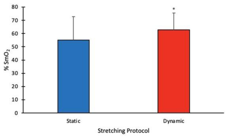
SmO2 values for the end of the initial rest period, immediately following the end of the stretching protocol, and at the end of the final rest are reported in Table 1. DS was significantly higher post-stretch (p = 0.04). The mean SmO2 values are reported in Table 1.
Table 1.
Mean SmO2 values for three key time periods during the stretching protocols.
| Protocol | Initial Rest (%) | Post-Stretch (%) | Final Rest (%) |
|---|---|---|---|
| SS | 59.3 ± 16.0 | 55.1 ± 17.8 | 53.1 ± 16.4 |
| DS | 55.8 ± 12.18 | 62.8 ± 12.6* | 54.0 ± 12.6 |
All values reported as means ± SD;
denotes p < 0.05.
There was a statistically significant two-way interaction between treatment and time (p < 0.05). A post-hoc paired-samples t-test showed significant differences between the two protocols (p < 0.05), with results of the dependent t-test indicating that the muscle oxygen saturation for DS was significantly higher than SS (Figure 1).
Figure 1.
SmO2 for DS was significantly higher than SS protocol (p = 0.04) at the end of stretching. All values are mean ± SD.
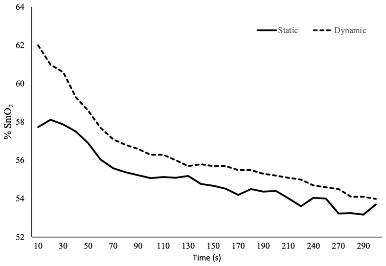
Additionally, the magnitude of change in SmO2 was interpreted as being large in effect size (d = 2.21). Trends in SmO2 were also measured in the five minutes after stretching (Fig. 2). SmO2 data for one participant could not be collected due to equipment malfunction and data from that participant was excluded from the analysis.
Figure 2.
Mean SmO2 levels for both protocols began decreasing immediately after stretching ended. Only DS remained elevated above baseline, and this effect persisted for 2 minutes post-stretch.
DISCUSSION
The primary purpose of this investigation was to determine if DS resulted in higher SmO2 compared to SS as measured in the rectus femoris muscle. SmO2 levels were significantly higher following DS (62.8%) compared with SS (55.1%). Additionally, a large effect size was seen, consistent with SmO2 effect sizes reported in the literature (9). SS did not result in an elevation of SmO2 levels, a finding that is consistent with the work of Kruse & Scheuermann (8), who reported an increase in SmO2 only with static stretches > 60 seconds. SmO2 levels with SS decreased from the initial baseline rest period and did not return to those baseline levels at any point during the SS period or during the second rest. Static stretches > 60 seconds appeared to produce a post-stretch hyperemia (8); an effect not seen with static stretching < 60 seconds. This effect may be due to shorter duration static stretches not occluding local tissues long enough to result in post-stretch hyperemia. The statistically significant increase in SmO2 seen with DS could be due to increasing heart rate and thus cardiac output, augmenting blood flow to the stretched muscle (1, 3). DS has also been shown to as raise core body temperature, which facilitates unloading of oxygen from hemoglobin (1, 3). The SS protocol was likely not at a sufficient intensity to elicit these cardiac responses. These combined effects are likely responsible for the increase in SmO2 seen immediately after the DS, as well as why this protocol took longer to return to baseline with this protocol.
The increased SmO2 levels with DS are likely to increase baseline oxygen consumption in the muscle as well, potentially augmenting exercise performance in later tasks. Bishop proposed that this effect could result in a decreased oxygen deficit, preserving anaerobic capacity and thus improving aerobic endurance performance (3). These effects have been seen in studies examining the effects of DS in trained male runners (15, 16). This suggests that techniques which increase SmO2 levels are a critical consideration when designing a warm-up, particularly for aerobic endurance performance.
Additionally, we sought to determine how long SmO2 levels remained elevated after stretching. In both protocols, SmO2 levels began decreasing as soon as the second rest began; however, after DS, SmO2 levels remained elevated until the 2-minute mark. These results are consistent with the literature, as performance improvements from DS appear to persist only for a few minutes after stretching is complete (1), which further supports the notion that SmO2 levels contribute to enhanced performance after DS. Clinically, this information is important as it would allow for more effective prescription of DS in a warm-up if the intended effect was increased muscle oxygenation. As this is a transient effect persisting only for a few minutes after DS, it is important to begin activity as soon as possible after the warm-up.
While our findings demonstrated a significant increase in SmO2 levels with DS, this study does have limitations. Limited demographic information was collected. Moreover, no data on body composition or cardiorespiratory function was recorded. The sample population in our study included primarily Caucasian college-aged students with males slightly outnumbering females. Though the subjects were active with observably healthy body composition and likely above average cardiorespiratory function, there may have been variance in body composition not accounted for in this study. Previous studies have reported that adipose tissue thickness >15 mm interferes with the NIRS signal (5). Thus, caution should be exercised when interpreting study results, and future studies should include measures of adipose tissue thickness. Additionally, this study only examined the SmO2 levels in one lower extremity muscle; results may differ for other muscle groups.
In conclusion, this study reports several important findings on the effects of static and dynamic stretching on muscle oxygen saturation in the rectus femoris. First, it was found that dynamic stretching significantly increases muscle oxygen saturation compared to static stretching, and second, that this effect only persisted for two minutes after stretching had ceased.
REFERENCES
- 1.Baechle TR, Earle RW. Essentials of strength training and conditioning. Champaign, IL: Human Kinetics; 2008. [Google Scholar]
- 2.Behm DG, Blazevich AJ, Kay AD, McHugh M. Acute effects of muscle stretching on physical performance, range of motion, and injury incidence in healthy active individuals: A systematic review. Appl Physiol Nutr Metab. 2016;41(1):1–11. doi: 10.1139/apnm-2015-0235. [DOI] [PubMed] [Google Scholar]
- 3.Bishop D. Warm up I: Potential mechanisms and the effects of passive warm up on exercise performance. Sports Med. 2003;33(6):439–54. doi: 10.2165/00007256-200333060-00005. [DOI] [PubMed] [Google Scholar]
- 4.Cohen J. Statistical power analysis for the behavioral sciences. London: Routledge; 2013. [Google Scholar]
- 5.Crum EM, O’Connor WJ, Van Loo L, Valckx M, Stannard SR. Validity and reliability of the Moxy oxygen monitor during incremental cycling exercise. Eur J Sport Sci. 2017;17(8):1037–1043. doi: 10.1080/17461391.2017.1330899. [DOI] [PubMed] [Google Scholar]
- 6.Feldmann A, Schmitz R, Erlacher D. Near-infrared spectroscopy-derived muscle oxygen saturation on a 0% to 100% scale: Reliability and validity of the Moxy monitor. J Biomed Opt. 2019;24(11):1–11. doi: 10.1117/1.JBO.24.11.115001. [DOI] [PMC free article] [PubMed] [Google Scholar]
- 7.Faul F, Erdfelder E, Buchner A, Lang AG. Statistical power analyses using G*Power 3.1: Tests for correlation and regression analyses. Behav Res Methods. 2009;41(4):1149–60. doi: 10.3758/BRM.41.4.1149. [DOI] [PubMed] [Google Scholar]
- 8.Kruse NT, Scheuermann BW. Effect of self-administered stretching on NIRS-measured oxygenation dynamics. Clin Physiol Funct Imaging. 2016;36(2):126–33. doi: 10.1111/cpf.12205. [DOI] [PubMed] [Google Scholar]
- 9.McManus CJ, Collison J, Cooper CE. Performance comparison of the MOXY and PortaMon near-infrared spectroscopy muscle oximeters at rest and during exercise. J Biomed Opt. 2018;23(1):1–14. doi: 10.1117/1.JBO.23.1.015007. [DOI] [PubMed] [Google Scholar]
- 10.Navalta JW, Stone WJ, Lyons TS. Ethical issues relating to scientific discovery in exercise science. Int J Exerc Sci. 2019;12(1):1–8. doi: 10.70252/EYCD6235. [DOI] [PMC free article] [PubMed] [Google Scholar]
- 11.Opplert J, Babault N. Acute effects of dynamic stretching on muscle flexibility and performance: An analysis of the current literature. Sports Med. 2018;48(2):299–325. doi: 10.1007/s40279-017-0797-9. [DOI] [PubMed] [Google Scholar]
- 12.Perrey S, Ferrari M. Muscle oximetry in sports science: A systematic review. Sports Med. 2018;48(3):597–616. doi: 10.1007/s40279-017-0820-1. [DOI] [PubMed] [Google Scholar]
- 13.Roberts JM, Wilson K. Effect of stretching duration on active and passive range of motion in the lower extremity. Br J Sports Med. 1999;33(4):259–63. doi: 10.1136/bjsm.33.4.259. [DOI] [PMC free article] [PubMed] [Google Scholar]
- 14.Su H, Chang NJ, Wu WL, Guo LY, Chu IH. Acute effects of foam rolling, static stretching, and dynamic stretching during warm-ups on muscular flexibility and strength in young adults. J Sport Rehabil. 2017;26(6):469–477. doi: 10.1123/jsr.2016-0102. [DOI] [PubMed] [Google Scholar]
- 15.Yamaguchi T, Takizawa K, Shibata K. Acute effect of dynamic stretching on endurance running performance in well-trained male runners. J Strength Cond Res. 2015;29(11):3045–52. doi: 10.1519/JSC.0000000000000969. [DOI] [PubMed] [Google Scholar]
- 16.Zourdos MC, Wilson JM, Sommer BA, Lee SR, Park YM, Henning PC, Panton LB, Kim JS. Effects of dynamic stretching on energy cost and running endurance performance in trained male runners. J Strength Cond Res. 2012;26(2):335–41. doi: 10.1519/JSC.0b013e318225bbae. [DOI] [PubMed] [Google Scholar]


