

Aortography revealed extravasation of contrast media from stentgraft–graft anastomosis.
Central Message.
A pseudoaneurysm due to stentgraft–graft anastomosis failure is a rare but life-threatening complication after 2-stage TAAA repair following a previous frozen elephant trunk technique.
Pseudoaneurysm after aortic surgery is a rare but life-threatening complication. In many cases, pseudoaneurysms after aortic surgery occur at the suture line between the native aorta and prosthetic graft. We report a rare case of a pseudoaneurysm caused by stentgraft–graft anastomosis failure after 2-stage thoracoabdominal aortic repair.
Clinical Summary
An 86-year-old woman was diagnosed with thoracoabdominal aortic aneurysm (TAAA) 3 years ago. We planned a 2-stage surgery for her TAAA. We first performed total arch replacement with a prosthetic graft (J Graft 26 mm; Japan Lifeline) and the frozen elephant trunk (FET) technique (J Graft FROZENIX 27 mm; Japan Lifeline) through median sternotomy. Second, we performed residual TAAA repair (J Graft 24 mm) 4 months after total arch replacement + FET. During residual TAAA repair, the prosthetic graft was anastomosed directly to the stentgraft with a partial aortic wall using a 4-0 monofilament and a peripheral felt strip at the proximal anastomosis (Figure 1, A). A computed tomography (CT) follow-up examination was performed (Figure 1, B) every 6 months or 1 year with no abnormal findings.
Figure 1.

A, The prosthetic graft was anastomosed directly to the stentgraft with the aortic wall using a 4-0 monofilament and a peripheral felt strip. B, Postoperative computed tomography image.
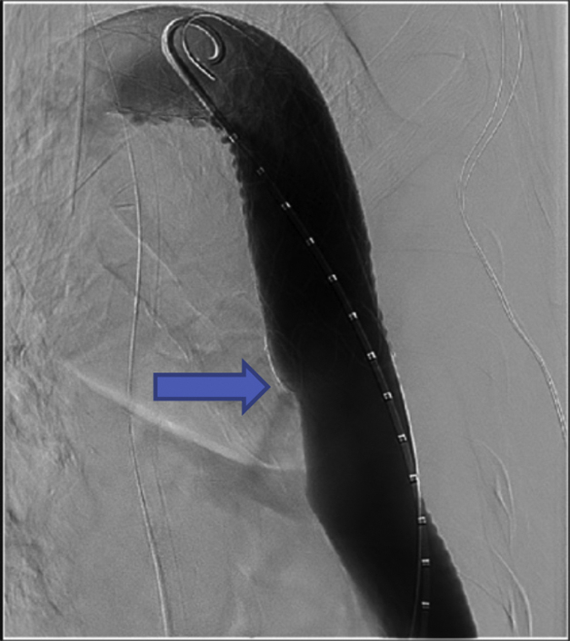
When the patient was referred to our hospital for renal failure and anemia, she had lower back and left chest pain that was persistent for 2 months. Body temperature was 36.2 °C, and the white blood cell count was normal. Contrast-enhanced CT demonstrated a pseudoaneurysm (81 mm in diameter) at the descending aorta (Figure 2, A and B). The stentgraft–graft anastomosis site was at the center of the pseudoaneurysm (Figure 2, A and C). We suspected stentgraft–graft anastomosis failure and impending pseudoaneurysm rupture.
Figure 2.

A-C, Preoperative contrast-enhanced computed tomography showed the presence of a pseudoaneurysm and extravasation (arrow) at the anastomosis site. D, Aortography before stentgraft implantation revealed extravasation (arrow) at the anastomosis site. E, Stent graft was implanted at the anastomosis site. F, Aortography after stentgraft implantation showed no residual extravasation.
We performed emergency thoracic endovascular aortic repair for the impending pseudoaneurysm rupture. Intraoperative aortography revealed extravasation from the stentgraft–graft anastomosis (Figure 2, D, and Video 1). The stentgraft–graft anastomosis site was covered using a GORE TAG Conformable Thoracic Stent Graft with ACTIVE CONTROL System (W. L. Gore & Associates, Inc) (Figure 2, E). The final aortography scan revealed no residual extravasation (Figure 2, F).
No adverse events occurred during or after the surgery. Plain CT performed on postoperative day (POD) 4 revealed no pseudoaneurysm enlargement. Magnetic resonance angiography performed on POD 6 revealed no blood flow in the pseudoaneurysm. Postoperative contrast-enhanced CT was not performed, considering her medical history suggesting chronic kidney disease (estimated glomerular filtration rate: 10 mL/min/1.73 m2). The patient was transferred to a rehabilitation hospital on POD 10.
Discussion
Pseudoaneurysm due to stentgraft–graft anastomosis failure is a rare but life-threatening complication after aortic surgery.1 To our knowledge, this is the first report of a case of pseudoaneurysm arising due to a stentgraft–graft anastomosis failure.
Recently, 2-stage TAAA repair after FET has emerged as a promising treatment option for TAAA. In second-stage surgery, the proximal graft is generally anastomosed directly to the end of the stentgraft. Folkmann and colleagues2 performed this technique and reported satisfactory outcomes. In our case, the prosthetic graft was anastomosed directly to the stentgraft using a 4-0 monofilament, and anastomosis failure occurred approximately 3 years postoperatively.
The cause of the pseudoaneurysm remains unclear. A possible mechanism for the pseudoaneurysm is the progressive thinning of the edematous and swollen aortic wall over the years, causing the sutures to loosen and leading to pseudoaneurysm formation.3 This change occurs between the native aorta and a prosthetic graft, but not between 2 prosthetic grafts, as in our case.
Another possible mechanism is the deterioration of the suture string, which may cause anastomotic pseudoaneurysm in the long-term postoperatively.4,5 In the present case, the prosthetic graft was anastomosed directly to stentgraft using a monofilament suture, ie, the monofilament suture was constantly in contact with stent strut. Moreover, the stentgraft was larger than the prosthetic graft (FROZENIX 27 mm vs J Graft 24 mm) and has self-expandability. These conditions (the friction between suture string and stentgraft and the stretching force of the suture line) may have strained the suture string and led to premature deterioration of string and to anastomosis failure. Macroscopic and histopathologic evaluations were not performed in this case.
To establish safer stentgraft–graft anastomosis, the “felt sandwich technique” and use of 2-0 or 3-0 braid suture may be useful to adjust size and protect the suture from friction. In conclusion, pseudoaneurysm due to stentgraft–graft anastomosis failure is a rare but life-threatening complication after 2-stage TAAA repair after FET. Regular close follow-up examination is required in such cases.
This case report was approved by the Ethical Committee in Kokura Memorial Hospital (ID: 22041101, date April 11,2022). Written informed consent was obtained from the patients for publication of this case report.
Footnotes
Disclosures: The authors reported no conflicts of interest.
The Journal policy requires editors and reviewers to disclose conflicts of interest and to decline handling or reviewing manuscripts for which they may have a conflict of interest. The editors and reviewers of this article have no conflicts of interest.
Supplementary Data
Aortography revealed extravasation of contrast media from stentgraft–graft anastomosis. Video available at: https://www.jtcvs.org/article/S2666-2507(22)00348-0/fulltext.
References
- 1.Gomibuchi T., Takano T., Wada Y., Terasaki T., Seto T., Fukui D. Pseudoaneurysm of graft-graft anastomosis of a hand-sewn branched graft: a case report. J Cardiothorac Surg. 2015;10:152. doi: 10.1186/s13019-015-0356-0. [DOI] [PMC free article] [PubMed] [Google Scholar]
- 2.Folkmann S., Weiss G., Pisarik H., Czerny M., Grabenwoger M. Thoracoabdominal aortic aneurysm repair after frozen elephant trunk procedure. Eur J Cardiothorac Surg. 2015;47:115–119. doi: 10.1093/ejcts/ezu096. [DOI] [PubMed] [Google Scholar]
- 3.Tanaka K., Makuuchi H., Naruse Y., Kobayashi T., Hayashi I., Takayama T., et al. False aneurysm due to suture loosening after aortic arch replacement. Asian Cardiovasc Thorac Ann. 2002;10:346–348. doi: 10.1177/021849230201000417. [DOI] [PubMed] [Google Scholar]
- 4.Kabasawa M., Takahara Y., Mogi K., Hatakeyama M. A case of surgical treatment for pseudoaneurysm 19 years after aortic root replacement. Jpn J Cardiovasc Surg. 2008;37:268–271. [Google Scholar]
- 5.Gaspar M.R., Movius H.J., Rosental J.J., Bell D.D., Lemire G.G., Odou M. Prolene sutures are not a significant factor in anastomotic false aneurysms. Am J Surg. 1983;146:216–219. doi: 10.1016/0002-9610(83)90376-8. [DOI] [PubMed] [Google Scholar]
Associated Data
This section collects any data citations, data availability statements, or supplementary materials included in this article.
Supplementary Materials
Aortography revealed extravasation of contrast media from stentgraft–graft anastomosis. Video available at: https://www.jtcvs.org/article/S2666-2507(22)00348-0/fulltext.