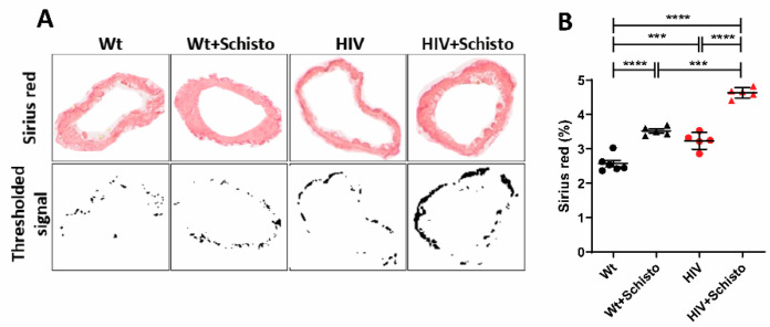
Figure 4.
HIV mice show augmented pulmonary perivascular fibrosis with marked exacerbation after exposure to schistosome eggs. (A) Representative images of pulmonary vessels stained with Sirius red (upper panels). Stained vessel areas are shown as thresholded signal (botton panels). (B) Quantification of Sirius red staining as percent Sirius red-positive area fraction (as shown in (A), bottom) in the vessel area analyzed. Mice and vessels analyzed per group were: Wt (6, 77), Wt+Schisto (5, 83), HIV (5, 53), and HIV+Schisto (5, 75). Results are expressed as mean ± SEM. *** p < 0.001 and **** p < 0.0001 as determined by one-way ANOVA analysis followed by Tukey’s post-hoc test.

