Abstract
Amid the coronavirus disease 2019 (COVID-19) pandemic, numerous publications of imaging findings in children have surfaced in a very short time. Publications discuss populations of overlapping age groups and describe different imaging patterns. We aim to present an overview of the quantity and type of literature available regarding COVID-19 chest imaging findings in children according to a 2020 publication timeline. We conducted a systematic review using the Preferred Reporting Items for Systematic Reviews and Meta-Analyses (PRISMA) guideline. We searched terminology related to COVID-19, chest, children and imaging modalities in PubMed and Embase. The included papers were published online in 2020 and described imaging findings specific to children and reported five or more cases. Two researchers reviewed each abstract to determine inclusion or exclusion, and a radiologist reconciled any disagreements. Then we reviewed full articles for the main analysis. Eligible study designs included original articles, case series (≥5 cases), systematic reviews and meta-analyses. We excluded non-English manuscripts, retracted articles, and those without available full text. The remaining articles were distributed to four pediatric radiologists (on the Society for Pediatric Radiology Thoracic Committee), who summarized chest imaging findings. Eighty-two articles were included in the final analysis — 28% in radiology journals and 71% in non-radiology journals; 71% contained original data and 29% were review-style papers. There was a disproportionate contribution of review-style papers in April (55%), considering the paucity of preceding publications with original data in March (5 papers). June had the highest number of publications (n=14), followed by April (n=11) and July (n=11). Most (52%) original papers were from China and most individual pediatric imaging descriptions were from China (57%), while the majority of review papers (83%) were international. Imaging descriptions were available for 2,199 children (1,678 CT descriptions and 780 chest radiography descriptions). Findings included a 25% normal CT scan reports vs. 40% normal chest radiography reports. Ground-glass opacification was the most common CT finding (33%) and was reported in only a minority of chest radiographs (9%). A significant amount of information on pediatric COVID-19 chest imaging has become rapidly available over a short period. Most publications in 2020 were original articles, but they were published more often in non-radiology journals. A disproportionate number of review articles were published early on and were based on little original pediatric imaging data. CT scan reports, which represent the standard, outnumbered radiographic reports and indicated that ground-glass opacification is the main imaging finding and that only a quarter of scans are normal in children with COVID-19.
Keywords: Chest, Children, Computed tomography, Coronavirus disease 2019, Lung, Pneumonia, Publications, Radiography
Introduction
The coronavirus disease 2019 (COVID-19) pandemic has impacted the world since early 2020. This global crisis took the world by surprise, including clinicians and scientists. In an effort to further understand the new disease, researchers across the globe scaled efforts from early on to publish what was witnessed and researched. From the first COVID-19-related publication in December 2019 [1] to September 2021, 175,017 COVID-19-related articles were added to the PubMed database [2] alone. Early in the pandemic, diagnostic imaging descriptions for adults became available in indexed databases as early as December 2019 [1] and pediatric imaging descriptions became available in January 2020 [3]. Since then, numerous publications have surfaced, with variable imaging descriptions for adults and children alike.
The majority of review articles and recommendations on pediatric imaging published in early 2020 were based on small patient populations, mostly consisting of case studies and short clinical reports. While there is now a wealth of information on pediatric diagnostic imaging of COVID-19, descriptions of imaging were initially authored by non-radiologist clinical specialists using variable terminology [4]. In fact, the majority of imaging descriptions of pediatric patients with COVID-19 have been published in non-radiology journals rather than radiology-focused journals.
This paper provides an overview of the publication timeline of imaging findings in children with COVID-19 to highlight lessons from the pandemic experience in 2020, especially regarding the need for a rapid radiologist response to meet the need for information. We performed a systematic review of all COVID-19 articles with radiologic imaging descriptions of pediatric patients published in 2020.
Materials and methods
Literature search and screening
We conducted this systematic review in compliance with the Preferred Reporting Items for Systematic Reviews and Meta-Analyses (PRISMA) guideline. We used appropriate terminology in the search to include terms related to COVID-19, chest, children and imaging modalities (computed tomography [CT], chest X-ray, radiography, ultrasonography [US], magnetic resonance [MR]) for the period of Jan. 1–Dec. 31, 2020, using PubMed and Embase.
Articles were screened by title, abstract and full text. We included studies if they were published online in the year 2020, and for those containing original data, only if they described imaging findings of five or more pediatric COVID-19 cases. We included original articles, case series, systematic reviews and meta-analyses. We excluded non-English manuscripts, retracted articles and those without available full text.
Publications analysis
Two researchers (M.M.-S. and K.I.R.-S., with 6 years and 3 years of experience, respectively) reviewed each abstract using the software Covidence (Veritas Health Innovation, Melbourne, Australia), a web-based platform that streamlines the production of systematic reviews and other research reviews. Conflicts in inclusion decision were solved by a senior radiologist (S.A., with 24 years of experience in pediatric radiology), and all selected articles were included for full-text review. Full-text articles were randomly assigned and divided among four subspecialist pediatric radiologists (S.A., J.B.R., D.S. and K.S.S., with 24 years, 4 years, 7 years and 22 years of experience in pediatric radiology, respectively) who are members of the Thoracic Committee of the Society for Pediatric Radiology (SPR) and were commissioned by the committee leadership to support this project. Each reviewer independently evaluated and summarized COVID-19 imaging findings of CT, chest radiography, US and MR.
Data management and analysis
Summaries of each article were collected and managed using Research Electronic Data Capture (REDCap) [5, 6]. Data collection included: the month when the final publication was available online; type of article (original data, case series, review, metanalysis, guideline); country of origin of the paper or country of origin of the patients described; patient age range; and, for papers with original data, a description of each imaging modality, number of patients per modality, as well as decision and reason for inclusion or exclusion. Included articles were classified in two broad categories: primary data (case series and original data), and reviews and guidelines (review articles or systematic reviews, meta-analyses and consensus recommendations). Publications in the latter categories were organized according to month of publication and proportional contribution for each month. We performed descriptive analysis of the included articles and figures using the software Stata 17 (StataCorp, College Station, TX) and JMP 15.0.0 (SAS Institute, Cary, NC).
Results
Literature findings
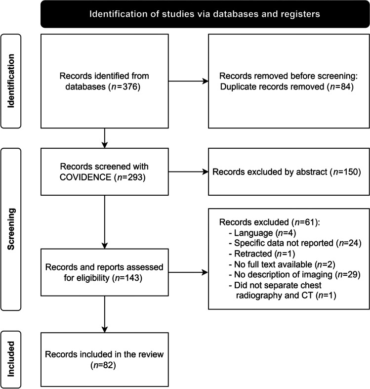
The initial search yielded 376 records from PubMed and Embase. Of these, we included 292 for title and abstract screening after removing duplicates. At least one of the four pediatric radiologists reviewed full text for 143 publications. After this review, a total of 82 articles remained for the final analysis. Details on screening and exclusion criteria are shown in the PRISMA table (Fig. 1).
Fig. 1.
Preferred Reporting Items for Systematic Reviews and Meta-Analyses (PRISMA) flow diagram of the inclusion and exclusion of eligible studies for systematic review. CXR chest radiograph, CT computed tomography. Records screened with COVIDENCE include an additional publication from the FLEISCHNER Society that was not retrieved from initial database search
Characteristics of publications
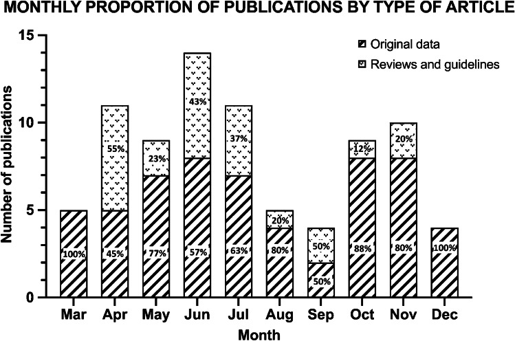
The 82 included articles containing imaging descriptions of children with COVID-19 were available online between March 2020 and December 2020 (10 months). Fifty-eight of these papers had primary data (70.7%) and 24 were reviews and guidelines (29.3%). The proportion of original papers versus reviews is detailed in Fig. 2. Overall, 45 (54.9%) were original articles (with >10 pediatric cases), 17 (20.7%) review articles, 13 (15.9%) case series (5–10 cases), 5 (6.1%) meta-analyses and 2 (2.4%) consensus recommendations.
Fig. 2.
Monthly proportion of publications by type of article for the year 2020 (total=82). Number and percentage of articles with original data (n=58) and reviews and guidelines (n=24) are depicted
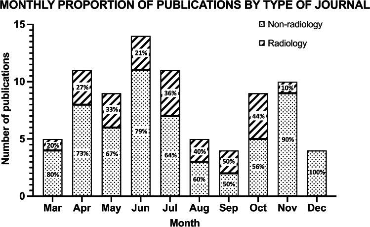
Of the 82 publications, 28% (23) were published in radiology journals and 72% (59) in non-radiology journals. Of note, the proportion of publications by journals specialized in radiology versus non-radiology varied each month but papers were consistently predominantly from non-radiology journals (Fig. 3).
Fig. 3.
Monthly proportion of publications by type of journal for the year 2020 (total=82). Number and percentage of radiology (n=23) vs. non-radiology publications (n=59) are depicted
Analysis by month
The earliest eligible articles were published in March, and those containing original data described imaging from a cumulative total of 65 patients with COVID-19 in 5 publications. Of these, one article was published in a radiology journal and the other four in non-radiology journals. In April there were six review articles and five original data publications, all in non-radiology journals. Proportionally, the number of publications containing original data versus review-style papers varied by month as summarized in Fig. 2.
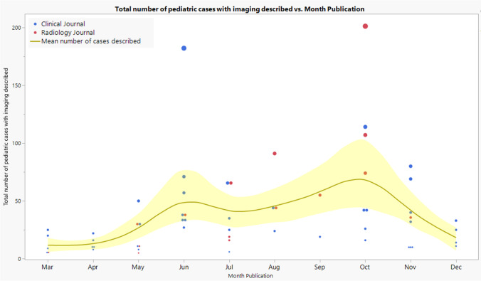
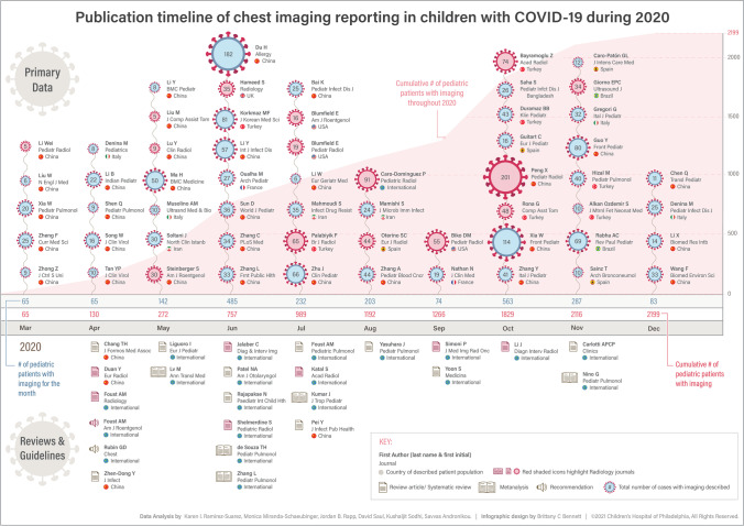
The publication timeline of the 82 included works is summarized by month in Fig. 4, where the 58 papers with original data are presented distinguishing non-radiology and radiology journals by color coding.
Fig. 4.
Graph reflects the mean number (uncertainty levels) of pediatric cases with imaging descriptions from original articles per month (yellow) from the year 2020, with point size reflecting each publication’s sample size. Red points reflect publications in radiology journals and blue points reflect publications in non-radiology journals. A total of 2,199 cases were described
June had the highest number of total publications with 14 articles (17.1%), followed by April with 11 (13.4%), July with 10 (12.2%) and November with 10 (12.2%). Most review articles and recommendations were published between April and June (14/24, 58%): 6 reviews were published in April (42.8%), 2 in May (14.3%) and 6 in June (42.9%). There were fewer reviews from radiology journals (5/14, 35.7%) than from non-radiology journals (9/14, 64.3%) from March to June.
The number of children with reported chest imaging findings of COVID-19 varied each month with no particular trend: June (485 patients) and October (563 patients) had the highest number of COVID-19 pediatric patients for whom imaging was described.
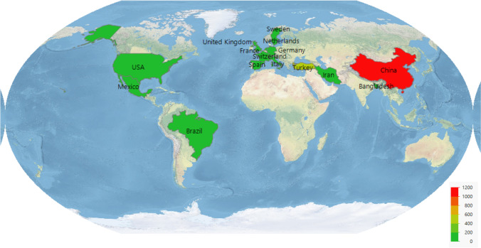
Overall, original data publications (n=58) originated from 15 countries. The country with the most publications was China with 30 (51.7%), followed by Turkey with 7 (12%) and Italy with 4 (6.9%). More than half of the children with reported COVID-19 imaging findings were from China, 1,203 (54.7%) (Fig. 5). The vast majority of reviews were international contributions (20/24, 83.3%), followed by reviews exclusively from China (4/24, 16.7%).
Fig. 5.
Diagram of globe shows cumulative number of pediatric patients with coronavirus disease 2019 (COVID-19) with imaging descriptions from all data-containing publications per country from 2020, displayed by color-graded frequencies. China had the largest number of patients (1,203) with imaging description (red), followed by Turkey (361) (light green), Spain (112), Brazil (103), Italy (103), USA (94), Iran (96), France (54), United Kingdom (36), Bangladesh (26), Germany (3), Switzerland (3), Netherlands (2), Sweden (2) and Mexico (1) (all dark green)
Imaging descriptions
Imaging descriptions were available for 2,199 children with COVID-19 across the 58 articles with original data. While most articles described specific imaging per patient, a considerable number presented generalized summarized descriptions for a group of patients. In 47 of these original data publications (81%), CT imaging was specifically described for each patient, 24 publications (41.4%) described chest radiography imaging findings, 9 articles (15.5%) lung US, and 1 article (1.7%) chest findings on MR.
Commonly reported features for CT and chest radiography can be found in Table 1. Overall, there were more than double the original pediatric imaging descriptions of COVID-19 for CT scans (1,678) than for chest radiography (780). Only one-quarter of CTs were reported as normal (24.7%), whereas cumulative chest radiography data indicated that 40% of these were normal. Ground-glass opacities were the most common finding on CT (32.5%) followed by consolidations (16.0%) and interstitial pattern/opacities (2.2%). CT reported higher frequencies of both ground-glass opacities and consolidation than chest radiographs. The main CT finding (ground-glass opacities) is illustrated in Fig. 6. The most common finding on chest radiography was interstitial pattern/opacities (11.8%), followed by consolidations (10.4%) and peribronchial thickening (9.1%). Ground-glass opacities were only reported in 8.6% of chest radiographs.
Table 1.
Imaging parameters specified in original articles and case series (total children with imaging descriptions is 2,199)
| Parameters | CT (n=1,678) | Radiographs (n=780) | ||
|---|---|---|---|---|
| Normal | 414 | 24.7% | 313 | 40.1% |
| Ground-glass opacities | 546 | 32.5% | 67 | 8.6% |
| Consolidations | 268 | 16.0% | 81 | 10.4% |
| Peribronchial thickening | 16 | 1.0% | 71 | 9.1% |
| Interstitial pattern/opacities | 37 | 2.2% | 92 | 11.8% |
| Pleural effusion | 26 | 1.6% | 36 | 4.6% |
| Atelectasis | 3 | 0.2% | 9 | 1.2% |
| Predominant distribution | ||||
| Unilateral | 5 | 0.3% | 2 | 0.3% |
| Bilateral | 10 | 0.6% | 7 | 0.9% |
| Reverse halo sign | 3 | 0.2% | ||
| Bilateral multifocal | 40 | 2.4% | ||
| Apical to basal gradient | 53 | 3.2% | ||
| Halo sign | 55 | 3.3% | ||
| Alveolar opacities | 35 | 4.5% | ||
| Hyperinflation | 2 | 0.3% | ||
Negative findings were not consistently reported
Fig. 6.
Schematic of three selected slices of the chest on CT imaging demonstrate the most common finding in children with coronavirus disease 2019 (COVID-19), which is ground-glass opacification. A gradient of increasing severity is demonstrated from apical to basal slices, which is not as notable in children as it is in adults
Terminology to describe imaging on CT and chest radiography varied across papers. We encountered the use of non-standard terminology that is not included in the Fleischner Society guidelines in multiple publications [7] (Table 2).
Table 2.
Imaging CT and chest radiography parameters with non-standardized terminology, according to Fleischner Society [7] (total imaging descriptions, n=2,199)
| CT | Chest radiography |
|---|---|
| Blurred bronchovascular bundle | Pulmonary abnormalities |
| Speckled shadow | Parenchymal breakdown |
| Fine mesh shadow | Increased/blurred texture of lung |
| Focal vascular engorgement | |
| Vascular enhancement sign | |
| Vasodilatation | |
| White lung |
Imaging with US was described for 178 children and included: B-lines 62 (34.8%), consolidations 30 (16.9%), pleural irregularities 13 (7.3%), white lung 2 (1.1%) and pleural effusion 2 (1.1%). Normal findings were described for 73 (41%) children. In one child, the US showed an increased B-line in the right lower lobe despite normal chest CT findings. Only one original publication described MRI findings, where peripheral nodular opacities were seen in one case [8].
Discussion
The COVID-19 pandemic has highlighted both weaknesses and strengths in our health systems and our ability or inability to respond rapidly to global health threats. Imaging played a significant role early in the pandemic where CT scans were used for diagnostic purposes in adults [9, 10], before adequate and abundant laboratory testing became available. Scientific information on imaging of children with suspected COVID-19 was limited early on in the year 2020 (no papers with five or more cases and no reviews in January and February 2020) and the role of imaging for diagnosis in children was unclear. The source of information of some reviews and guidelines early in 2020 was largely based on anecdotal experience, adult data or unedited submissions or proofs of original data. The appropriateness of publishing guidelines and recommendation for children under such conditions, when advice is widely sought, remains the subject of some debate [11].
We have interrogated the results of our literature review of the year 2020, specifically in relation to the publication timeline, to establish what lessons can be learned from the publication of imaging findings in children with COVID-19 during the global pandemic, and to make recommendations regarding a scientific publication response for any future global health threats.
A publication timeline
Included papers spanned the period March 2020 to December 2020. These reflect a dynamic shift from a higher proportion of review papers and small series early in 2020 to a large number of original data papers later in the year with a tapering off of review articles and guidelines. There was a disproportionate contribution of review-style papers in April, considering the paucity of preceding final publications with original data. Preprint servers including publication of unedited submissions or proofs were not considered for our review; however, an immense number of COVID-19 studies were first (and in many cases only) published on a preprint server, and these might be the data source of many of the early reviews. Also, the turnaround time for publication of review papers might have been faster than the turnaround time for publication of papers with original data, which might also contribute to this disproportion.
An overview of the timeline of events summarized in Fig. 7 suggests that as more original data were being published through large patient cohorts over the year, fewer reviews and guidelines were being produced, despite mounting evidence that early reviews misrepresented the findings in children.
Fig. 7.
Infographic demonstrates the publication timeline of papers with chest imaging descriptions in children with coronavirus disease 2019 (COVID-19) over the year 2020. The timeline extends from March 2020 on the left to December 2020 on the right. The upper half of the page summarizes papers with original patient data reflected by month and by sample size, with larger COVID-19 organism schematics representing larger samples and red reflecting radiology journals vs. blue representing non-radiology journals. The middle horizontal bars reflect monthly total as well as cumulative patient numbers reported. The lower half of the page summarizes review articles and guidelines with the same color coding
Original data papers vs. reviews
A number of review articles regarding COVID-19 imaging features in children [12–15], including an international expert consensus on chest imaging [16], were published as early as April 2020 despite the limited original data for children at the time. Studies with larger samples of pediatric patients only started being published in May 2020 [17] and over the year, the original imaging descriptions of more than 2,199 children with COVID-19 were published in the scientific literature.
Our review and timeline plot show that in April 2020, the imaging of 65 pediatric cases had been reported in final publications with cohorts of greater than 5 children, and 4 review articles were already available in that month. As an example, the information regarding the imaging findings of COVID-19 in children reported in one guideline published in April was sourced from case reports, small case series, adult data and anecdotal experiences of authors [16].
Source of publications regarding journal type
Closer scrutiny of original dates of submission to the journal Pediatric Radiology shows that although the editors tried to fast-track COVID-19-related articles and authors were quick to respond with revisions, there was still a substantive time gap from the time of submission to availability of articles online.
Our data also indicate that the majority of papers published on the pediatric imaging features of COVID-19 were in non-radiology journals. The radiology journal publication distribution over the year 2020 also points to a relatively slow publication response by the radiology, and especially the pediatric radiology, community (Figs. 3 and 4).
Source of publications regarding country of origin over the timeline
It is concerning, however, that guidelines and review papers early in the pandemic leaned on adult data and anecdotal evidence rather than original data originating in China. The previous section on the timelines for publishing in radiology journals as well as the upcoming section on terminology might give some clues as to why this occurred. Figure 5 indicates overall publications by country where patients with imaging descriptions were seen. Not surprisingly, the data are mainly derived from China in 2020 because that was where the pandemic began.
Caro-Dominguez et al. [4] are to be commended for producing international cohort data by soliciting cases internationally through a societal call-out and for reporting these centrally for consistency.
Diagnostic imaging findings in children with coronavirus disease 2019
Coronavirus disease 2019 does not impact adults and children equally. There is not only a discrepancy in the severity of the clinical presentation, but also in imaging findings between adults and children [18, 19]. The imaging findings in children seem less severe than those described in adults.
Our systematic review showed there is more cumulative imaging data in children with COVID-19 from CT (1,678) than from chest radiography (780). Regardless of whether CT is appropriate for pediatric practice relating to COVID-19, it should be accepted that data from CT are more accurate and reliable than data from chest radiography interpretations. If one accepts this, then many findings reported on chest radiography are likely to be invalid. It is, however, possible that there is a selection bias in that only those with more severe disease presentation are referred for CT. Despite this unavoidable selection bias that is present from retrospectively gathered data, the scenario early on in the COVID-19 pandemic is that CT was being used as a diagnostic tool in adults and children alike because of the lack of available laboratory testing options. The reported rate of normal exams varies widely for chest radiographs, e.g., the paper by Biko and colleagues [8] in Pediatric Radiology reported 50% of chest radiographs were normal despite a 75% incidence of co-morbidities in their cohort, while the paper by Caro-Dominguez and colleagues [4] reported that only 10% of chest radiograph were normal. This is attributed by the Caro-Dominguez group to their recognition of para-hilar peribronchial thickening, which is not described in patients in the Biko et al. [8] paper. However, the general CT data do not support the findings of Caro-Dominguez et al. [4] and neither does the lack of air-trapping reported in any significant portion of radiographs (0.25%) by any group (Table 1).
The more important imaging finding is the presence of ground-glass opacities in a third of children on CT. Unsurprisingly, chest radiography showed a much lower frequency of ground-glass opacities (8.6%) because radiographs are less useful for detecting these. Peribronchial thickening was reported in 9.1% of chest radiographs and 1.0% on CTs. The reporting of an interstitial pattern in approximately 12% of chest radiographs is also not borne out on CT scans (2%), the gold standard for detecting interstitial disease.
In fact, CT showed that signs other than ground-glass opacities and consolidation occurred in less than 5% of children. Several commonly reported imaging findings in adult CTs were rare in our pediatric data. Examples of these are the reverse halo sign (0.2%), the halo sign (3.3%), apical-to-basal gradient of ground-glass opacities (3.2%). The associated hyperinflation expected for viral infections on chest radiography was only reported in 0.3% of children. It is possible that the low percentages for unilateral, bilateral, reverse halo sign, bilateral multifocal, apical-to-basal gradient and halo sign reflect reporting practices for the presence or absence of these findings.
Terminology
The reporting of chest imaging findings using non-standard terminology is considered a major limitation for collating data. This was highlighted by Caro-Dominguez et al. [4], who reported findings prior to their submission date of April 2020 and attributed the use of non-standard terminology to clinicians putting out early imaging reports without radiologist input. This is more pertinent for the COVID-19 imaging data from the first half of the timeline. It is expected that papers coming from radiology journals undergo more stringent peer review of radiologic aspects compared to papers published in non-radiology journals. It is also likely that papers from non-English-first-language countries have a disadvantage in this regard. Radiologists are not immune to terminology differences, and radiologist groups use several different terms that reflect the same or overlapping pathology (e.g., alveolar opacity vs. consolidation).
Radiologist interpretational differences
Other important concerns when reviewing this literature include an apparent philosophical difference in the interpretation of chest radiography findings, the reliance on chest radiography findings (over CT scan) and personal perception of subjective skills.
A comparison of papers written by radiologists Biko et al. [8] and Caro-Dominguez et al. [4], both published in the journal Pediatric Radiology, reflects:
Likely philosophical differences in the use of term ground-glass opacity on chest radiography, where Caro-Dominguez et al. [4] used the term ground-glass opacity for chest radiography and Biko et al. [8] did not use the term, likely reserving it for CT descriptions.
Caro-Dominguez et al. [4] chose to report para-hilar peribronchial cuffing in the absence of air-trapping whereas Biko et al. [8] chose to report only on the lack of air-trapping. Interestingly, the former group attributed the high percentage of normal chest radiography in other studies to chest radiography readers being inexperienced in interpreting para-hilar peribronchial cuffing.
The collated CT data over 2020 (1,678 CT scans vs. 780 chest radiographs) becomes relevant in deciding which of the above author groups is more likely correct, even though there may well be selection bias in who underwent CT scanning. Only one-quarter of CTs were reported as normal (24.7%), while 40% of chest radiographs were reported as normal, and ground-glass opacities were the most common finding on CT (32.5%), followed by consolidations (16.0%). The reported high frequency of para-hilar peribronchial cuffing on chest radiography by Caro-Dominguez et al. [4] was not borne out in the CT data, which demonstrated very few cases with this finding (1.0%), despite being the gold standard for detecting it.
Challenges in collating data
Collating data from multiple sources for metanalysis or review, when terminology and interpretation vary, is challenging.
Our work highlights the importance of centralized data collection through societies and shows that continued collation of high-quality radiologic data with consensus terminology is necessary to formulate expert opinion consensus guidelines for pediatric chest imaging of COVID-19.
Recommendations for imaging children with COVID-19 from both early and late reports are largely similar: chest imaging is necessary neither as a diagnostic tool in children with suspected COVID-19, nor for clinical management. However, CT is indicated for children with comorbidities or suspected complications to aid in decision-making. Chest radiography could be used in symptomatic children, but considering that ground-glass opacity is the main finding and it is not easily detected with chest radiography, CT should be the preferred imaging technique unless transportation of the child to the scanner is difficult or where access to CT imaging is limited, such as in low-resource environments.
Lessons learned
Turnaround of radiology journals was too long. We would encourage radiology journals to fast-track related articles during a pandemic while maintaining review quality.
Variable imaging findings/terminology not included in the Fleischner Society guidelines emerged primarily from non-radiology journals. Standardization of radiology terminology is key for data collection and interpretation.
Pediatric radiologists must reflect on the limitations of chest radiography rather than assume we can interpret these better than others.
The pediatric radiology community needs to mobilize quickly to collect data in future pandemics by centralizing through societies, as achieved by the Caro-Dominguez group [4].
Pediatric radiologists must be involved with our clinical colleagues to publish early and also influence the accuracy of imaging reports.
More than half of the reported information on pediatric imaging of COVID-19 is based on articles published in China. We need a mechanism to bring data from China into the mainstream, such as translation services within journals with the support of the subspecialty. Partnerships with non-English-speaking countries need to be fostered through our global societies. In addition to improving access there needs to be some assessment of quality.
Early reviews and guidelines need to be more transparent about the source of the information: anecdotal, adult- vs. pediatric-based, sources of data. Journals need to apply strict review criteria of early reports that do not contain original data because these might be biased by studies with small samples and findings in adult populations.
Given the wide availability of CT findings in children with COVID-19, these should be given appropriate weight, being the gold standard technique for imaging the chest, and these should inform on whether chest radiography findings are valid.
Conclusion
A large quantity of information on pediatric COVID-19 chest imaging became rapidly available over a short period of time in the year 2020. While most publications in 2020 were original articles in non-radiology journals, the early review and guideline publications available from April 2020 were not based on enough pediatric data. Broadly, Pediatric Radiology and other radiology journals published later in the pandemic than non-radiology journals, which led to reliance on papers from non-radiology journals using non-standardized terminology to inform clinical management. The pediatric radiology community and leading publishers need to be able to set global data collection in motion more quickly through societies early in any future pandemic and influence our journals through editorial positions to fast-track important material.
Last, pediatric radiologists need to work on standardizing terminology and reporting by creating appropriateness criteria for reporting chest radiographs based on accuracy and reliability of data, where possible, using a gold standard such as CT.
Acknowledgments
Special thanks to Brittany Bennett, MA, for her skillful work and dedication to illustrate this article.
Declarations
Conflicts of interest
None
Footnotes
Publisher’s note
Springer Nature remains neutral with regard to jurisdictional claims in published maps and institutional affiliations.
References
- 1.Zhu N, Zhang D, Wang W, et al. A novel coronavirus from patients with pneumonia in China, 2019. N Engl J Med. 2020;382:727–733. doi: 10.1056/NEJMoa2001017. [DOI] [PMC free article] [PubMed] [Google Scholar]
- 2.No authors listed (2020) Public health emergency COVID-19 initiative. National Institutes of Health National Library of Medicine. https://www.ncbi.nlm.nih.gov/pmc/about/covid-19/. Accessed 22 Jun 2021
- 3.Chan JF-W, Yuan S, Kok K-H, et al. A familial cluster of pneumonia associated with the 2019 novel coronavirus indicating person-to-person transmission: a study of a family cluster. Lancet. 2020;395:514–523. doi: 10.1016/S0140-6736(20)30154-9. [DOI] [PMC free article] [PubMed] [Google Scholar]
- 4.Caro-Dominguez P, Shelmerdine SC, Toso S, et al. Thoracic imaging of coronavirus disease 2019 (COVID-19) in children: a series of 91 cases. Pediatr Radiol. 2020;50:1354–1368. doi: 10.1007/s00247-020-04747-5. [DOI] [PMC free article] [PubMed] [Google Scholar]
- 5.Harris PA, Taylor R, Thielke R, et al. Research electronic data capture (REDCap) — a metadata-driven methodology and workflow process for providing translational research informatics support. J Biomed Inform. 2009;42:377–381. doi: 10.1016/j.jbi.2008.08.010. [DOI] [PMC free article] [PubMed] [Google Scholar]
- 6.Harris PA, Taylor R, Minor BL, et al. The REDCap consortium: building an international community of software platform partners. J Biomed Inform. 2019;95:103208. doi: 10.1016/j.jbi.2019.103208. [DOI] [PMC free article] [PubMed] [Google Scholar]
- 7.Hansell DM, Bankier AA, MacMahon H, et al. Fleischner Society: glossary of terms for thoracic imaging. Radiology. 2008;246:697–722. doi: 10.1148/radiol.2462070712. [DOI] [PubMed] [Google Scholar]
- 8.Biko DM, Ramirez-Suarez KI, Barrera CA, et al. Imaging of children with COVID-19: experience from a tertiary children’s hospital in the United States. Pediatr Radiol. 2021;51:239–247. doi: 10.1007/s00247-020-04830-x. [DOI] [PMC free article] [PubMed] [Google Scholar]
- 9.Kovács A, Palásti P, Veréb D, et al. The sensitivity and specificity of chest CT in the diagnosis of COVID-19. Eur Radiol. 2021;31:2819–2824. doi: 10.1007/s00330-020-07347-x. [DOI] [PMC free article] [PubMed] [Google Scholar]
- 10.Schalekamp S, Bleeker-Rovers CP, Beenen LFM, et al. Chest CT in the emergency department for diagnosis of COVID-19 pneumonia: Dutch experience. Radiology. 2021;298:E98–E106. doi: 10.1148/radiol.2020203465. [DOI] [PMC free article] [PubMed] [Google Scholar]
- 11.Desoky SM, Andronikou S, Brody AS, Hirsch FW (2020) Re: “international expert consensus statement on chest imaging in pediatric COVID-19 patient management: imaging findings, imaging study reporting and imaging study recommendations.” Radiol Cardiothorac Imaging 2:e200305 [DOI] [PMC free article] [PubMed]
- 12.Duan Y-N, Zhu Y-Q, Tang L-L, Qin J. CT features of novel coronavirus pneumonia (COVID-19) in children. Eur Radiol. 2020;30:4427–4433. doi: 10.1007/s00330-020-06860-3. [DOI] [PMC free article] [PubMed] [Google Scholar]
- 13.Yasuhara J, Kuno T, Takagi H, Sumitomo N. Clinical characteristics of COVID-19 in children: a systematic review. Pediatr Pulmonol. 2020;55:2565–2575. doi: 10.1002/ppul.24991. [DOI] [PubMed] [Google Scholar]
- 14.Foust AM, Winant AJ, Chu WC, et al. Pediatric SARS, H1N1, MERS, EVALI, and now coronavirus disease (COVID-19) pneumonia: what radiologists need to know. AJR Am J Roentgenol. 2020;215:736–744. doi: 10.2214/AJR.20.23267. [DOI] [PubMed] [Google Scholar]
- 15.Zhen-Dong Y, Gao-Jun Z, Run-Ming J, et al. Clinical and transmission dynamics characteristics of 406 children with coronavirus disease 2019 in China: a review. J Inf Secur. 2020;81:e11–e15. doi: 10.1016/j.jinf.2020.04.030. [DOI] [PMC free article] [PubMed] [Google Scholar]
- 16.Foust AM, Phillips GS, Chu WC, et al. International expert consensus statement on chest imaging in pediatric COVID-19 patient management: imaging findings, imaging study reporting, and imaging study recommendations. Radiol Cardiothorac Imaging. 2020;2:e200214. doi: 10.1148/ryct.2020200214. [DOI] [PMC free article] [PubMed] [Google Scholar]
- 17.Ma H, Hu J, Tian J, et al. A single-center, retrospective study of COVID-19 features in children: a descriptive investigation. BMC Med. 2020;18:123. doi: 10.1186/s12916-020-01596-9. [DOI] [PMC free article] [PubMed] [Google Scholar]
- 18.Cui X, Zhang T, Zheng J, et al. Children with coronavirus disease 2019: a review of demographic, clinical, laboratory, and imaging features in pediatric patients. J Med Virol. 2020;92:1501–1510. doi: 10.1002/jmv.26023. [DOI] [PMC free article] [PubMed] [Google Scholar]
- 19.Dong Y, Mo X, Hu Y et al (2020) Epidemiological characteristics of 2,143 pediatric patients with 2019 coronavirus disease in China. Pediatrics. 10.1542/peds.2020-0702


