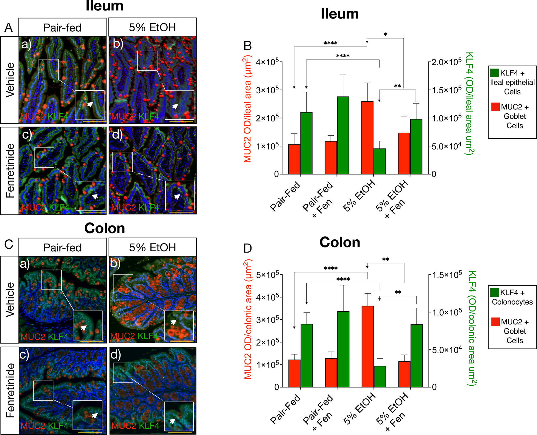
Fig. 5. Fenretinide increases ileal and colonic KLF4 positive epithelial cells.

A) and C) Representative double immunofluorescence microscopy (IFM) images of ileal, and colonic tissue double stained with antibodies against MUC2 (red channel) and KLF4 (green channel, white arrows). Magnification: ×100 (inset 200X), scale bar = 100 μm; B) and D) Quantification of ileal and colonic IFM optical intensity for MUC2 (red bars) and KLF4 (green bars). All data error bars represent ±SD, with **p < 0.01, ****p < 0.0001 and ns = not significant.
