Key message
Transesophageal ultrasound‐guided bronchoscopic aspiration (EUS‐B‐FNA) allowed for minimally invasive and simultaneous diagnosis and evaluation of the degree of invasion by echocardiography. EUS‐B‐FNA may be useful for the evaluation and diagnosis of tumours with cardiac invasion.
Keywords: cardiac invasion, DLBCL, EUS‐B‐FNA, mediastinal tumour, transesophageal approach
EUS‐B‐FNA may be used to evaluate and diagnose tumours with cardiac invasion. Safe, minimally invasive procedures were sufficient to confirm the diagnosis in our patient and assess the degree of invasion, which also allowed for chemotherapy to be initiated promptly.

CLINICAL IMAGE
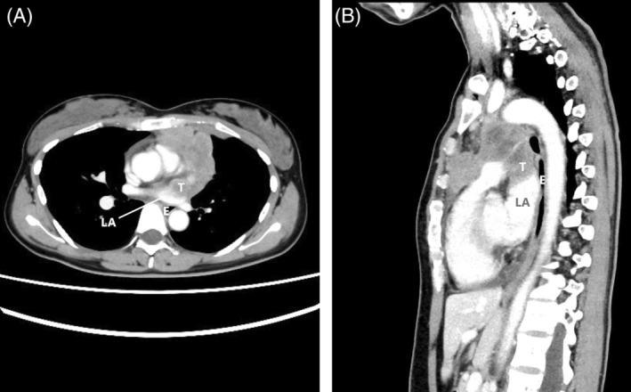
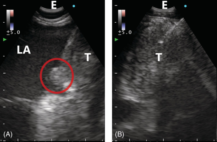
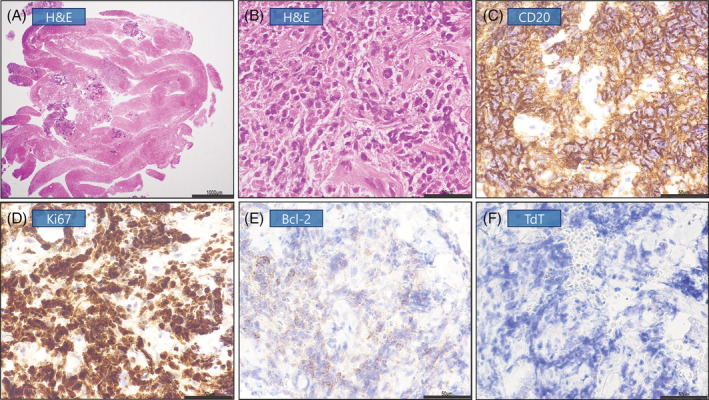
A 34‐year‐old woman presented with dyspnoea and a 1‐month history of coughing. Chest computed tomography showed a mass in the anterior mediastinum with suspected partial cardiac invasion (Figure 1). Transesophageal ultrasound‐guided bronchoscopic aspiration (EUS‐B‐FNA) confirmed tumour invasion of the left atrium (Video 1). Transesophageal needle biopsy of the tumour adjacent to the heart was performed while simultaneously visualizing the cardiac invasion (Figure 2). Diffuse large B‐cell lymphoma (DLBCL) diagnosis was confirmed through immunohistochemical staining (Figure 3). No arrhythmia, bleeding, extravasation, pericardial tamponade or other adverse events occurred. The patient received rituximab, etoposide, prednisolone, vincristine, cyclophosphamide and doxorubicin (R‐EPOCH), and the tumour shrank enough without developing tumour embolism. EUS‐B‐FNA enables safe real‐time sampling of lung tumours and mediastinal lymph nodes adjacent to the oesophagus. 1 While a percutaneous needle biopsy could have been performed to confirm left atrial invasion, EUS‐B‐FNA allowed for minimally invasive and simultaneous diagnosis and evaluation of the degree of invasion by echocardiography, which resulted in prompt initiation of chemotherapy. Thus, EUS‐B‐FNA may be useful for evaluating and diagnosing tumours with cardiac invasion. 2
FIGURE 1.

Enhanced computed tomography findings. Chest CT reveals suspected partial invasion of tumour adjacent to the heart into the left atrium. CT, computed tomography; LA, left atrium; T, tumour; E, oesophagus
VIDEO 1.
EUS‐B‐FNA findings. Tumours invading the endocardium of the left atrium can be observed visible and hidden with the heartbeat.
FIGURE 2.

EUS‐B‐FNA findings. EUS‐B‐FNA findings show tumour invasion into the left atrium (A) and fine‐needle aspiration (B) of the mediastinal tumour adjacent to the heart. The red circle indicates tumour invasion. EUS‐B‐FNA, transesophageal ultrasound‐guided bronchoscopic aspiration
FIGURE 3.

Pathological examination of specimens. H&E staining of specimens and immunohistochemistry for CD20, Ki67, Bcl‐2 and TdT. The tumour cells expressed CD20 and Ki67 (index 90%–100%) but not Bcl‐2 and TdT. The scale bar represents 1000 μm (A), 50 μm (B–F). H&E, haematoxylin and eosin
AUTHOR CONTRIBUTION
Conceptualization: Toshiyuki Sumi. Data curation: Yusuke Tanaka, Haruhiko Michimata and Daiki Nagayama. Formal analysis: Yoshiko Keira and Hiroki Watanabe. Investigation: Yoshiko Keira and Yuichi Yamada. Roles/Writing – original draft: Toshiyuki Sumi. Writing – review & editing: Hirofumi Chiba.
CONFLICT OF INTEREST
None declared.
ETHICS STATEMENT
The authors declare that appropriate written informed consent was obtained for the publication of this manuscript and accompanying images.
ACKNOWLEDGMENT
The authors thank Editage (www.editage.jp) for English language editing.
Nagayama D, Sumi T, Keira Y, Tanaka Y, Michimata H, Koshino Y, et al. Diffuse large B‐cell lymphoma with cardiac invasion diagnosed using transesophageal ultrasound‐guided bronchoscopic aspiration. Respirology Case Reports. 2022;10:e01022. 10.1002/rcr2.1022
Associate Editor: Jennifer Ann Wi
DATA AVAILABILITY STATEMENT
Data sharing not applicable to this article as no datasets were generated or analysed during the current study.
REFERENCES
- 1. Kuijvenhoven JC, Leoncini F, Crombag LC, Spijker R, Bonta PI, Korevaar DA, et al. Endobronchial ultrasound for the diagnosis of centrally located lung tumors: a systematic review and meta‐analysis. Respiration. 2020;99:441–50. 10.1159/000500363 [DOI] [PubMed] [Google Scholar]
- 2. Martinez HA, Kuijvenhoven JC, Annema JT. Intracardiac EUS‐B‐guided FNA for diagnosing cardiac tumors. Respiration. 2021;100:918–22. 10.1159/000516504 [DOI] [PMC free article] [PubMed] [Google Scholar]
Associated Data
This section collects any data citations, data availability statements, or supplementary materials included in this article.
Data Availability Statement
Data sharing not applicable to this article as no datasets were generated or analysed during the current study.


