Abstract
Osteoarthritis (OA) is the most commonly occurring disease of middle and elderly population, which is characterized by focal loss of joint articular cartilage, osteophyte formation and sub chondral bone remodeling. Classical risk factors of OA include age, gender, weight, joint injury, trauma, however hereditary component is one of the main crucial factors. Several genome wide association studies and candidate gene approaches have identified genetic variants involved in the influence and association of OA. In the current study influence of Methylene tetra hydro folate reductase MTHFR C677T (rs1801133) gene with early primary knee OA was evaluated.
In this study 400 samples were included (200 cases & 200 controls). DNA was extracted & processed for PCR- RFLP evaluation and genotype analysis. Statistical analysis was performed & results indicated a lack of association between MTHFR gene polymorphism and early primary KOA. The stratification was done based on age & gender and also both. Individual's i.e females below the age of 40 years are more prone to the disease when compared with males. MTHFR gene polymorphism showed a lack of association with early primary knee osteoarthritis. To the best of our knowledge this is the first study from south India.
Keywords: Polymorphism, MTHFR gene, Osteoarthritis, molecular analysis
Introduction
Osteoarthritis (OA) is one of the most common types of arthritis associated with musculoskeletal disorders, affecting an estimated 302 million people in the middle and elderly age groups globally1. There is a greater proportion of women in favor of OA than men which is predicted to be attributable to the aging2. The disease is caused by both environmental and genetic factors. However, the genetic risk factors can have an impact on whether or not the disease develops, how rapidly the disease progresses, and how severe the symptoms appear. It can have an effect on obesity as well as bone structure. There may be genetic factors that influence the disease's development as well as its likelihood of occurrence. Obesity and having a natural or ideal body weight are also risk factors3. The genetics of OA are complex and not completely understood. Several epidemiological studies have suggested a genetic contribution. The turnover is synergistic between a gene-activated mechanism complex involving injury and its response, body weight, muscle mass, bone, and cartilage structure4, 5. OA affects the knee, hands, hip, and spine, but has the greatest effect on the vertebrae. In the patient, it inflicts severe pain and disability, which means that OA has a tremendous effect on the population6. The prevalence of chronic knee pain over the past couple of decades has risen by nearly 65% and now affects about 25% of all adults7. Knee Osteoarthritis (KOA) is known for the degenerative joint disease is caused by cartilage and underlying bone breakdown8. Age, gender and ethnicity are the prime causes for the incidents of KOA. However, there are numerous sources for arthritis; the clinically most common knee in our population9. Around 30% of older people over the age of 45 have radiographic confirmation of KOA, with about half of those experiencing knee symptoms. Obese people have a higher lifetime risk of symptomatic KOA than non-obese subjects10.
Despite significant advances in molecular biology techniques, the precise mechanism of the disease remains unclear 11. Twin studies, analysis of segregation and linkage and studies of candidate genes have provided important knowledge on patterns of inheritance and the genome location of possible causative mutations in the human diseases12. It is apparent that genetic predisposition from genome wide associations studies (GWAS) influences OA susceptibility. Many loci are relevant to the creation of OA in established literature13. Candidate genes were investigated due to their critical roles in the pathogenesis of OA. Several candidate genes have been linked to the phenotypic manifestation of early KOA in various population-based studies. Among them, Methylene Tetra HydrofolateReductase (MTHFR; OMIM:607093) is a regulatory enzyme of folate and homocysteine metabolism, is one of the candidate genes for osteoarthritis growth14. MTHFR gene has been located on chromosome 1p36.22. A common C to T transition in the MTHFR gene at nucleotide 677 (C677T) results in the replacement of alanine by valine in the protein structure. This genetic polymorphism is found in exon 5 of the MTHFR gene, which corresponds to the protein's folate binding site. The existence of this polymorphism has been linked to increased MTHFR thermo lability and decreased specific activity. This mutation is thought to be the most common genetic cause of high homocysteine levels15–17. MTHFR is a good candidate gene for OA because this polymorphism has been linked to the inflammatory process. In the Indian population and with KOA disease, limited studies were documented and none of the studies were carried out with C677T polymorphism and KOA. Hence, the current study aims to investigate the genetic relation between C677T polymorphism in KOA patients diagnosed in South Indian population.
Materials and methods
Participants involved in this study
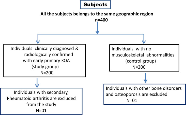
This case-control study was implemented after the grant of ethical approval from institutional committee within the premises of Kamineni Hospitals, capital city of Telangana, India (KHL No. e374/13). Simultaneously, the participants who were recruited in this study has signed the consent form. In this study, based on inclusion and exclusion criteria, we have opted 200 KOA cases and 200 healthy controls (Figure-1). Individuals that have been clinically diagnosed with early primary KOA and have had radiological tests support their diagnosis. In terms of KL grading, all patient grades were considered as the inclusion criteria for KOA cases. The exclusion criteria of KOA cases are infectious, rheumatoid, and secondary osteoarthritis. The healthy controls are selected randomly, with a normal body mass index (BMI) and no personal and family history of any form of arthritis, and who have visited the master health check-up for routine tests. The control subjects with other diseases, family history, and abnormal BMI were excluded from this study. The age and gender matching KOA cases and controls were recruited. The mean age of the KOA cases were 44 years and 43 years was found to be the mean age of healthy controls involved in this study.
Figure 1.
Selection criteria of the study subjects
Sample collection
3ml of peripheral blood was obtained from study participants in an EDTA vacutainer for DNA isolation and molecular analysis at the department of Genetics and Molecular Medicine, Kamineni Life Sciences, Moula -Ali, Hyderabad.
Involvement of anthropmetric measurements and other details
Age and gender information was recorded for both cases and controls. Height was measured in centimeters, weight was measured in kilograms, and BMI was determined using WHO guidelines 18. KOA cases were allocated as per KL classification based on the nature of their symptoms and X-ray observations. Information on co-morbidities such as hypertension, type 2 diabetes, and thyroid dysfunction were obtained from both cases and controls. A family history of osteoarthritis was also obtained in the family pedigree.
Molecular analysis
According to the previous research incorporated by our lab, the C677T polymorphism of the MTHFR gene was chosen from the candidate gene approach to test in our population15. DNA was extracted from both cases and controls using the same salting out technique used in our routine lab, and samples were stored at -20o C before further use19.
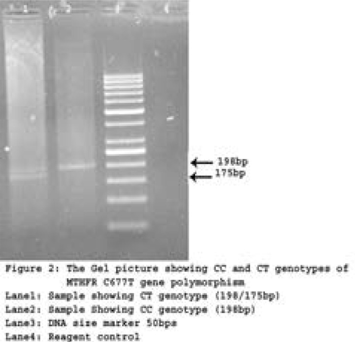
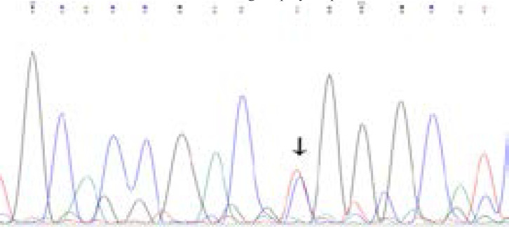
Initially, a 25-µl reaction containing 50-100ng of genomic DNA, 10pmoles of primers, 10X buffer, 2mM Mgcl2, 0.5mM dNTPs mix, and 5 units of Taq DNA polymerase (Ferments) is adjusted with distilled water for the final volume. PCR conditions for the reaction are as follows: initial denaturation (95°C-5mins), denaturation (95°C -30sec), annealing (65°C -45sec), extension (72°C -45sec), and final extension (72°C -7mins), with a hold at 4°C after 35 cycles20. The PCR amplification for MTHFR gene of C677T; specific primers were used, with an amplicon scale of 198 base pairs 15. Table-1 contains the details of C677T polymorphism. The restriction enzyme Hinf1 was used to perform RFLP to understand the nucleotide transition at position 677 that causes the amino acid to shift from Alanine to Valine. The digested products were then electrophoresed on a 2% ethidium bromide stained gel (Figure-2). Prior to genotyping study subjects, a few samples were validated with Sanger sequencing parallel to RFLP (Figure-3).
Table 1.
Details of SNP evaluated in the study with early primary knee osteoarthritis and controls
| S. No | SNPedia | Orientation Details |
| 1 | Gene | MTHFR |
| 2 | Reference sequence number | rs1801133 |
| 3 | Condition | Early primary knee Osteoarthritis |
| 4 | Organism | Homo sapiens |
| 5 | Mutation | Non-synonymous single nucleotide polymorphism |
| 6 | Amnio acid substitution | C677T (Ala 677 Val) |
| 7 | Single Nucleotide Polymorphism | C-T |
| 8 | Exon position | Exon 5 |
| 9 | Chromosome Region | Chromosome-1 p36.22 |
| 10 | 5′-3′ Primer sequence | TGAAGGAGAAGGTGTCTGCGGGA |
| 11 | 3′-5′ Primer Sequence | GGACGGTGCGGTGAGAGTG |
| 12 | PCR Amplicon size | 198bp |
| 13 | Restriction Enzyme | Hinf1 |
| 14 | Substitution of Nucleotide band size | 176bp |
| 15 | Digested amplicon size | CC: 198bp ; CT:198/176/282bp ; TT176/22bp |
| 16 | Genotype effect | CC: Normal or no risk CT: Increased risk TT: Increased risk |
Figure 2 A.
Representative gel picture showing CC & CT genotypes for the MTHFR C677T gene polymorphism
Figure 2B.
Chromatogram indicating CT (Heterozygous) genotype for the MTHFR C677T gene polymorphism after Sanger sequencing
Statistical analysis
We used openepi software (Openepi, version 2.3.1, Atlanta, USA) for the statistical analysis. Independent sample t-test analysis was used to analyse the clinical data between KOA cases and controls (Table-2). Clinical data are expressed as mean ± standard deviation (M±SD). Hardy Weinberg equilibrium (HWE) analysis was performed with C677T genotypes of control subjects as described by Khan et al21. Genotype and allele frequency (Table-3) were calculated between KOA cases and controls using chi square test. Odds ratios (ORs) and 95% confidence intervals are calculated to estimate the association of genotypes by logistic regression analysis. All the p values less than 0.05 (p<0.05) was considered as statistically significant and association between cases and controls19.
Table 2.
Demographic and clinical findings of the study group
| Individual Characteristics | Cases N=200 (Mean±SD) |
Controls N=200 (Mean±SD) | P Value |
| Age | 44.04±6.77 | 43.03±6.09 | 0.11 |
| Gender | |||
| a. Females | 119 (59.5%) | 114 (57%) | NA |
| b. Males | 81 (40.5%) | 86 (43%) | NA |
| Height | 155.135±4.53 | 155.59±3.9 | 0.28 |
| Weight | 73.275±9.61 | 61.815±7.5 | 0.0001 |
| BMI | 30.44±3.8 | 25.5±3.29 | 0.0001 |
| Average age of onset | 41.125±6.28 | NA | NA |
| KL Grades | |||
| Grade 2 | 120 (60%) | NA | NA |
| Grade 3 | 80 (40%) | NA | NA |
| Co-morbidities | |||
|
Hypertensive a.– Present |
91 (45.5%) | 34 (17%) | |
| b.-Absent | 109 (54.5%) | 166 (83%) | 0.0001 |
|
Type 2 Diabetes a.– Present |
60 (30%) | 26 (13%) | |
| b.-Absent | 140 (70%) | 174 (87%) | 0.0001 |
| Thyroid Dysfunction a.– Present |
51 (25.5%) | 28 (14%) | |
| b. -Absent | 149 (74.5%) | 172 (86%) | 0.153 |
| Family History of Osteoarthritis a.– Present |
57 (28.5%) | NA | |
| b.-Absent | 143 (71.5%) | NA |
Table 3.
Distribution of genotypes and allele frequencies of MTHFR (C677T) gene polymorphism on early primary knee Osteoarthritis cases and controls
| Genotypes | Cases N=200 (%) |
Controls N=200 (%) |
Chi square |
P Value | OR (95% CI) |
| CC | 176 (88%) | 181 (90%) | |||
| CT | 24 (12%) | 19 (10%) | 0.65 | 0.4206 | OR=0.769 95% CI (0.407–1.455) |
| TT | 0 | 0 | |||
| CT vs CC+TT | 24 vs 177 | 19 vs 182 | 0.4207 | OR: 1.2988 95% CI (0.687–2.454) |
|
| CT+TT vs CC | 25 vs 176 | 20 vs 181 | 0.4298 | OR: 1.2855 95% CI (0.689–2.398) |
|
| Allele Frequency | |||||
| C | 376 (0.94) | 381 (0.953) | |||
| T | 24 (0.06) | 19 (0.047) | 0.61 | 0.434 | OR: 1.2800 95% CI (0.689–2.375) |
Results
Baseline and Anthropometric details of study group
Baseline characteristics and anthropometric details of both KOA cases and controls were provided in Table 2. The mean age ±standard deviation (SD) of cases was 44.04±06.77yrs, and in control group 43.03±06.09yrs. The percentage of females and males were 59.5%, 40.5% respectively in cases whereas in controls 57% & 43%. The ratio of females was higher than males. The mean age of onset ±standard deviation of cases was 41.12 ± 6.28 years. In cases 60% of them were of KL grade 2 and the remaining 40% were KL grade3. The percentage of cases with Hypertension, Type 2 Diabetes, Thyroid dysfunction were 45.5%, 30%, 25.5% and in controls 17%, 13%, 14% respectively. Cases with positive family history accounts for 28.5%, indicating genetic predisposition of the disease. Anthropometric measurements like weight, BMI, hypertension, type 2 Diabetes were strongly associated in early primary KOA subjects compared to controls [p=.0001] (Table-2).
Genotype of C677T -MTHFR gene analysis in cases and controls
The HWE analysis revealed that the genotype frequencies of C677T polymorphism in the control subjects were in the agreement (χ2=0.49; p=0.48). In the current study the genotypes of cases and controls were evaluated for C677T, MTHFR gene polymorphism. The study deviates from the Hardy Weinberg Equilibrium with the complete absence of variant TT genotype in both cases and controls. The percentage of CC, CT and TT genotypes in cases and controls were found to be 88%,12%, 0% and 90%, 10%, 0% respectively. The C allele frequency found to be 0.94 and T allele is 0.06 in cases, whereas in controls is 0.953 and 0.047 respectively. There is no significant difference between genotypes of cases and controls. Dominant, recessive and co-dominant models could not show any statistical significance [OR=1.2855, 95% CI (0.6891–2.3981) p=0.429] (Table 3). MTHFR C677T gene polymorphism lacks the association with early primary knee osteoarthritis.
Genotype analysis based on gender in KOA cases
Cases were stratified based on the gender and genotype. The percentage of CC genotype in KOA females and males found to be 59.6% and 40.4% respectively. Whereas the percentage of CT genotype in females and males found to be 58% and 42% indicating that there is no significant difference based on gender.
Genotype analysis based on age in KOA cases
The percentage of CC and CT genotypes in < 40 years age group was 91.5% and 8.5% where as in > 40 years age group it was 86.5% and 13.5%. There is a slight increase in the percentage of CT genotype in >40years age group however; it is not statically significant.
Genotype analysis based on age and gender in KOA cases
The percentage of CC genotype in <40 years females was 97% where as in males 85%. The percentage of CC genotype in > 40 years females was 85% where as in males 89%. The percentage of CT genotype in females of > 40 years (15%) was higher compared to < 40 years females (3%) and chi square test showed significant difference between females of below and above 40 years of age [chi square=8.79; p=0.003]. However, in males the CT genotype was almost similar in both the age groups and not significantly different.
Discussion
Osteoarthritis is one of the common musculoskeletal diseases, creating major burden influencing life style in middle and elderly population. The molecular genetics research of OA has been substantially bolstered in the recent years and advancement of the powerful genome wide scans that have revealed a larger number of novel risk loci associated with the disease22. Furthermore, the knowledge of candidate gene association studies reveals an avenue for genetic predisposition of the disease. KOA is most prevalent type of arthritis and genetic factors are associated with the occurrence and development of OA 23, 24. In the last decades, many researchers tried to identify the causation variants through GWAS, Meta-analysis, and candidate gene analysis and there by understanding more about the insights of the disease with potential implications to predict predisposition of disease by identifying risk alleles. This in turn paves way to development of novel treatment and management of the disease.
MTHFR gene is considered to be one of the candidate genes for the knee osteoarthritis. The enzyme MTHFR inhibition is responsible for the increase in homocysteine levels. From the earlier studies it was evident that the plasma homocysteine levels increase in individuals with arthritis25. Hence, the current study was planned to analyse MTHFR gene polymorphism in south Indian population to assess its possible role in Osteoarthritis.
In the current study, the findings suggest that the MTHFR (C677T) rs1801133 gene polymorphism lack association with early primary knee Osteoarthritis in the South Indian population; however, inclusion of other ethnic groups and larger sample size are needed to fully analyse the role of these polymorphisms with KOA risk prediction. In this study, gender and age-based stratification of genotypes were also categorized however, we could not find any statistical significance between them. But, when we had performed chi square based on age and gender, females below and above the age of 40 yrs showed a significant difference indicating early onset of the disease in presence of risk allele than males of same age group.
Weight is considered as one of the robust risk factors for osteoarthritis and in this study also weight and BMI showed a significant difference between cases and controls, hence, the treating clinicians/surgeons should consider weight reduction also in the treatment plans of OA26.
Using regression analysis odds ratio was calculated for genotypes and BMI as cofounder. However, it is not statistically significant. [OR=2.178, 95% CI (0.5580–8.5029) p=0.2625]. It was reported that increased blood pressure is associated with low bone mass and also high risk for fractures. In T2DM patients bone quality was also reduced with high chances of risk for fractures. From the literature it was evident that co morbidities like hypertension and T2DM, interact with subchondral bone remodeling and aggravate the severity of osteoarthritis27. In the current study: hypertension, T2DM showed a significant association indicating these play an internal role in the disease mechanism.
According to a study conducted on a Turkish population, the C allele of the MTHFR C677T mutation may be associated with an increased risk of developing osteoarthritis 14. There were only a few studies that focused at the role of the MTHFR C677T gene polymorphism in osteoarthritis. Tasbas et al28 previously investigated MTHFR gene polymorphism in Rheumatoid arthritis (RA) patients and discovered that the frequency of MTHFR C677T variant was comparable in Turkish RA patients and control group. Another study from the same population investigated the correlation of MTHFR gene mutation in Alopecia Areata and confirmed that this mutation could be associated with an increased risk of Alopecia Areata 29. A meta-analysis of 16 study populations found that the MTHFR gene C677T SNP has a strong potential association with Rheumatoid Arthritis risk30. As there were paucity of studies in different ethnic groups with the MTHFR C677T gene polymorphism and osteoarthritis, this study makes an impact to the existing literature. To the best of our knowledge this is the first study from South India which evaluated the association of MTHFR C677 T gene polymorphism and early primary knee osteoarthritis.
One of the limitations of this study group is opting the control subjects without the radiologic screening. The other limitation of our study is the low sample size and all the cases were from the same ethnic group. Hence, larger study with a greater number of samples from different ethnic groups will help us to understand the influence of this gene polymorphism in predisposing to Osteoarthritis.
Conclusion
The current study evaluated the association of MTHFR C677T (rs1801133) gene polymorphism with early primary KOA and the results showed a lack of association. Knee Osteoarthritis is multifactorial and also anticipated to be a result of multiple gene involvement and gene-gene interaction. To the best of our knowledge this is the first study from South India which evaluated the association of MTHFR gene polymorphism with early primary knee osteoarthritis.
Conflict of interest
Authors declare that there is no conflict of interest for the study.
References
- 1.Pavone V, Vescio A, Turchetta M, et al. Injection-Based Management of Osteoarthritis of the Knee: A Systematic Review of Guidelines. Frontiers in Pharmacology. 2021;12:741. doi: 10.3389/fphar.2021.661805. [DOI] [PMC free article] [PubMed] [Google Scholar]
- 2.Sharma L. Osteoarthritis of the Knee. New England Journal of Medicine. 2021;384:51–59. doi: 10.1056/NEJMcp1903768. [DOI] [PubMed] [Google Scholar]
- 3.Valdes AM, Spector TD. Genetic epidemiology of hip and knee osteoarthritis. Nature Reviews Rheumatology. 2011;7:23. doi: 10.1038/nrrheum.2010.191. [DOI] [PubMed] [Google Scholar]
- 4.He Y, Li Z, Alexander PG, et al. Pathogenesis of osteoarthritis: risk factors, regulatory pathways in chondrocytes, and experimental models. Biology. 2020;9:194. doi: 10.3390/biology9080194. [DOI] [PMC free article] [PubMed] [Google Scholar]
- 5.Warner SC, Valdes AM. The genetics of osteoarthritis: A review. Journal of Functional Morphology and Kinesiology. 2016;1:140–153. [Google Scholar]
- 6.Kloppenburg M, Berenbaum F. Osteoarthritis year in review 2019: epidemiology and therapy. Osteoarthritis and cartilage. 2020;28:242–248. doi: 10.1016/j.joca.2020.01.002. [DOI] [PubMed] [Google Scholar]
- 7.Gilat R, Haunschild ED, Patel S, et al. Understanding the difference between symptoms of focal cartilage defects and osteoarthritis of the knee: a matched cohort analysis. International Orthopaedics. 2021:1–6. doi: 10.1007/s00264-020-04919-w. [DOI] [PubMed] [Google Scholar]
- 8.Zhou M, Chen J, Wang D, et al. Combined effects of reproductive and hormone factors and obesity on the prevalence of knee osteoarthritis and knee pain among middle-aged or older Chinese women: a cross-sectional study. BMC Public Health. 2018;18:1–9. doi: 10.1186/s12889-018-6114-1. [DOI] [PMC free article] [PubMed] [Google Scholar]
- 9.Dantas LO, de Fátima Salvini T, McAlindon TE. Knee osteoarthritis: key treatments and implications for physical therapy. Brazilian Journal of Physical Therapy. 2020 doi: 10.1016/j.bjpt.2020.08.004. [DOI] [PMC free article] [PubMed] [Google Scholar]
- 10.Katz JN, Arant KR, Loeser RF. Diagnosis and Treatment of Hip and Knee Osteoarthritis: A Review. JAMA. 2021;325:568–578. doi: 10.1001/jama.2020.22171. [DOI] [PMC free article] [PubMed] [Google Scholar]
- 11.Inanır A, Sogut E, Ayan M, et al. Evaluation of Pain Intensity and Oxidative Stress Levels in Patients with Inflammatory and Non-Inflammatory Back Pain. European Journal of General Medicine. 2013;10 [Google Scholar]
- 12.Khan IA, Vattam KK, Jahan P, et al. Importance of glucokinase -258G/A polymorphism in Asian Indians with post-transplant and type 2 diabetes mellitus. Intractable Rare Dis Res. 2016;5:25–30. doi: 10.5582/irdr.2015.01040. 2016/03/19. [DOI] [PMC free article] [PubMed] [Google Scholar]
- 13.Tong Z, Liu Y, Chen B, et al. Association between MMP3 and TIMP3 polymorphisms and risk of osteoarthritis. Oncotarget. 2017;8:83563. doi: 10.18632/oncotarget.18745. [DOI] [PMC free article] [PubMed] [Google Scholar]
- 14.Inanir A, Yigit S, Tural S, et al. MTHFR gene C677T mutation and ACE gene I/D polymorphism in Turkish patients with osteoarthritis. Disease markers. 2012;34:17–22. doi: 10.3233/DMA-2012-00939. [DOI] [PMC free article] [PubMed] [Google Scholar]
- 15.Khan IA, Shaik NA, Kamineni V, et al. Evaluation of Gestational Diabetes Mellitus Risk in South Indian Women Based on MTHFR (C677T) and FVL (G1691A) Mutations. Front Pediatr. 2015;3:34. doi: 10.3389/fped.2015.00034. 2015/05/23. [DOI] [PMC free article] [PubMed] [Google Scholar]
- 16.Matam K, Khan IA, Hasan Q, et al. Coronary artery disease and the frequencies of MTHFR and PON1 gene polymorphism studies in a varied population of Hyderabad, Telangana region in south India. Journal of King Saud University-Science. 2015;27:143–150. [Google Scholar]
- 17.Derar TMO, Gader AGMA, Kordofani AAY, et al. 2017. Role of cystathionine β synthase gene variant in Sudanese population. [Google Scholar]
- 18.Alshammary AF, Khan IA. Screening of Obese Offspring of First-Cousin Consanguineous Subjects for the Angiotensin-Converting Enzyme Gene with a 287-bp Alu Sequence. Journal of obesity & metabolic syndrome. 2021;30:63. doi: 10.7570/jomes20086. [DOI] [PMC free article] [PubMed] [Google Scholar]
- 19.Khan IA, Jahan P, Hasan Q, et al. Genetic confirmation of T2DM meta-analysis variants studied in gestational diabetes mellitus in an Indian population. Diabetes Metab Syndr. 2019;13:688–694. doi: 10.1016/j.dsx.2018.11.035. 2019/01/16. [DOI] [PubMed] [Google Scholar]
- 20.Alharbi KK, Abudawood M, Khan IA. Amino-acid amendment of arginine-325-tryptophan in rs13266634 genetic polymorphism studies of the SLC30A8 gene with type 2 diabetes-mellitus patients featuring a positive family history in the Saudi population. Journal of King Saud University-Science. 2021;33:101258. [Google Scholar]
- 21.Khan IA, Movva S, Shaik NA, et al. Investigation of Calpain 10 (rs2975760) gene polymorphism in Asian Indians with Gestational Diabetes Mellitus. Meta Gene. 2014;2:299–306. doi: 10.1016/j.mgene.2014.03.001. 2015/01/22. [DOI] [PMC free article] [PubMed] [Google Scholar]
- 22.Rice SJ, Beier F, Young DA, et al. Interplay between genetics and epigenetics in osteoarthritis. Nature Reviews Rheumatology. 2020;16:268–281. doi: 10.1038/s41584-020-0407-3. [DOI] [PubMed] [Google Scholar]
- 23.Liu J, Wang G, Peng Z. Association between the MMP-1-1607 1G/2G polymorphism and osteoarthritis risk: a systematic review and meta-analysis. BioMed Research International. 2020;2020 doi: 10.1155/2020/5190587. [DOI] [PMC free article] [PubMed] [Google Scholar]
- 24.Zhao T, Zhao J, Ma C, et al. Evaluation of relationship between common variants in FGF18 gene and knee osteoarthritis susceptibility. Archives of medical research. 2020;51:76–81. doi: 10.1016/j.arcmed.2019.12.007. [DOI] [PubMed] [Google Scholar]
- 25.Van Ede A, Laan R, Blom H, et al. Homocysteine and folate status in methotrexate-treated patients with rheumatoid arthritis. Rheumatology. 2002;41:658–665. doi: 10.1093/rheumatology/41.6.658. [DOI] [PubMed] [Google Scholar]
- 26.Zheng H, Chen C. Body mass index and risk of knee osteoarthritis: systematic review and meta-analysis of prospective studies. BMJ Open. 2015;5 doi: 10.1136/bmjopen-2014-007568. [DOI] [PMC free article] [PubMed] [Google Scholar]
- 27.Wen C, Chen Y, Tang H, et al. Bone loss at subchondral plate in knee osteoarthritis patients with hypertension and type 2 diabetes mellitus. Osteoarthritis and Cartilage. 2013;21:1716–1723. doi: 10.1016/j.joca.2013.06.027. [DOI] [PubMed] [Google Scholar]
- 28.Taşbaş Ö, Borman P, Karabulut HG, et al. The frequency of A1298C and C677T polymorphisms of the methylentetrahydrofolate gene in Turkish patients with rheumatoid arthritis: relationship with methotrexate toxicity. The Open Rheumatology Journal. 2011;5:30. doi: 10.2174/1874312901105010030. [DOI] [PMC free article] [PubMed] [Google Scholar]
- 29.Kalkan G, Yigit S, Karakuş N, et al. Methylenetetrahydrofolate reductase C677T mutation in patients with alopecia areata in Turkish population. Gene. 2013;530:109–112. doi: 10.1016/j.gene.2013.08.016. [DOI] [PubMed] [Google Scholar]
- 30.Bagheri-Hosseinabadi Z, Imani D, Yousefi H, et al. MTHFR gene polymorphisms and susceptibility to rheumatoid arthritis: A meta-analysis based on 16 studies. Clinical rheumatology. 2020;39:2267–2279. doi: 10.1007/s10067-020-05031-5. [DOI] [PubMed] [Google Scholar]


