Fig. 1.
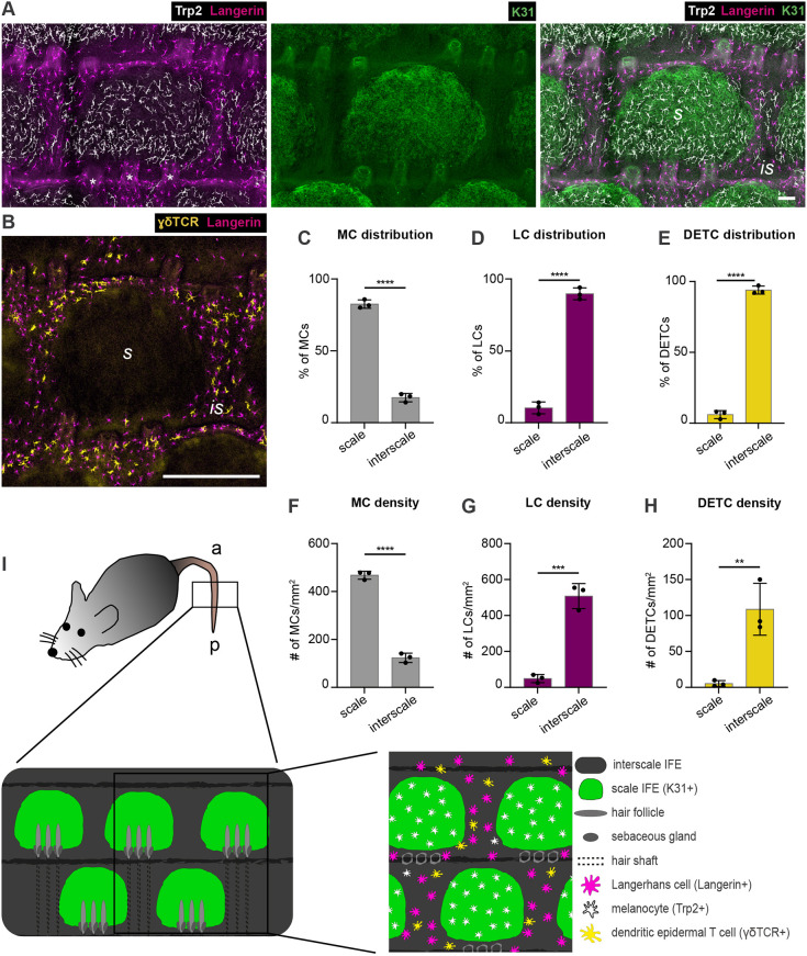
Mutually exclusive localization of MCs and epidermis-resident immunocytes to scale and interscale IFE compartments of murine tail epidermis. (A) Micrographs of tail epidermis wholemounts from 3-month-old wild-type C57BL/6 mice, immunostained for markers of LCs (langerin), MCs (Trp2), and scale IFE (K31). Representative for n=4. (B) Immunostaining of tail epidermis wholemounts from 3-month-old wild-type C57BL/6 mice for langerin and DETC marker γδTCR. Representative for n=3. (C) Quantification of A: MC distribution per scale:interscale unit (% of MCs) in tail epidermis. MC numbers in each compartment (scale, interscale) were normalized to the total MC number per scale:interscale unit. (D) Quantification of A: LC distribution per scale:interscale unit (% of LCs) in tail epidermis. LC numbers in each compartment (scale, interscale) were normalized to the total LC number per scale:interscale unit. (E) Quantification of B: DETC distribution per scale:interscale unit (% of DETCs) in tail epidermis. DETC numbers in each compartment (scale, interscale) were normalized to the total DETC number per scale:interscale unit. (F) Quantification of A: MC density (MC number/mm2) in tail epidermis. (G) Quantification of A: LC density (LC number/mm2) in tail epidermis. (H) Quantification of B: DETC density (DETC number/mm2) in tail epidermis. Data are mean±s.d.; n=3; ****P<0.0001, ***P=0.0004, **P=0.0079; unpaired Student's t-test. (I) Schematic of scale:interscale IFE patterns and associated structures and cell types in murine tail epidermis; a, anterior; is, interscale; p, posterior; s, scale. Scale bars: 75 µm (A); 300 µm (B).

