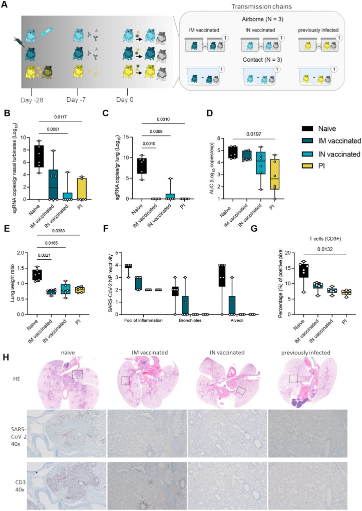
Figure 3. Reduction of disease severity and shedding through pre-existing immunity.
A. Schematic. Hamsters were either vaccinated IN or IM against lineage A or experienced a previous infection with Delta through contact exposure to IN inoculated hamsters. Immune status was confirmed after 21 days. Transmission competitiveness in these populations was investigated at least 28 days post vaccination or infection. Donor animals (N = 6 for each group) were inoculated with a total of 104 TCID50 of Delta and Omicron via the IN route (1:1 ratio), and sentinels 1 (N = 6) were exposed 24h later. For each transmission event, a naïve control animal was also exposed. Half were exposed by direct contact (housed in the same cage), and half at 16.5 cm distance (airborne exposure). Animals were exposed at a 1:1:1 ratio, and exposure occurred on day 1 and lasted for 48 hours. B.C. Tissue samples were collected at 5 DPI/DPE for donors. Donor sgRNA in lungs and nasal turbinates. Whisker-plots depicting median, min and max values, and individual values, N = 6, ordinary two-way ANOVA, followed by Šídák’s multiple comparisons test. D. Cumulative shedding. Area under the curve (AUC) of sgRNA measured in oral swabs taken on 2,3, and 5 DPI. Whisker-plots depicting median, min and max values, and individual values, N = 6, ordinary two-way ANOVA, followed by Šídák’s multiple comparisons test. E. Lung weights (lung:body weight ratio). F. SARS-CoV-2 reactivity measured by immunohistochemistry targeting SARS-CoV-2 nucleoprotein (NP) in upper and lower respiratory tract. Whisker-plots depicting median, min and max values, and individual values, N = 6, ordinary two-way ANOVA, followed by Šídák’s multiple comparisons test. G. T-cell infiltration into the lung, measure by CD3 antigen presence and positive pixel quantification. Whisker-plots depicting median, min and max values, and individual values, N = 6, Kruskal-Wallis test. black = naïve, dark blue = IM vaccinated, light blue = IN vaccinated, yellow = PI. P-values stated were significant (<0.05). H. Lung pathology. top = HE stains, middle = IHC for nucleoprotein, bottom = IHC for CD3. Squares indicate area of magnification.

