



Summary
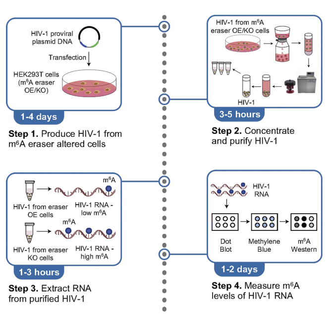
N6-methyladenosine (m6A) modification of human immunodeficiency virus type 1 (HIV-1) RNA plays a critical role in regulating viral replication and evasion of innate immunity. Here, we describe a protocol for the production of HIV-1 with altered m6A levels by manipulating the expression of m6A demethylases in HIV-1 producer cells. RNA from purified virions is analyzed by northern blot and dot blot for m6A levels prior to use in downstream assays to determine the function of m6A modification of viral RNA.
For complete details on the use and execution of this protocol, please refer to Chen et al. (2021).
Subject areas: Microbiology, Molecular biology
Graphical abstract
Highlights
-
•
Procedures to generate m6A eraser knockout cells
-
•
Procedures to generate m6A eraser overexpressing cells
-
•
Protocols for HIV-1 production using m6A eraser modulated cells
-
•
Protocols for analysis of HIV-1 RNA and detecting m6A levels
Publisher’s note: Undertaking any experimental protocol requires adherence to local institutional guidelines for laboratory safety and ethics.
N6-methyladenosine (m6A) modification of human immunodeficiency virus type 1 (HIV-1) RNA plays a critical role in regulating viral replication and evasion of innate immunity. Here, we describe a protocol for the production of HIV-1 with altered m6A levels by manipulating the expression of m6A demethylases in HIV-1 producer cells. RNA from purified virions is analyzed by northern blot and dot blot for m6A levels prior to use in downstream assays to determine the function of m6A modification of viral RNA.
Before you begin
The protocol below describes the production of concentrated HIV-1 with altered levels of m6A in the viral genomic RNA. Manipulation of m6A levels is accomplished by knockout (KO) or overexpression (OE) of a single m6A demethylase (eraser), either fat mass and obesity-associated protein (FTO) or α-ketoglutarate-dependent dioxygenase AlkB homolog 5 (ALKBH5). This protocol uses HEK293T cell lines and HIV-1 proviral plasmid DNA (pNL4-3) to produce replication-competent virus. While we have not tested other strains of HIV-1, the procedures described here should also be applicable to any virus strain that can be produced in HEK293T cells, including single-cycle HIV-1 vectors. We describe measuring relative levels of m6A in the resultant viral genomes using RNA dot blot in conjunction with Western blot using an m6A-specific antibody. Other methods for measuring m6A levels such as quantitative ELISA or m6A RNA immunoprecipitation followed by RT-qPCR would also be appropriate.
Institutional permissions
Biosafety precautions
Infection of replication-competent HIV-1 can cause AIDS in humans. In general, performing experiments that involve using infectious HIV-1 should carefully follow the Virology Manual for HIV Laboratories (Janas and Wu, 2009). This manual was developed by the Division of AIDS, National Institute of Allergy and Infectious Diseases, National Institutes of Health (NIH), USA.
All experiments using lentiviral vectors and replication competent HIV-1 should only be performed by trained personnel and with the appropriate Institutional Biosafety Committee approvals in place.
Key resources table
| REAGENT or RESOURCE | SOURCE | IDENTIFIER |
|---|---|---|
| Antibodies | ||
| Anti-HIV-1 p24 (mouse) | NIH HIV Reagent Program | Cat.#ARP-4121, Clone AG3.0 |
| Anti-FTO (rabbit) | Abcam | Cat.#ab124892, Clone EPR6895, RRID: AB_10972698 |
| Anti-ALKBH5 (rabbit) | Sigma | Cat.#HPA007196, RRID: AB_1850461 |
| Anti-GAPDH (rabbit) | Bio-Rad | Cat.#AHP1628, RRID: AB_1604986 |
| Anti-m6A (rabbit) | Synaptic Systems | Cat.#202 003, RRID: AB_2279214 |
| Goat anti-mouse HRP | Promega | Cat.#W4021, RRID: AB_430834 |
| Goat anti-rabbit HRP | Promega | Cat.#W401B, RRID: AB_430833 |
| Bacterial and virus strains | ||
| HIV-1NL4-3 | This study | Generated from pNL4-3 (see Recombinant DNA section) |
| Chemicals, peptides, and recombinant proteins | ||
| Dulbecco’s Modified Eagle Medium (DMEM) | Gibco | Cat.#11965-092 |
| Fetal Bovine Serum (FBS) | R&D Systems | Cat.#S1150H |
| Penicillin/Streptomycin | Gibco | Cat.#15140-122 |
| Polyethyleneimine (PEI) | Polysciences | Cat.#24313-2 |
| OPTI-MEM® serum-free media | Gibco | Cat.#31985-062 |
| Hexadimethrine bromide (polybrene) | Sigma | Cat.#H9268 |
| Puromycin | Sigma | Cat.#P8833 |
| Dulbecco’s phosphate-buffered saline (DPBS) | Gibco | Cat.#14190-144 |
| RIPA Buffer | Sigma | Cat.#R0278 |
| Protease inhibitor cocktail | Sigma | Cat.#P8340 |
| Sucrose | Fisher | Cat.#S5-3 |
| Tris-base | RPI | Cat.#T60040 |
| 0.5 M EDTA pH 8.0 | Thermo Fisher Scientific | Cat.#AM9261 |
| NaCl | RPI | Cat.#S23025 |
| TURBO™ DNase I | Invitrogen | Cat.#AM2238 |
| RNAseOut™ | Thermo | Cat.#10777019 |
| TRIzol™ | Invitrogen | Cat.#15596026 |
| Chloroform | Sigma | Cat.#288306 |
| Glycogen | Fermentas | Cat.#R0561 |
| 2-propanol | Sigma | Cat.#34863 |
| Ethyl alcohol | Sigma | Cat.#E7023 |
| RNase ZAP™ (optional) | Thermo Fisher Scientific | Cat.#AM9780 |
| Urea | RPI | Cat.#U20200 |
| 6× Orange-G | RPI | Cat.#O21200 |
| RNA Millenium™ Markers | Thermo Fisher Scientific | Cat.#AM7150 |
| SeaKem agarose | Lonza | Cat.#50004 |
| SYBR Gold | Thermo Fisher Scientific | Cat.#S11494 |
| 20× saline-sodium citrate (SSC) | Sigma | Cat.#S6639 |
| 37% formaldehyde | Sigma | Cat.#252549 |
| 10× Tris-buffered saline (TBS) | Bio-Rad | Cat.#1706435 |
| Tween-20 | RPI | Cat.#P20370 |
| Methylene blue | Molecular Research Center | Cat.#MB119 |
| Blotting-Grade Blocker (nonfat dry milk) | Bio-Rad | Cat.#1706404 |
| Critical commercial assays | ||
| Nucleobond Xtra Midi Kit | MACHEREY-NAGEL | Cat.#740410 |
| Pierce™ BCA Protein Assay | Thermo Fisher Scientific | Cat.#23225 |
| NorthernMax™ Kit | Thermo Fisher Scientific | Cat.#AM1940 |
| Chemiluminescent Nucleic Acid Detection Module | Thermo Fisher Scientific | Cat.#89880 |
| SuperSignal™ West Femto Maximum Sensitivity Substrate | Thermo Fisher Scientific | Cat.#34094 |
| Experimental models: Cell lines | ||
| HEK293T cell line | ATCC | Cat.#CRL-3216 |
| Oligonucleotides | ||
| Northern blot HIV-1 LTR U3 probe: 5′-biotin-agttctgccaatcagggaagtagccttgtg-3' | This study | N/A |
| Recombinant DNA | ||
| pNL4-3 | NIH HIV Reagent Program | Cat.#ARP-114 |
| pCMV6 Vector | (Wei et al., 2018) | N/A |
| pCMV6-FTO | (Wei et al., 2018) | N/A |
| pCMV-ALKBH5 | (Zheng et al., 2013) | N/A |
| CRISPR-Cas9 sgControl | (Winkler et al., 2019) | N/A |
| CRISPR-Cas9 sgFTO | (Winkler et al., 2019) | N/A |
| CRISPR-Cas9 sgALKBH5 | (Winkler et al., 2019) | N/A |
| pMD2.G | Addgene | Addgene plasmid #12259 |
| psPAX2 | Addgene | Addgene plasmid #12260 |
| Software and algorithms | ||
| ImageJ software | National Institutes of Health | https://imagej.nih.gov/ij/download.html |
| Other | ||
| 10-cm diameter culture dishes | Corning | Cat.#353003 |
| 15 mL conical tube | Dot Scientific | Cat.#416-PG |
| 50 mL conical tube | Dot Scientific | Cat.#451-PG |
| 1.5 mL microcentrifuge tube | USA Scientific | Cat.#1615-5500 |
| Syringe Filter (0.45 μm) | VWR | Cat.#76479-020 |
| 10 mL syringe | BD | Cat.#309604 |
| 6-cm diameter culture dishes | Corning | Cat.#430166 |
| 96-well culture dishes | Corning | Cat.#3596 |
| 12-well culture dishes | Corning | Cat.#3513 |
| Cell lifter | Corning | Cat.#3008 |
| Tube revolver rotator | Thermo Fisher Scientific | Cat.#88881001 |
| Nylon membrane | Roche | Cat.#11209299001 |
| Dot-blot apparatus | Bio-Rad | Cat.#1706545 |
| Beckman Coulter Thinwall Ultra-Clear Tube | Beckman | Cat.#344058 |
| UV Crosslinker | Stratagene | Model: UV Stratalinker 1800 |
| SW32Ti Rotor | Beckman Coulter | Cat.#342194 |
| Compatible ultracentrifuge | Beckman Coulter | Various |
| Amersham Biosciences Imager 600 | GE Healthcare |
Model: 600 |
Materials and equipment
HEK293T cell complete medium
| Reagent | Final concentration |
|---|---|
| DMEM | N/A |
| Fetal bovine serum (FBS) | 10% |
| Penicillin/streptomycin | 50 U/mL |
Store at 4°C during the shelf-life of the product.
Control, FTO, and ALKBH5 KO HEK293T cell complete medium
| Reagent | Final concentration |
|---|---|
| DMEM | N/A |
| Fetal bovine serum (FBS) | 10% |
| Penicillin/streptomycin | 50 U/mL |
| Puromycin | 1 μg/mL |
Store at 4°C during the shelf-life of the product.
25% sucrose in TNE buffer
| Reagent | Amount | Final concentration |
|---|---|---|
| 1 M Tris pH 7.4 | 50 mL | 50 mM |
| 0.5 M EDTA pH 8.0 | 0.2 mL | 0.1 mM |
| NaCl | 5.844 g | 100 mM |
| Sucrose | 250 g | 25% |
| ddH2O | To 1,000 mL | N/A |
Store at 4°C for up to 3 months.
50× Tris acetate electrophoresis buffer (TAE)
| Reagent | Amount | Final concentration |
|---|---|---|
| Tris base | 242 g | 2 M |
| Acetic acid (glacial) | 57.1 mL | 1 M |
| 0.5 M EDTA pH 8.0 | 100 mL | 50 mM |
| RNase-free water | To 1,000 mL | N/A |
Store at 22°C for up to 6 months.
1× TAE
| Reagent | Amount | Final concentration |
|---|---|---|
| 50× TAE | 20 mL | 1× |
| RNase-free water | 980 mL | N/A |
Store at 22°C for up to 6 months.
1 M urea electrophoresis buffer
| Reagent | Amount | Final concentration |
|---|---|---|
| urea | 42 g | 1 M |
| 1× TAE | To 700 mL | 1× |
Store at 4°C for up to 24 h.
8 M urea loading buffer
| Reagent | Amount | Final concentration |
|---|---|---|
| urea | 0.24 g | 8 M |
| 1× TAE | To 500 μL | 1× |
Store at 4°C for up to 24 h.
Tris-buffered saline + Tween-20 (TBST)
| Reagent | Amount | Final concentration |
|---|---|---|
| 10× TBS | 100 mL | 1× |
| Tween-20 | 1 mL | 0.1% |
| ddH2O | 899 mL | N/A |
Store at 22°C for up to 3 months.
CRITICAL: All solutions for RNA work should be prepared using certified RNase-free reagents.
Step-by-step method details
Generation of FTO or ALKBH5 KO HEK293T cells
Timing: 5 weeks
The purpose of this step is to generate HIV-1 producer cells that do not express one of the m6A erasers, allowing for the production of HIV-1 containing elevated levels of m6A.
-
1.Produce LentiCRISPR v2 lentiviral particles.
-
a.Prepare plasmids using a commercially available kit such as Nucleobond Xtra Midi Kit according to the manufacturer’s protocol which can be found here: NuceloBond Xtra Midi. Dissolve DNA pellet in 10 mM Tris, 1 mM EDTA pH 8.0.
-
b.Seed 5 × 10⁶ HEK293T cells per 10-cm diameter culture dish in 10 mL of complete medium. Seed one dish per lentiviral vector. Cells should be ∼70% confluent at the time of transfection (Figure 1A).
-
c.Incubate cells at 37°C, 5% CO2 for 16 h.
-
d.Transfect cells.
-
i.Dilute DNA by adding three plasmids (maximum 15 μL) to 1.5 mL OPTI-MEM:
Plasmid Amount (μg)
(Optimized ratio)pLentiCRISPR v2 (sgControl, sgFTO, or sgALKBH5) 8 psPAX2 (gag-pol) 4.5 pMD2.G (vesicular stomatitis virus G protein [VSV-G]) 1.5 -
ii.Dilute 1 mg/mL PEI solution by adding 70 μL PEI to 1.5 mL OPTI-MEM.
-
iii.Add diluted DNA to diluted PEI, mix and incubate for 15 min at 22°C.
-
i.
-
e.Transfer cells from incubator to biosafety cabinet.
-
f.Gently add DNA:PEI mix dropwise to cells. Swirl gently to disperse the mixture evenly without disturbing the cell monolayer.
-
g.Incubate cells at 37°C, 5% CO2 for 6 h.
-
h.Remove the cell culture medium containing the transfection mixture and carefully replace with fresh complete medium.
-
i.Incubate cells at 37°C, 5% CO2 for 48 h.
-
j.Collect culture medium and transfer to a 15 mL conical tube.
-
k.Centrifuge at 1,000 × g for 10 min at 22°C.
-
l.Pass supernatant through 0.45 μm syringe filter to remove remaining cellular debris.
-
m.Use medium containing lentiviral particles immediately for transduction or aliquot 1 mL medium containing lentiviral particles per screw cap cryotube to freeze on dry ice for storage at −80°C.Alternatives: There may be batch-to-batch variability of transfection efficiency for different preparations of PEI. Therefore, the appropriate PEI:DNA (μg:μg) ratio should be determined empirically. Alternatively, any transfection reagent may be used. If desired, culture supernatants containing lentivirus may be collected at 48 and 72 h post-transfection and stored either separately or combined. If combining, supernatant collected at 48 h can be stored at 4°C and combined with culture supernatant collected at 72 h post-transfection.
 Pause point: Lentivirus can be stored at −80°C until proceeding to the next step.
-
a.
-
2.Transduce HEK293T cells with LentiCRISPR v2 lentiviral particles and select transduced cells.
-
a.Seed 2 × 10⁶ HEK293T cells per 6-cm diameter culture dish in 5 mL of complete medium. Cells should be ∼50% confluent at the time of transduction. Include a dish for mock transduction to be used as a selection control.
-
b.Incubate cells at 37°C, 5% CO2 for 16 h.
-
c.Remove culture medium.
-
d.Add 1 mL medium with lentiviral particles containing 8 μg/mL polybrene (or complete medium for mock).
-
e.Incubate cells at 37°C, 5% CO2 for 2 h with occasional rocking back and forth.
-
f.Add 4 mL complete medium.
-
g.Incubate cells at 37°C, 5% CO2 for 24 h.
-
h.Carefully replace culture medium.
-
i.Incubate cells at 37°C, 5% CO2 for 24 h.
-
j.Replace culture medium with complete medium containing the appropriate concentration of puromycin. In our hands, 1 μg/mL puromycin kills all untransduced cells within 5 days. We recommend performing a kill curve using your HEK293T cells prior to selecting transduced cells to determine the optimal concentration for selection (0.5, 0.8, 1.0, 1.5, and 2.0 μg/mL puromycin). If cells are confluent, split the cells so that they are ∼80% confluent the next day and then add selective medium.
-
k.Continue selection, replacing medium every other day with fresh puromycin until all cells in the mock transduced control dish are dead. Ideally selection will take 3–5 days.
-
l.Once selection is complete, freeze a batch of the polyclonal cell population prior to clonal selection.
-
a.
-
3.Select clonal populations of KO cells.
-
a.Dilute cells to 5 cells/mL in complete growth medium containing puromycin.
-
b.Seed each well of four 96-well dishes with 100 μL of the cell suspension.
-
c.As a positive control, seed one well with 1 × 10⁴ cells. This will aid in focusing the microscope for quick scanning of wells that contain clones.
-
d.Allow clones derived from single cells to grow out for ∼2 weeks; change the medium every 3–5 days if medium becomes yellow.CRITICAL: Single clones should be monitored routinely and transferred to a larger well once confluence is reached. In addition to microscopic examination of cells, changes in the color of the culture medium can indicate clones that are ready to be transferred.
-
e.Once cells are confluent, expand each clone to one well of a 12-well dish.
-
f.When 12-well is confluent, use half of the cells for Western blot analysis of m6A eraser expression. Keep the other half in culture and continue to amplify.
-
g.Aim to screen ≥20 individual clones for KO of FTO or ALKBH5.
-
a.
-
4.Screen clonal populations of KO cells. (See step 8 for more details).
-
a.Perform Western blot using cell lysate from each clone. Run control lysates from parental HEK293T cells on each gel to determine expression levels of erasers in unedited cells.
-
b.Discard clones that retain eraser expression.
-
c.Prepare several vials of low passage frozen stocks of confirmed KO clones.
-
a.
Figure 1.

Optimal cell confluency at the time of transfection
(A) HEK293T cells were plated at a density of 5 × 10⁶ cells per 10-cm diameter culture dish, resulting in ∼70% confluency 16 h after plating.
(B) HEK293T cells were plated at a density of 3 × 10⁶ cells per 10-cm diameter culture dish, resulting in ∼40% confluency 16 h after plating. Scale bars represent 100 μm.
Generation of FTO or ALKBH5 OE HEK293T cells
The purpose of this step is to generate HIV-1 producer cells that overexpress one of the m6A erasers, allowing for the production of HIV-1 containing reduced levels of m6A.
-
5.Transfect cells with FTO or ALKBH5 expression plasmids or vector control.
-
a.Seed twenty 10-cm diameter cell culture dishes with 3 × 10⁶ HEK293T cells in 10 mL of complete medium. Half of the cells will be transfected with vector control plasmid and half with eraser overexpression plasmid. Cells should be ∼40% confluent at the time of transfection (Figure 1B).
-
b.Incubate cells at 37°C, 5% CO2 for 16 h.
-
c.Transfect cells. Refer to above section 1c for details. The numbers below are per 10-cm diameter dish. Make a master mix for multiple dishes. The number of dishes can be scaled up or down as necessary.
-
i.Dilute each plasmid DNA by adding 12 μg plasmid to 1.5 mL OPTI-MEM.
-
ii.Dilute PEI by adding 60 μL PEI to 1.5 mL OPTI-MEM.
-
iii.Add diluted DNA to diluted PEI and incubate for 15 min at 22°C.
-
i.
-
d.Proceed immediately to step 6.
-
a.
HIV-1 production using FTO/ALKBH5 KO or FTO/ALKBH5 OE HEK293T cells
The purpose of this step is to produce HIV-1 containing altered levels of m6A.
Use the Biosafety Precautions after transfection of cells with HIV-1 proviral DNA plasmid (pNL4-3).
-
6.Transfect cells (5 × 10⁶ per 10-cm diameter dish) with pNL4-3 proviral plasmid.
-
a.Repeat steps 5c–5g with 12 μg pNL4-3 per dish. Cells should be ∼70% confluent at the time of pNL4-3 transfection.
-
b.Incubate cells at 37°C, 5% CO2 for 48 h.
-
a.
-
7.Collect and clarify the virus-containing supernatant.
-
a.Collect the media from cells and transfer to a 50 mL conical tube. Combine up to 40 mL supernatant (from four 10-cm diameter dishes) in a single tube.
-
b.Centrifuge at 1,500 × g for 5 min at 22°C.
-
c.Pass supernatant through a 0.45 μm syringe filter into a fresh 50 mL conical tube.
-
d.Remove 5 mL supernatant and save for p24 measurement and HIV-1 titer determination. For detailed protocols, please refer to (Janas and Wu, 2009).
-
a.
-
8.Collect protein from one dish of cells per condition for Western blot analysis.
-
a.Wash cells once gently with 10 mL cold PBS.
-
b.Completely remove PBS wash.
-
c.Add 1 mL radioimmunoprecipitation assay (RIPA) buffer with protease inhibitor cocktail directly to dish.Alternatives: Commercially available or other detergent-based cell lysis buffers compatible with Western blot may be used as an alternative to RIPA buffer.
-
d.Use cell lifter to scrape cells to one side of dish.
-
e.Transfer cell lysate to a 1.5 mL tube on ice.
-
f.Rotate lysate at 10 rpm for 20 min at 4°C.
-
g.Centrifuge at top speed (at least 13,200 × g) for 30 min at 4°C.
-
h.Transfer supernatant to fresh 1.5 mL tube.Pause point: Cell lysate can be stored at −80°C until performing bicinchoninic acid (BCA) protein assay and Western blot analysis.
-
i.Perform BCA Protein Assay according to manufacturer’s protocol which can be found here: Pierce BCA Protein Assay.
-
j.Use 20 μg total protein per lane for Western blot analysis using standard protocol.Recommended antibody concentrations
Antibody [Stock] mg/mL Dilution [Final] μg/mL HIV-1 p24 N/A 1:500 N/A FTO 0.15 1:1,000 0.15 ALKBH5 0.4 1:1,000 0.4 GAPDH 1.0 1:5,000 0.2 Goat anti-mouse HRP 1.0 1:5,000 0.2 Goat anti-rabbit HRP 1.0 1:5,000 0.2
-
a.
-
9.Pellet the virus through a sucrose cushion to purify and concentrate.
-
a.Transfer 3 mL of 25% Sucrose in TNE buffer to the bottom of a 38.5 mL thinwall ultracentrifuge tube.
-
b.Very carefully add up to 35 mL of supernatant containing HIVNL4-3 (from step 7c) slowly down the side of the tube to the top of the sucrose cushion using a pipet aid. There should be a sharp demarcation between the layers of sucrose and supernatant. Handle tubes carefully to avoid mixing of the layers.
-
c.Place the ultracentrifuge tubes carefully in SW32Ti rotor buckets and affix lids. Weigh each bucket and record the weight to the 1/100th gram.
-
d.Balance the weight of each tube using DMEM (without additives).
-
e.Place the tubes in sealed bucket and hang in pre-chilled SW32Ti rotor.
-
f.Centrifuge at 28,000 rpm for 1.5 h at 4°C using slow acceleration and deceleration setting of the centrifuge to avoid disturbing the layers/pellet.CRITICAL: Carefully read the manual for the ultracentrifuge and rotor prior to use. Ultracentrifuge tubes need to contain enough liquid volume to avoid collapsing during ultracentrifugation. Tubes containing less than 35 mL of virus-containing supernatant should be brought to 35 mL with DMEM without additives so that the total volume of sucrose and supernatant is 38 mL. If using a different ultracentrifuge rotor, compatible thinwall tubes must be used with the appropriate fill volume, adjusting the volume of sucrose cushion accordingly. Tubes contain infectious virus and should never be open outside of an appropriate biosafety cabinet. Tubes should be weighed on a balance while inside the swinging bucket with the lid on. Weight of opposing buckets should be exactly the same to avoid damage to the ultracentrifuge.
-
g.Carefully remove the buckets from the rotor and transfer them to the biosafety cabinet.
-
h.Discard the supernatant by vacuum or decanting.CRITICAL: Supernatant should be decontaminated in a final concentration of 10% bleach for 30 min (or according to institutional regulations) prior to disposal.
-
i.Invert the tube on a paper towel for 1–2 min to completely remove all supernatant.
-
j.Add 150 μL DMEM (without additives) to the pellet.Pause point: Samples can be stored at 4°C for 16–24 h. Do not exceed 24 h incubation.
-
k.Combine rehydrated virions from identical samples (if applicable).
-
l.Remove 100 μL for p24 measurement and HIV-1 infection assay (Janas and Wu, 2009).
-
a.
-
10.Digest the virus with TURBO™ DNase for 30 min at 37°C.
Reagent Final concentration Amount Purified HIV-1NL4-3 N/A 160 μL 10 ×TURBO™ DNase buffer 1× 20 μL 2 U/μL TURBO DNase I 0.1 U/μL 10 μL ddH2O N/A 10 μL Total N/A 200 μL -
a.Add 80 U (2 μL) RNaseOut™.
-
b.Store at −80°C or use directly for RNA extraction.Pause point: Samples can be stored at −80°C until proceeding to the next step.Alternatives: To confirm successful transfection and virus production, we routinely collect protein from one dish of HEK293T producer cells per condition for Western blot analysis of erasers and HIV-1 p24 expression (step 8). If the purified virus will be used for preparation of viral RNA only, DNase treatment can be performed after TRIzol™ purification rather than after resuspension of the virus pellet. In this case, TRIzol™ can be added directly to the ultracentrifuge tubes and all identical samples combined in 1 mL of TRIzol™. If generated HIV-1 stocks will be used for infections, perform DNase treatment as described in step 10 and freeze on dry ice and store in 20 μL aliquots at −80°C for future use. The virus titer decreases when using the stock after multiple freeze and thaw cycles. Thus, it is best to avoid multiple freeze and thaw cycles of virus stocks.
-
a.
HIV-1 RNA extraction
The purpose of this step is to extract HIV-1 genomic RNA from purified virions.
Biosafety Precautions for work with HIV-1NL4-3 may be discontinued after addition of TRIzol™ to concentrated virus.
-
11.Purify viral RNA using TRIzol™.
-
a.Add 1 mL of TRIzol™ Reagent to the centrifuge tube containing DNase-treated virions, pipet up and down several times to mix.
-
b.Incubate for 5 min at 22°C.
-
c.Add 0.2 mL of chloroform and mix well by vigorous shaking for 15 s. Do not vortex.
-
d.Incubate at 22°C for 15 min.
-
e.Centrifuge at 13,200 × g for 15 min at 4°C.
-
f.Transfer the colorless upper aqueous phase to a new tube.
-
g.Add 1 μL 20 mg/mL glycogen, mix.
-
h.Add 400 μL isopropanol, mix.
-
i.Incubate for 10 min at 22°C.
-
j.Centrifuge at 13,200 × g for 20 min at 4°C.
-
k.Carefully discard the supernatant without disturbing the pellet.
-
l.Wash the pellet in 1 mL 75% ethanol.
-
m.Centrifuge at 13,200 × g for 10 min at 4°C.
-
n.Discard the supernatant by decanting.
-
o.Pulse spin the tube to collect residual ethanol and remove using pipette.
-
p.Air dry the RNA pellet for 5 min.
-
q.Rehydrate the RNA in 50 μL RNase-free H2O.
-
r.Determine the concentration of RNA by Nanodrop and store RNA in −80°C.
-
a.
HIV-1 RNA analysis by denaturing urea gel electrophoresis
The purpose of this assay is to directly visualize and examine the integrity of the full-length extracted HIV-1 RNA.
-
12.Prepare electrophoresis reagents the day before electrophoresis.
-
a.Prepare 700 mL 1 M urea electrophoresis buffer. See recipe in “materials and equipment” section.
-
b.Dissolve at ≤60°C for 3 h with mixing at 400 rpm.
-
c.Prepare 8 M urea loading buffer. See recipe in “materials and equipment” section.
-
d.Vortex until dissolved.
-
e.Store at 4°C for 16–24 h or use immediately if performing same day electrophoresis.
-
a.
-
13.Prepare 1 M urea/0.8% agarose gel the day of electrophoresis.
-
a.Add 0.8 g agarose to 94 mL 1× TAE. See recipe in “materials and equipment” section.
-
b.Dissolve in microwave and allow to cool to 60°C.
-
c.Add 6 g urea and mix 15 min to dissolve. The final volume will be 100 mL.
-
d.Pour into gel casting apparatus and allow to solidify.
-
a.
-
14.Prepare samples for electrophoresis.
-
a.Bring 500 ng RNA sample to 5 μL using RNase-free H2O. Sample volume can be scaled up if necessary, based on RNA concentration (See Alternatives below).
-
b.Prepare Millenium™ ssRNA ladder by mixing 2 μL ladder with 3 μL RNase-free H2O.
-
c.To each sample and ladder add 20 μL 8 M urea loading buffer and 5 μL 6× Orange-G loading dye.
-
d.Denature samples for 5 min at 90°C and place immediately on ice.
-
a.
-
15.Run denaturing urea agarose gel.
-
a.Place gel in buffer tank and immerse in electrophoresis buffer.
-
b.Load samples and run the gel until the loading dye (Orange-G) reaches 3/4 of the gel. (approximately 1.6 h at 85 V).
-
c.Remove gel and wash twice for 5 min each in 1× TAE buffer.
-
d.Immerse gel in ∼175 mL (1/4 volume of electrophoresis buffer used at step 12a) of 100 mM NaCl for 30 min.
-
e.Stain gel in SYBR Gold diluted 1:10,000 in 1× TAE for 30 min with gentle rocking and protected from light.
-
f.Image the gel using 300 nm UV transilluminator.
-
a.
Alternatives: If visualization of RNA is the end-point assay, 500 ng RNA per well is sufficient. If the gel will also be used for Northern blot, 3 μg RNA should be loaded per well.
HIV-1 RNA analysis by northern blot (optional)
The purpose of this assay is to specifically detect HIV-1 genomic RNA and its length using Northern blot. This is an optional step depending on the use of HIV-1 RNA.
-
16.
Perform denaturing urea gel electrophoresis according to the previous section using 3 μg RNA per well.
-
17.
Perform Northern blot using the NorthernMax™ Kit (Thermo Fisher Scientific) according to the manufacturer’s protocol which can be found here: NorthernMax Kit.
-
18.
Perform Nonisotopic probe detection using the Chemiluminescent Nucleic Acid Detection Module (Thermo Fisher Scientific) according to the manufacturer’s protocol which can be found here: Chemiluminescent Nucleic Acid Detection Module.
Alternatives: Once the protocol is well-established it is not strictly necessary to perform Northern blotting for every experiment.
HIV-1 RNA m6A detection by dot blot
The purpose of this assay is to specifically detect m6A modifications of HIV-1 genomic RNA using a semi-quantitative dot blot assay.
-
19.Immobilize RNA to a nylon membrane.
-
a.Bring 220 ng RNA to 55 μL volume using of 1 mM EDTA pH 8.0.
-
b.Add 33 μL of 20× SSC buffer and 22 μL of 37% formaldehyde.
-
c.Mix and heat the samples at 65°C for 30 min. Transfer samples immediately to ice.
-
d.Use dot blot apparatus as a guide to cut a nylon membrane to the appropriate size for the required number of wells.
-
e.Soak the nylon membrane in 10× SSC buffer for 10 min.
-
f.Assemble the membrane in the dot blot apparatus following the manufacturer’s instructions.
-
g.Add 200 μL of RNase-free water to each well to be used and pull through using vacuum.
-
h.Add 200 μL of 10× SSC buffer to each well to be used and pull through using vacuum.
-
i.Load 100 μL of each sample and pull through using vacuum.
-
j.Wash each well twice with 200 μL 10× SSC buffer.
-
k.Keep the vacuum on and disassemble the instrument.
-
l.Place the membrane face up on a paper towel dampened with 10× SSC.
-
m.Place in the Stratalinker and perform autocrosslink (120,000 μJ/cm2) three times.
-
a.
-
20.Stain membrane with methylene blue to visualize RNA samples, which serves as a loading control for quantification of m6A levels.
-
a.Transfer the membrane to a small plastic dish.
-
b.Rinse the membrane in methylene blue stain for 1 min.
-
c.Rinse the membrane several times in ddH2O for ∼10 s each until background is de-stained and signal is appropriate for imaging.
-
d.Image the membrane.
-
a.
-
21.Perform Western blot for m6A detection.
-
a.Block the membrane in 5% non-fat dry milk (NFDM) TBST for 60 min at 22°C.
-
b.Remove blocking solution and replace with 2.5% NFDM containing m6A primary antibody at a final concentration of 0.1 μg/mL (1:10,000 dilution).
-
c.Incubate for 60 min at 22°C or for 16–24 h at 4°C.Pause point: Blot may be incubated with primary antibody for 16–24 h for convenience.
-
d.Wash the membrane in TBST three times for 5 min at 22°C.
-
e.Incubate with goat anti-rabbit HRP-conjugated secondary antibody in 2.5% NFDM TBST at a final concentration of 0.2 μg/mL (1:5000 dilution).
-
f.Wash the membrane in TBST five times for 5 min at 22°C.
-
g.Develop the membrane using SuperSignal™ West Femto Maximum Sensitivity Substrate according to manufacturer’s protocol which can be found here: SuperSignal West Femto Maximum Sensitivity Substrate.
-
h.Image the membrane.
-
a.
-
22.
Use ImageJ to quantify the relative levels of m6A signal for each sample, using methylene blue signal as a loading control for normalization.
Alternatives: Antibody concentrations are for the specific antibodies listed in the key resources table. Any anti-rabbit HRP-conjugated secondary may be used; however, the optimal concentration would need to be determined empirically. Similarly, any chemiluminescent substrate system will work for development of signal from HRP-conjugated secondary antibodies. However, some substrates may not provide sufficient sensitivity to accurately quantify the m6A signal.
Expected outcomes
Using lentiCRISPR v2 vector transduction for generation of KO cells, we consistently observe complete loss of protein expression in approximately ∼20% of screened HEK293T KO clones by Western blot (data not shown). We obtained ∼5 clones from four plates. The transduction efficiency may affect the yield of the KO cell clone selection. It is thus essential to carry out the limiting dilution step to isolate single clones from the pooled population. Successful KO clones should be confirmed by sequencing and will show no detectable protein expression (Figure 2A).
Figure 2.

Western blot analysis of m6A erasers (FTO or ALKBH5) and HIV-1 Gag in HEK293T producer cells transfected with pNL4-3
(A) Clonal vector control, FTO, or ALKBH5 knockout (KO) HEK293T cells were transfected with HIV-1 proviral plasmid pNL4-3. Protein lysates were prepared for Western blot analysis 48 h post-transfection.
(B and C) HEK293T cells were transfected with vector control or FTO expressing plasmid (B) or vector control or ALKBH5 expressing plasmid (C). After 24 h, cells were transfected with pNL4-3. Protein lysates were prepared for Western blot analysis 48 h post-transfection of pNL4-3. OE, overexpression; CA, HIV-1 capsid. GAPDH was used as a loading control. This figure is adapted from (Chen et al., 2021).
There should be significant overexpression of eraser proteins in producer cells transfected with overexpression plasmids (Figures 2B and 2C.) Viral Gag proteins should be readily detectable in producer cells transfected with proviral plasmid. Efficient virus production will be demonstrated by abundant levels of both processed capsid (p24) and unprocessed full-length Gag (p55) (Figures 2A–2C).
In a typical experiment, the average yield of purified viral RNA should be ∼1 μg per 10-cm diameter dish of transfected HEK293T cells. Viral RNA purified from sucrose-pelleted virions should appear as a single full-length species that migrates at approximately 9.7 kb in a denaturing agarose gel (Figure 3A). Confirmation of the identity of the viral RNA can be obtained by Northern blot using an HIV-1-specific oligonucleotide probe (Figure 3B).
Figure 3.

Visualization of full-length HIV-1 genomic RNA
(A) HIV-1 RNA purified from sucrose-pelleted virions was visualized using denaturing agarose gel electrophoresis followed by SYBR Gold staining. Total cellular RNA was run in parallel to demonstrate specific purification of full-length HIV-1 RNA, with the major 18S and 28S ribosomal RNA species indicated (arrows).
(B) The RNA from (A) was transferred to a positively charged membrane and Northern blot was performed using a 30-mer biotin-labeled DNA probe complementary to the HIV-1 genome U3 region. The full-length HIV-1 RNA size is ∼9.7 kb (arrow).
The level of m6A modification of viral RNA should be inversely correlated to the level of eraser expression in producer cells lines. Therefore, viral RNA produced in eraser KO cells will contain elevated levels of m6A compared to viral RNA from control cells (Figure 4). Conversely, viral RNA produced in cells overexpressing erasers will contain lower levels of m6A compared to vector control cells (Figure 4).
Figure 4.

Dot blot analysis of relative m6A levels of viral RNA
HIV-1 RNA (200 ng) purified from virions produced in the indicated cell lines was subjected to dot blot analysis as described. Methylene blue (MB) stain is used as a loading control (top panels). The membrane was then subjected to Western blot using an m6A-specific antibody (bottom panels). Relative m6A levels normalized to MB staining were determined using ImageJ. This figure is adapted from (Chen et al., 2021).
Limitations
Depending on the downstream assay(s) to be performed with the viral RNA, the viral production step may need to be significantly scaled up to obtain the required yield of RNA. In this case, the desired number of dishes can be transfected, and the resulting virions from multiple dishes can be combined after ultracentrifugation. This approach will increase the yield per condition from TRIzol™ RNA purification. We have not attempted to increase the scale of virus production to 15-cm diameter or larger cell culture dishes. Scaling to larger culture dishes should be possible but may require additional optimization of plasmid amounts and transfection conditions.
Troubleshooting
Problem 1
Low titer of concentrated virus (step 7d) or low yield of viral RNA from purified virions (step 11r).
Potential solution
The most important factor for determining virus yield is the efficiency of transfection of the proviral plasmid. It is critical to use healthy HEK293T cells at the optimal confluence for transfection. Cells should ideally be 60%–70% confluent at the time of proviral plasmid transfection (Figure 1A). Therefore, if sequential transfections are to be performed as in steps 5 and 6, the first transfection of eraser OE plasmids should be carried out when the cells are about 30%–40% confluent (Figure 1B). The cells should then be at the optimal 60%–70% confluence at the time of proviral plasmid transfection. We recommend always including an extra dish of cells to be transfected with a fluorescent reporter such as GFP to monitor transfection efficiency for every experiment. If the cell numbers listed in this protocol do not give rise to the desired confluence, adjust the number of cells seeded per dish.
Care should be taken to ensure that proviral plasmid preparations are free of recombination mediated by the viral long terminal repeat (LTR) sequences. Plasmids containing homologous LTRs should always be propagated in recombination-deficient bacteria such as Stbl3™ or an analogous strain. We recommend performing a diagnostic restriction digest of every new preparation of HIV-1 proviral plasmid with an enzyme such as HindIII, which will give rise to a distinct and readily identifiable pattern of fragments when analyzed by agarose gel electrophoresis.
Problem 2
HIV-1 RNA is not detectable or appears as a smear after SYBR gold stain of urea-agarose gel (step 15f).
Potential solution
It is critical to maintain an RNase-free environment when working with viral RNA. We recommend always including a high-quality control RNA sample known to be intact when performing electrophoresis of RNA. If RNA degradation is observed, remake all solutions using RNase-free reagents and treat all plastic and glassware to remove any contaminating RNases.
Problem 3
No band observed on Northern blot (step 18).
Potential solution
Confirm efficient transfer of RNA from gel to positively charged membrane. Refer to manufacturer protocols for Northern blot and probe visualization for additional troubleshooting.
Problem 4
No or weak signal for methylene blue stain or m6A Western blot (steps 20d and 21h).
Potential solution
Crosslinking time can be optimized based on the energy output of individual crosslinkers (the output of UV bulb goes down due to aging) by starting with 30 s and comparing several increasing times by determining which condition gives the strongest signal for RNA stain and m6A signal.
Problem 5
No or little difference in m6A levels between eraser KO or OE conditions (step 21h).
Potential solution
The most likely reason for observing the same levels of m6A in viral RNA from control or eraser OE cells is low transfection efficiency. Each cell that is producing virus must also be overexpressing FTO or ALKBH5 to give rise to HIV-1 RNA with low relative levels of m6A. Perform optimization of transfection efficiency for both sequential transfections (OE plasmid and proviral plasmid).
If there are no differences in m6A levels in RNA produced in control and eraser KO cells, it is possible that after serial passage in culture there has been compensatory up or down-regulation of other m6A pathway enzymes. Make sure to freeze down sufficient stocks of freshly cloned KO cells and use low passage stocks for virus production.
Resource availability
Lead contact
Further information and requests for resources and reagents should be directed to and will be fulfilled by the lead contact, Li Wu (li-wu@uiowa.edu).
Materials availability
All reagents generated in this study are available from the lead contact upon request.
Acknowledgments
This work was supported by grants from the National Institutes of Health (NIH, AI150343, AI141495, and AI169659), USA, to L.W. and (HG010318 and HG010108) to S.K. L.W. is also partially supported by NIH grants AI159546 and CA086862-22S1. S.C. is partially supported by the National Natural Science Foundation of China (82172258). The funders had no role in study design, data collection and analysis, decision to publish, or preparation of the manuscript. We thank the NIH HIV Reagent Program for pNL4-3 and anti-HIV-1 p24. We also thank Xiaoli Mou for her kind assistance in preparing the graphical abstract and Mike Cahill for assisting with the key resources table.
Author contributions
Conceptualization: S.C., S.P., and L.W.; methodology: S.C., S.P., and A.B.; validation: L.W.; investigation: S.C. and A.B.; resources: L.W. and S.K.; writing – original draft: S.C. and S.P.; writing – review and editing: S.C., S.P., L.W., A.B., and S.K.; visualization: S.C. and S.P.; project Administration: L.W. and S.P.; funding acquisition: S.C., L.W., and S.K.
Declaration of interests
The authors declare no competing interests.
Contributor Information
Shuliang Chen, Email: chen-shuliang@whu.edu.cn.
Li Wu, Email: li-wu@uiowa.edu.
Data and code availability
This protocol does not report any datasets or codes.
References
- Chen S., Kumar S., Espada C.E., Tirumuru N., Cahill M.P., Hu L., He C., Wu L. N6-methyladenosine modification of HIV-1 RNA suppresses type-I interferon induction in differentiated monocytic cells and primary macrophages. PLoS Pathog. 2021;17:e1009421. doi: 10.1371/journal.ppat.1009421. [DOI] [PMC free article] [PubMed] [Google Scholar]
- Janas A.M., Wu L. HIV-1 interactions with cells: from viral binding to cell-cell transmission. Curr. Protoc. Cell Biol. 2009;Chapter 26:Unit26.5. doi: 10.1002/0471143030.cb2605s43. [DOI] [PMC free article] [PubMed] [Google Scholar]
- Wei J., Liu F., Lu Z., Fei Q., Ai Y., He P.C., Shi H., Cui X., Su R., Klungland A., et al. Differential m(6)A, m(6)Am, and m(1)A demethylation mediated by FTO in the cell nucleus and cytoplasm. Mol. Cell. 2018;71:973–985.e5. doi: 10.1016/j.molcel.2018.08.011. [DOI] [PMC free article] [PubMed] [Google Scholar]
- Winkler R., Gillis E., Lasman L., Safra M., Geula S., Soyris C., Nachshon A., Tai-Schmiedel J., Friedman N., Le-Trilling V.T.K., et al. m(6)A modification controls the innate immune response to infection by targeting type I interferons. Nat. Immunol. 2019;20:173–182. doi: 10.1038/s41590-018-0275-z. [DOI] [PubMed] [Google Scholar]
- Zheng G., Dahl J.A., Niu Y., Fedorcsak P., Huang C.M., Li C.J., Vågbø C.B., Shi Y., Wang W.L., Song S.H., et al. ALKBH5 is a mammalian RNA demethylase that impacts RNA metabolism and mouse fertility. Mol. Cell. 2013;49:18–29. doi: 10.1016/j.molcel.2012.10.015. [DOI] [PMC free article] [PubMed] [Google Scholar]
Associated Data
This section collects any data citations, data availability statements, or supplementary materials included in this article.
Data Availability Statement
This protocol does not report any datasets or codes.