Abstract
Background
To evaluate the clinical outcomes and second-look arthroscopic findings after intra-articular adipose-derived regenerative cell (ADRC) injection as treatment for knee osteoarthritis (OA).
Methods
ADRCs were administered to 11 patients (19 knees; mean age, 61.7 years) with knee OA. Subcutaneous adipose tissue was harvested by liposuction from both thighs, and arthroscopic lavage was performed, followed by ADRC injection (mean dose, 1.40 × 10⁷ cells) into the synovial fluid. Outcome measures included the Knee Injury and Osteoarthritis Outcome Score, Lysholm score, and visual analog scale score. Arthroscopic examinations were performed to assess the International Cartilage Repair Society cartilage injury grade preoperatively and overall repair postoperatively. Noninvasive assessments were performed at baseline and at 1-, 3-, and 6-month follow-ups; arthroscopic assessments were performed at baseline and at 6 months.
Results
All outcome measures significantly improved after treatment. This improvement was evident 1 month after treatment and was sustained until the 6-month follow-up. Data from second-look arthroscopy showed better repair in low-grade cartilage lesions than in lesions with a greater degree of damage. No patients demonstrated worsening of Kellgren-Lawrence grade, and none underwent total knee arthroplasty during this period.
Conclusions
Clinical outcomes were improved in patients with knee OA after ADRC administration. Cartilage regeneration was more effective in smaller damaged lesions than in bigger lesions.
Keywords: Cartilage disease, Knee osteoarthritis, Regenerative medicine, Adipose-derived regenerative cells, Second-look surgery
Osteoarthritis (OA) of the knee due to cartilage degeneration is the most common musculoskeletal disorder.1) Synovial inflammation can disrupt joint homeostasis and is associated with pain and progression of knee OA.2) Current treatment usually includes nonsteroidal anti-inflammatory drugs (NSAIDs), weight loss, intra-articular hyaluronic acid injection, and rehabilitation, but none of these treatments can reverse the degenerative damage brought on by the disease.3) There are a number of conventional cartilage repair surgery techniques, such as microfracture, but they are indicated only in patients with localized cartilage damage or mild OA, not in many elderly patients with moderate to severe OA. As a result, total knee arthroplasty (i.e., replacement of the dysfunctional joint with an artificial implant) may be warranted. However, there are many elderly patients with moderate to severe OA who refuse to undergo total knee arthroplasty and prefer conservative treatment.
Regenerative therapy is a new approach that harnesses the natural ability of the body’s stem cells to promote healing of organs or tissues that do not have the capacity for self-repair, such as cartilage. Recently, adipose tissue has been proven to be a rich source of cells capable of promoting regeneration.4,5) These adipose-derived stem cells are expected to have the potential to differentiate into cartilage. Adipose-derived regenerative cell (ADRC) transplantation includes separation and concentration of stromal vascular fraction (SVF) cells from subcutaneous adipose tissue and subsequent injection into the intra-articular space.6) ADRCs can also modulate low-level chronic inflammation and synovitis present in OA, thereby potentially increasing cartilage formation by reducing cartilage breakdown.4) The relative abundance of these cells in adipose tissue and the limited morbidity of adipose tissue collection enable treatment of knee OA patients with their own cells without the need for cell culture, thereby eliminating risks associated with donor tissue rejection, contamination, and other potential culture-related problems.
Intra-articular ADRC injection has been used to reduce chronic pain and improve clinical outcomes in elderly patients with early to late knee OA.7) However, whether this approach will also be effective in improving articular cartilage status remains unclear. Although several studies have applied second-look arthroscopy after regenerative therapy, most of them are reports of mesenchymal stromal cell (MSC) transplantation,8,9,10) and only a few studies have applied this method in patients treated by ADRC injection. We prefer to treat elderly patients without a long postoperative therapeutic period, and we administer a single dose of ADRC injection instead of MSC transplantation. However, there are still few studies reporting second-look arthroscopy after ADRC injection. Herein, we report the clinical outcomes and second-look arthroscopic findings in patients with knee OA treated by a single intra-articular ADRC injection.
METHODS
Patient Recruitment and Characteristics
This study was reviewed and approved by the Ethics Committee of Takatsuki General Hospital (IRB No. 2021-31). The study was conducted in accordance with the Declaration of Helsinki, and all patients provided written informed consent.
The inclusion criteria were diagnosis of idiopathic knee OA (in single or multiple compartments, including the medial or lateral tibiofemoral joint compartments and patellofemoral compartment), persistent knee joint pain despite a minimum of 3 months of nonsurgical treatment (such as physical therapy and non-steroidal anti-inflammatory drugs), and refusal to undergo arthroplasty. Diagnosis was confirmed by radiography using the Kellgren-Lawrence (KL) classification criteria, and magnetic resonance imaging (MRI) was used to exclude non-OA diseases, such as spontaneous osteonecrosis, and to evaluate cartilage damage in detail.
The exclusion criteria were diagnosis of inflammatory or post-infectious arthritis, previous arthroscopic treatment for knee OA, previous major knee trauma, secondary knee OA, intra-articular hyaluronic acid or corticosteroid injection within the past 3 months, mechanical pain caused by meniscal tears, inability to provide informed consent, and refusal to undergo preoperative and second-look arthroscopy.
Between October 2017 and June 2018, 19 patients with knee OA were treated by intra-articular ADRC injections. Among these patients, 8 were excluded because of refusal to undergo arthroscopy. The remaining 11 patients were enrolled in this study and comprised 6 men and 5 women (mean age, 61.7 years; range, 52–75 years; mean preoperative body mass index [BMI], 28.5 kg/m2; range, 21.2–38.5 kg/m2). Three patients were treated on the isolated knee, whereas the other 8 patients had simultaneous bilateral knee treatment (total knees treated: 19). The KL classification of the enrolled subjects was grade 1 in 1 knee, grade 2 in 2 knees, grade 3 in 4 knees, and grade 4 in 12 knees. The minimum follow-up time for all patients in the study was 6 months.
Collection of Subcutaneous Adipose Tissue
On the day of surgery, subcutaneous adipose tissues were harvested from the patient’s bilateral thighs by liposuction, as described previously.11) First, the patient was placed in the supine position under general anesthesia. Next, a hollow cannula with a blunt tip was introduced into the subcutaneous space through a 5-mm incision. The subcutaneous adipose tissue was then impregnated with a tumescent solution (500 mL of lactated Ringer’s solution supplemented with 20 mL 1% lidocaine and 1 g epinephrine) to prevent pre-suction blood loss and contamination with peripheral blood cells. Finally, liposuction was performed by gentle aspiration to collect > 130 mL of adipose tissue for injection.
Isolation of ADRCs from Subcutaneous Adipose Tissue
Adipose tissue was processed in the operating room, using the Celution System and Celase reagent (Cytori Therapeutics K.K., Tokyo, Japan) according to the manufacturer’s instructions. Briefly, the system washed the aspirated tissue with sterile lactated Ringer’s solution to remove blood and free lipids. The extracellular matrix was then digested to release the buoyant adipocytes and SVF cells. The SVF cells were concentrated and washed with the closed fluid pathway of the Celution Disposable Set to prepare a final ADRC product of 5 mL. The number and viability of ADRCs were measured three times using Nucleocounter NC-200 (ChemoMetec, Allerod, Denmark). Only ADRC products with a total cell yield of at least 1.0 × 10⁶ cells and a viability of ≥ 80% were used for treatment.12) All procedures were performed in the same operating room, and all open procedures were performed in a sterile hood of A-class.
Arthroscopic Examination
Patients underwent arthroscopic examination under general anesthesia without the use of a tourniquet. The same orthopedic surgeon (TH) evaluated the following: (1) medial femoral condyle, (2) medial tibial condyle, (3) lateral femoral condyle, (4) lateral tibial condyle, (5) patella, (6) femoral trochlea, (7) medial meniscus, (8) lateral meniscus, (9) anterior cruciate ligament, and (10) posterior cruciate ligament. The sizes of the cartilage injury lesions were measured using a probe. The articular lesions were graded according to the International Cartilage Repair Society (ICRS) Cartilage Injury Evaluation Package13) as follows: grade 1 is a superficial injury (soft indentation and/or superficial fissures and cracks), grade 2 has an injury involving < 50% of cartilage depth, grade 3 includes defects of > 50% of cartilage depth but not reaching the subchondral bone, and grade 4 involves the subchondral bone. The following treatments were not performed: (1) aggressive synovectomy, (2) accurate debridement of all unstable and damaged cartilage, (3) meniscectomy, (4) osteophyte removal, (5) microfracture surgery, (6) subchondral drilling, or (7) abrasion arthroplasty of chondral defects.
ADRC Injection
ADRC products were completed approximately 2 hours after liposuction, the time during which the patient was transferred to a bed and kept in the supine position upon emergence from general anesthesia. The knee joints were disinfected using the standard sterile technique. After aspiration of a small amount of synovial fluid, ADRCs were gently injected into the intra-articular space through a 23-G/1.5-inch needle. Three patients with isolated knee involvement received 5 mL of ADRC product, while 8 patients with bilateral involvement received simultaneous administration of 2.5 mL ADRC product for each knee. The needle was then removed, and direct pressure was applied to the injection site for a few seconds to ensure hemostasis. The injection site was cleaned with an alcohol wipe and covered with a sterile bandage.
Postoperative Instructions
After ADRC injection, the affected knee was not immobilized in any case. Patients were discharged from the hospital 1 day postoperatively, allowing them to walk without any weight-bearing limitation or range of motion restriction. The voluntary training programs, including muscle strengthening and stretching, were instructed by a physiotherapist as an integrated rehabilitation program, and their accomplishment was evaluated at 1, 3, and 6 months postoperatively by the therapist. Sports or high-impact activities were allowed after 3 months, but full return to them depended on individual recovery courses. The NSAID or acetaminophen was administered orally if required.
Clinical Assessment
Clinical outcomes were evaluated using the Knee Injury and Osteoarthritis Outcome Score (KOOS),14) the Lysholm score,15) and knee pain (using the visual analog scale [VAS] on a 100-point scale, where 0 indicates no pain and 100 indicates the worst possible pain). In addition, the patient’s orthopedic status was evaluated preoperatively and 1, 3, and 6 months postoperatively.
Knee joints were radiographically classified according to the KL criteria.16) MRI assessments were evaluated according to the Whole-Organ Magnetic Resonance Imaging Score (WORMS).17) If there are multiple lesions in the same area, cartilage, marrow abnormality, and bone cysts were evaluated by summing the size of the lesions, and bone attrition, osteophytes, menisci, ligaments, and synovitis were subjected to the worst value. These imaging evaluations were performed at baseline and 6 months postoperatively.
Second-Look Arthroscopy
All second-look arthroscopic procedures were performed by a single surgeon (TH) 6 months after ADRC administration. The cartilage healing status was evaluated using the ICRS overall repair assessment,13) which is a reliable macroscopic assessment of cartilage repair after microfracture or autologous chondrocyte implantation.18) This system consists of three criteria: (1) degree to which a defect is filled with repair tissue, (2) degree of integration between repair tissue and adjacent articular cartilage, and (3) macroscopic surface appearance of the repair part. Each criterion was assigned a score of up to 4 points, which were combined to obtain an overall repair assessment. Knees with a score of 12 points were classified as “normal,” 8–11 points as “nearly normal,” 4–7 points as “abnormal,” and 1–3 points as “severely abnormal.”
Statistical Analysis
Statistical analysis was performed using SPSS ver. 12.0.1 (SPSS Inc., Chicago, IL, USA), with significance defined as p < 0.05. Descriptive statistics were calculated as mean ± standard deviation. The main clinical outcomes were KOOS, Lysholm score, VAS, and WORMS evaluated pre- and postoperatively. Since the number of patients was small, nonparametric methods were performed. The Wilcoxon signed-rank test was conducted to evaluate changes in the preoperative and serial follow-up values. Mann-Whitney U-test was performed to analyze the association between several patient factors (age, sex, BMI, and number of ADRCs) and clinical outcomes. Spearman’s correlation coefficient by rank test was used to determine the association between preoperative articular cartilage status and postoperative cartilage repair assessment at second-look arthroscopy.
RESULTS
Clinical Outcomes at Follow-up
The clinical outcomes from preoperative evaluation to various follow-up time points are summarized in Table 1. The mean KOOS, Lysholm score, and VAS score at all the postoperative follow-up time points showed significant improvement (p < 0.01) compared with baseline scores.
Table 1. Clinical Outcomes in This Study.
| Follow-up time | KOOS | Lysholm score | VAS |
|---|---|---|---|
| Preoperative | 51.1 ± 19.2 | 49.6 ± 18.0 | 69.3 ± 23.2 |
| Postoperative 1 mo | 63.8 ± 19.1* | 64.1 ± 20.7* | 35.8 ± 25.4* |
| Postoperative 3 mo | 71.1 ± 17.4* | 72.6 ± 19.0* | 31.2 ± 18.3* |
| Postoperative 6 mo | 67.7 ± 17.6* | 71.7 ± 18.8* | 34.7 ± 24.6* |
Values are presented as mean ± standard deviation.
KOOS: Knee Injury and Osteoarthritis Outcome Score, VAS: visual analog scale.
*Denotes p < 0.001 from the Wilcoxon signed-rank test for comparison of scores with preoperative scores.
No patient exhibited a worse KL grade on radiography from baseline over the course of the study. MRI-based WORMS data have revealed no significant change from baseline to the 6-month follow-up. However, significant improvements were observed in the cartilage signal and morphology (p < 0.05) and bone marrow abnormality (p < 0.05) (Table 2). None of the patients underwent an additional operation, such as total knee arthroplasty.
Table 2. WORMS Changes from Baseline to Postoperative 6 Months.
| Variable | Preoperative | Postoperative 6 mo | p-value |
|---|---|---|---|
| Cartilage | 30.2 ± 9.4 | 25.5 ± 9.6 | 0.014 |
| Marrow abnormality | 7.4 ± 5.9 | 4.3 ± 4.1 | 0.023 |
| Bone cysts | 1.2 ± 1.5 | 1.3 ± 1.5 | 0.330 |
| Bone attrition | 9.1 ± 5.4 | 9.3 ± 5.4 | 0.082 |
| Osteophytes | 35.1 ± 15.0 | 35.3 ± 15.0 | 0.082 |
| Menisci | 3.3 ± 1.6 | 3.5 ± 1.2 | 0.297 |
| Ligaments | 0.3 ± 0.5 | 0.4 ± 0.5 | 0.330 |
| Synovitis | 2.4 ± 0.8 | 1.8 ± 0.7 | 0.004 |
| WORMS total | 85.7 ± 24.9 | 81.8 ± 26.7 | 0.294 |
Values are presented as mean ± standard deviation.
WORMS: Whole-Organ Magnetic Resonance Imaging Score.
Relationship between Patient Characteristics and Clinical Outcomes
Table 3 shows the associations between various baseline factors and changes in clinical outcomes from baseline to the 6-month follow-up.
Table 3. Associations between Various Factors and Changes in Clinical Outcome Scores from Baseline to the 6 Months of Follow-up.
| Factor | n | ΔKOOS | ΔLysholm score | ΔVAS | ||||
|---|---|---|---|---|---|---|---|---|
| Mean ± SD | p-value | Mean ± SD | p-value | Mean ± SD | p-value | |||
| Age* (yr) | 0.934 | 0.401 | 0.795 | |||||
| ≤ 60 | 5 | 16.9 ± 8.2 | 18.6 ± 11.7 | –32.4 ± 28.8 | ||||
| > 60 | 6 | 16.4 ± 11.7 | 25.0 ± 12.4 | –36.3 ± 16.1 | ||||
| Sex | 0.908 | 0.508 | 0.017 | |||||
| Male | 6 | 16.3 ± 8.8 | 24.3 ± 14.2 | –21.2 ± 15.5 | ||||
| Female | 5 | 17.0 ± 11.9 | 19.4 ± 9.4 | –50.6 ± 16.6 | ||||
| BMI* (kg/m2) | 0.755 | 0.307 | 0.582 | |||||
| ≤ 27.5 | 5 | 17.7 ± 11.5 | 26.4 ± 12.7 | –38.6 ± 16.9 | ||||
| > 27.5 | 6 | 15.7 ± 9.1 | 18.5 ± 11.1 | –31.2 ± 25.9 | ||||
| Number of ADRCs* | 0.986 | 0.947 | 0.057 | |||||
| ≤ 1.0 × 10⁷ | 5 | 16.7 ± 9.8 | 21.8 ± 14.2 | –21.4 ± 17.3 | ||||
| > 1.0 × 10⁷ | 6 | 16.6 ± 10.7 | 22.3 ± 11.1 | –45.5 ± 19.4 | ||||
The unpaired t-test was performed to determine statistical significance.
Δ: the amount of change from preoperative to 6 months postoperative, KOOS: Knee Injury and Osteoarthritis Outcome Score, VAS: visual analog scale, SD: standard deviation, BMI: body mass index, ADRC: adipose-derived regenerative cell.
*Median values are used as standard values for dividing the groups.
Neither age (stratified by ≤ 60 and > 60 years) nor BMI (stratified by ≤ 27.5 and > 27.5 kg/m2) was associated with a difference in response to ADRC treatment as assessed by KOOS, Lysholm score, or VAS scores. In contrast, a statistically significant difference was observed between sex and mean improvement in VAS (pain), with female patients having superior improvement than male patients. Further, no significant association was observed between sex and change in Lysholm score or KOOS.
Relationship between ADRC Count and Clinical Outcomes
The lipoaspirate harvests had a mean volume of 211.7 ± 67.0 mL (range, 130–340 mL) with an average viable ADRC count of 2.42 × 10⁷. Half of the total ADRC volume was administered to each knee in patients with bilateral knee involvement (8 patients), while the total amount of ADRC volume was administered to patients with unilateral involvement (3 patients). The mean number of ADRCs administered to each knee was 1.40 × 10⁷ (range, 0.23–4.03 × 10⁷). There were no significant associations between the count of administered ADRCs (cutoff, 1.0 × 10⁷, median) and mean improvements in KOOS, Lysholm scores, and VAS scores (Table 3).
Preoperative Arthroscopic Findings and Second-Look Arthroscopic Cartilage Repair Grades
All 11 patients, corresponding to a total of 19 knees treated, underwent preoperative arthroscopic diagnosis and second-look arthroscopy 6 months after ADRC administration. There were 66 lesions: (1) 19 in the medial femoral condyles, (2) 19 in the medial tibial condyles, (3) 7 in the lateral femoral condyles, (4) 6 in the lateral tibial condyles, (5) 8 in the patellar lesions, and (6) 7 in the femoral trochlea. At preoperative arthroscopy, the mean size of the cartilage lesions was 2.4 ± 2.0 cm2 (range, 0.3–8.0 cm2). Preoperative arthroscopic classification based on ICRS revealed 27 (41%) grade 1 lesions, 18 (27%) grade 2 lesions, 21 (32%) grade 3 lesions, and 0 grade 4 lesions.
On second-look arthroscopy, the cartilage lesion size was reduced to 1.9 ± 1.8 cm2 (range, 0–8.0 cm2). The overall ICRS cartilage repair assessment showed that 6 lesions (9%) healed to “normal,” 19 (29%) were repaired to “nearly normal,” 39 (60%) were scored “abnormal,” and 2 (3%) were “severely abnormal.”
There was a significant correlation between preoperative cartilage status and cartilage repair on second-look arthroscopy (p < 0.001) (Table 4). Specifically, 74% of preoperative grade 1 lesions (20/27) exhibited improvement on second-look arthroscopy. Of these lesions, 22% (6/27) and 52% (14/27) were repaired to “normal” and “nearly normal,” respectively.
Table 4. Association between Preoperative Arthroscopic Findings and Second-Look Arthroscopic Repair.
| Preoperative ICRS cartilage injury classification | Second-look arthroscopic ICRS repair assessment | ||||
|---|---|---|---|---|---|
| Normal | Nearly normal | Abnormal | Severely abnormal | ||
| Grade 1 | 27 (41) | 6 (22) | 14 (52) | 7 (26) | 0 |
| Grade 2 | 18 (27) | 0 | 3 (17) | 15 (83) | 0 |
| Grade 3 | 21 (32) | 0 | 2 (10) | 17 (81) | 2 (10) |
| Total | 66 (100) | 6 (9) | 19 (29) | 39 (59) | 2 (3) |
Values are presented as number (%). Spearman’s correlation coefficient by rank test has shown a significant difference between preoperative cartilage injury and postoperative cartilage repair (p < 0.001).
ICRS: International Cartilage Repair Society.
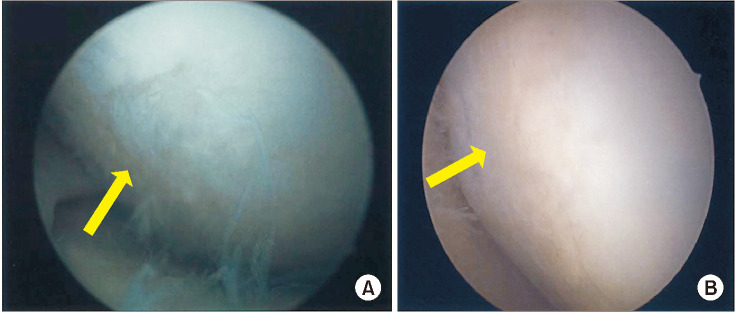
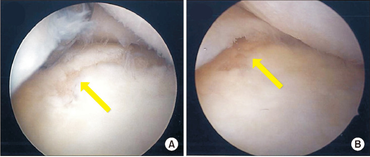
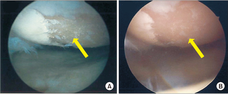
In contrast, only 17% of grade 2 lesions (3/18) and 10% of grade 3 lesions (2/21) were improved to “nearly normal.” However, 10% of grade 3 lesions (2/21) worsened to “severely abnormal” (Figs. 1, 2, 3).
Fig. 1. Arthroscopic evaluation of articular cartilage regeneration in a case of International Cartilage Repair Society grade 1. (A) The preoperative cartilage injury area (arrow). (B) The lesion was completely covered by regenerated cartilage (arrow). This was scored as “normal” by second-look arthroscopy.
Fig. 2. Arthroscopic evaluation of articular cartilage regeneration in a case of International Cartilage Repair Society grade 2. (A) The preoperative cartilage injury area (arrow). (B) The lesion was partially covered by regenerated cartilage and the cartilage defect area became smaller (arrow). This was scored as “nearly normal” by second-look arthroscopy.
Fig. 3. Arthroscopic evaluation of articular cartilage regeneration in a case of International Cartilage Repair Society grade 3. (A) The preoperative cartilage injury area (arrow). (B) There was a little fibrillation but not enough regenerated cartilage (arrow). This was scored as “severely abnormal.”.
Safety
No severe adverse events associated with arthroscopic lavage or liposuction were observed either intraoperatively or postoperatively.
DISCUSSION
The present study investigated the clinical outcomes and second-look arthroscopic findings after intra-articular ADRC injection for the treatment of knee OA, and the results showed that 38% of cartilage lesions were repaired 6 months after ADRC administration. Previous studies reported that 23%–65% of cartilage lesions were repaired after MSC therapy for knee OA (KL 1–2).9,10,18) For better cartilage repair, MSC implantation was proposed to be more desirable than MSC injection (65% vs. 35%),9) and MSC implantation with scaffold was preferred over that without scaffold (58% vs. 23%).18) Some studies have shown that injected cells are mostly located in other parts of the OA joint (such as the synovium) rather than at the site of cartilage lesion, limiting cartilage formation via chondrogenic differentiation.19,20) Kim et al.9) reported that MSC implantation with fibrin glue for knee OA provided better clinical outcomes and second-look arthroscopic findings than MSC injection. Efficient delivery of MSCs to the cartilage lesion site may be important for adequate cartilage repair. However, it is difficult for elderly people to undergo this time-consuming implantation and long postoperative mobilization, limiting weight-bearing and restricting walking. Thus, we treated our patients by intra-articular ADRC injection without any confusing restrictions. Despite the allowance of full weight-bearing immediately postoperatively, the results were satisfactory. Improved clinical outcomes and pain reduction were observed. Although the integrated rehabilitation could contribute to this because several reports have shown that rehabilitation can improve clinical outcome,21) ADRC injection could improve the arthroscopic findings and further restrain the symptom.
In the present study, efficient cartilage repair was more evident in lesions with less overall damage at baseline (ICRS grade 1) than in those with more severe damage (ICRS grade 2 or 3). This is consistent with the result reported by Koh et al.8) This may be a result of better cell survival, proliferation, differentiation, or matrix synthesis in patients with more intact cartilage microenvironment. This consideration is supported by previous studies,22,23) wherein chondrogenic effects were observed if a scaffold was used along with MSC implantation. Some studies suggest that MSCs derived from adipose tissue have a low chondrogenic potential.24,25) In the present study, there were not so many cases of cartilage repair, and ADRCs may not be fully satisfactory in terms of chondrogenesis. However, since some cartilage repair was observed, ADRCs may be indicated for future cartilage therapy.
In this study, although 62% of the lesions were not adequately repaired arthroscopically (“abnormal” or “severely abnormal”), the majority of patients demonstrated improvements in clinical outcomes. As early as 1 month after ADRC injection, significant improvements in clinical outcomes were achieved, as evaluated by KOOS, Lysholm score, and VAS. Other reports also investigated the clinical use of ADRC for knee OA by delivering these cells to the joint either via simple injection or implantation with or without a scaffold and showed satisfactory outcomes.10,18) MSCs can regulate inflammation, suppress apoptosis, stimulate endogenous cell proliferation and repair, and improve blood flow in damaged joints.26) In OA joints, these cells are also known to synthesize extracellular matrix needed to repair damaged articular cartilage and stimulate the proliferation of chondrocytes.27,28) Given the presence of cells with properties similar to MSCs within the adipose tissue,29) we hypothesized that the paracrine effects of ADRCs might also support the normal repair capabilities of chondrocytes and prohibit destructive processes in all stages of knee OA. Moreover, the effect may last > 6 months after ADRC injection. Our results showed that the clinical outcomes 6 months postoperatively were slightly lower than those 3 months postoperatively; however, this difference was not significant. We thought that this may be due to the increase in physical activity postoperatively.
With regard to sex, female patients had significantly better improvement on VAS than male patients. Although no other report has described the reason for this finding, we speculated that this may be due to the lower activity level of female patients. However, further study with a larger number of patients is required to clarify this hypothesis.
In terms of BMI, there was no significant difference in clinical outcomes observed between both high- and low-BMI groups (cutoff, 27.5 kg/m2). Previous studies revealed that MSCs from overweight patients had slower proliferation rate, increased cell senescence, and poor ability to differentiate into multiple lineages, including chondrogenesis.27) Koh et al.10) showed that overweight patients (BMI > 27.5 kg/m2) had significantly worse outcomes, as assessed by the International Knee Documentation Committee score, Tegner activity scale score, and ICRS grade on second-look arthroscopy than non-overweight patients (BMI ≤ 27.5 kg/m2). Further investigation of the role of BMI on the properties of ADRC, such as proliferation and multipotency, is required to reveal the accurate association between high BMI and clinical results of ADRC therapy.
In terms of ADRC dose, there was no significant difference in the clinical results between patients receiving high doses (> 1.0 × 10⁷) and those receiving low doses (≤ 1.0 × 10⁷) of ADRCs. However, radiologic, arthroscopic, and histologic evaluations by Jo et al.30) reported a regeneration of hyaline-like articular cartilage, which reduced the articular cartilage defects in the high-dose group. They performed intra-articular injections of MSCs for knee OA in three dose-escalation cohorts: low dose (1.0 × 10⁷ cells), medium dose (5.0 × 10⁷ cells), and high dose (1.0 × 10⁸ cells). High doses of ADRCs may lead to good results; however, the optimal number remains unknown. Further investigations are required to determine the optimal number of ADRC to achieve the best clinical outcomes for articular cartilage regeneration.
Recently, some comparative studies have shown that heterogeneous cellular composition of SVF (ADRCs) may be responsible for a better therapeutic outcome than adipose-derived stem cells (ADSCs). ADRCs contain only approximately 5% ADSCs, and the other cells in ADRCs have anti-inflammatory and angiogenic potential. In the present study, patients with severe cartilage damage did not show much cartilage repair, but their clinical outcomes improved. This may be due to the strong anti-inflammatory effect of ADRCs, suggesting that ADRCs may have a better anti-inflammatory effect than cartilage regeneration.
This study has some limitations. First, the data were collected retrospectively from a relatively small number of patients, and more than half of them had severe OA (KL grade 4). Second, this study did not perform histological analysis, only clinical and arthroscopic evaluations. Histologic assessment after biopsy is the most reliable method for examining the biomechanical properties of regenerated cartilage; however, we could not conduct biopsies because of ethical issues that may affect the patients negatively. Third, the follow-up period was relatively short. Given the slow metabolic rate of chondrocytes within cartilage, visible repair of severe lesions might require additional time or multiple treatments. Therefore, studies with longer follow-up periods or those assessing repeated treatment may be required.
In conclusion, this study revealed significant improvement in clinical outcomes 6 months after treatment with ADRCs. Furthermore, repair of cartilage defects at 6 months was more evident for lesions that had less overall damage at baseline.
Footnotes
CONFLICT OF INTEREST: No potential conflict of interest relevant to this article was reported.
References
- 1.Buckwalter JA, Martin JA. Osteoarthritis. Adv Drug Deliv Rev. 2006;58(2):150–167. doi: 10.1016/j.addr.2006.01.006. [DOI] [PubMed] [Google Scholar]
- 2.Scanzello CR, Plaas A, Crow MK. Innate immune system activation in osteoarthritis: is osteoarthritis a chronic wound? Curr Opin Rheumatol. 2008;20(5):565–572. doi: 10.1097/BOR.0b013e32830aba34. [DOI] [PubMed] [Google Scholar]
- 3.Coleman CM, Curtin C, Barry FP, O’Flatharta C, Murphy JM. Mesenchymal stem cells and osteoarthritis: remedy or accomplice? Hum Gene Ther. 2010;21(10):1239–1250. doi: 10.1089/hum.2010.138. [DOI] [PubMed] [Google Scholar]
- 4.Kesten S, Fraser JK. Autologous adipose derived regenerative cells: a platform for therapeutic applications. Surg Technol Int. 2016;29:38–44. [PubMed] [Google Scholar]
- 5.Lindroos B, Suuronen R, Miettinen S. The potential of adipose stem cells in regenerative medicine. Stem Cell Rev Rep. 2011;7(2):269–291. doi: 10.1007/s12015-010-9193-7. [DOI] [PubMed] [Google Scholar]
- 6.O’Sullivan J, D’Arcy S, Barry FP, Murphy JM, Coleman CM. Mesenchymal chondroprogenitor cell origin and therapeutic potential. Stem Cell Res Ther. 2011;2(1):8. doi: 10.1186/scrt49. [DOI] [PMC free article] [PubMed] [Google Scholar]
- 7.Fodor PB, Paulseth SG. Adipose derived stromal cell (ADSC) injections for pain management of osteoarthritis in the human knee joint. Aesthet Surg J. 2016;36(2):229–236. doi: 10.1093/asj/sjv135. [DOI] [PubMed] [Google Scholar]
- 8.Koh YG, Choi YJ, Kwon SK, Kim YS, Yeo JE. Clinical results and second-look arthroscopic findings after treatment with adipose-derived stem cells for knee osteoarthritis. Knee Surg Sports Traumatol Arthrosc. 2015;23(5):1308–1316. doi: 10.1007/s00167-013-2807-2. [DOI] [PubMed] [Google Scholar]
- 9.Kim YS, Kwon OR, Choi YJ, Suh DS, Heo DB, Koh YG. Comparative matched-pair analysis of the injection versus implantation of mesenchymal stem cells for knee osteoarthritis. Am J Sports Med. 2015;43(11):2738–2746. doi: 10.1177/0363546515599632. [DOI] [PubMed] [Google Scholar]
- 10.Koh YG, Choi YJ, Kwon OR, Kim YS. Second-look arthroscopic evaluation of cartilage lesions after mesenchymal stem cell implantation in osteoarthritic knees. Am J Sports Med. 2014;42(7):1628–1637. doi: 10.1177/0363546514529641. [DOI] [PubMed] [Google Scholar]
- 11.Khan MH. Update on liposuction: clinical pearls. Cutis. 2012;90(5):259–265. [PubMed] [Google Scholar]
- 12.Fraser JK, Hicok KC, Shanahan R, Zhu M, Miller S, Arm DM. The Celution System: automated processing of adipose-derived regenerative cells in a functionally closed system. Adv Wound Care (New Rochelle) 2014;3(1):38–45. doi: 10.1089/wound.2012.0408. [DOI] [PMC free article] [PubMed] [Google Scholar]
- 13.Brittberg M, Peterson L. Introduction of an articular cartilage classification. ICRS Newsletter. 1998;1(1):5–8. [Google Scholar]
- 14.Roos EM, Roos HP, Lohmander LS, Ekdahl C, Beynnon BD. Knee Injury and Osteoarthritis Outcome Score (KOOS): development of a self-administered outcome measure. J Orthop Sports Phys Ther. 1998;28(2):88–96. doi: 10.2519/jospt.1998.28.2.88. [DOI] [PubMed] [Google Scholar]
- 15.Kocher MS, Steadman JR, Briggs KK, Sterett WI, Hawkins RJ. Reliability, validity, and responsiveness of the Lysholm knee scale for various chondral disorders of the knee. J Bone Joint Surg Am. 2004;86(6):1139–1145. doi: 10.2106/00004623-200406000-00004. [DOI] [PubMed] [Google Scholar]
- 16.Kellgren JH, Lawrence JS. Radiological assessment of osteoarthrosis. Ann Rheum Dis. 1957;16(4):494–502. doi: 10.1136/ard.16.4.494. [DOI] [PMC free article] [PubMed] [Google Scholar]
- 17.Peterfy CG, Guermazi A, Zaim S, et al. Whole-Organ Magnetic Resonance Imaging Score (WORMS) of the knee in osteoarthritis. Osteoarthritis Cartilage. 2004;12(3):177–190. doi: 10.1016/j.joca.2003.11.003. [DOI] [PubMed] [Google Scholar]
- 18.Kim YS, Choi YJ, Suh DS, et al. Mesenchymal stem cell implantation in osteoarthritic knees: is fibrin glue effective as a scaffold? Am J Sports Med. 2015;43(1):176–185. doi: 10.1177/0363546514554190. [DOI] [PubMed] [Google Scholar]
- 19.Agung M, Ochi M, Yanada S, et al. Mobilization of bone marrow-derived mesenchymal stem cells into the injured tissues after intraarticular injection and their contribution to tissue regeneration. Knee Surg Sports Traumatol Arthrosc. 2006;14(12):1307–1314. doi: 10.1007/s00167-006-0124-8. [DOI] [PubMed] [Google Scholar]
- 20.Matsumoto T, Cooper GM, Gharaibeh B, et al. Cartilage repair in a rat model of osteoarthritis through intraarticular transplantation of muscle-derived stem cells expressing bone morphogenetic protein 4 and soluble Flt-1. Arthritis Rheum. 2009;60(5):1390–1405. doi: 10.1002/art.24443. [DOI] [PMC free article] [PubMed] [Google Scholar]
- 21.Allaeys C, Arnout N, Van Onsem S, Govaers K, Victor J. Conservative treatment of knee osteoarthritis. Acta Orthop Belg. 2020;86(3):412–421. [PubMed] [Google Scholar]
- 22.Grigolo B, Lisignoli G, Desando G, et al. Osteoarthritis treated with mesenchymal stem cells on hyaluronan-based scaffold in rabbit. Tissue Eng Part C Methods. 2009;15(4):647–658. doi: 10.1089/ten.TEC.2008.0569. [DOI] [PubMed] [Google Scholar]
- 23.Sato M, Uchida K, Nakajima H, et al. Direct transplantation of mesenchymal stem cells into the knee joints of Hartley strain guinea pigs with spontaneous osteoarthritis. Arthritis Res Ther. 2012;14(1):R31. doi: 10.1186/ar3735. [DOI] [PMC free article] [PubMed] [Google Scholar]
- 24.Sakaguchi Y, Sekiya I, Yagishita K, Muneta T. Comparison of human stem cells derived from various mesenchymal tissues: superiority of synovium as a cell source. Arthritis Rheum. 2005;52(8):2521–2529. doi: 10.1002/art.21212. [DOI] [PubMed] [Google Scholar]
- 25.Bornes TD, Adesida AB, Jomha NM. Mesenchymal stem cells in the treatment of traumatic articular cartilage defects: a comprehensive review. Arthritis Res Ther. 2014;16(5):432. doi: 10.1186/s13075-014-0432-1. [DOI] [PMC free article] [PubMed] [Google Scholar]
- 26.Veronesi F, Giavaresi G, Tschon M, Borsari V, Nicoli Aldini N, Fini M. Clinical use of bone marrow, bone marrow concentrate, and expanded bone marrow mesenchymal stem cells in cartilage disease. Stem Cells Dev. 2013;22(2):181–192. doi: 10.1089/scd.2012.0373. [DOI] [PubMed] [Google Scholar]
- 27.Barry F, Murphy M. Mesenchymal stem cells in joint disease and repair. Nat Rev Rheumatol. 2013;9(10):584–594. doi: 10.1038/nrrheum.2013.109. [DOI] [PubMed] [Google Scholar]
- 28.Vezina Audette R, Lavoie-Lamoureux A, Lavoie JP, Laverty S. Inflammatory stimuli differentially modulate the transcription of paracrine signaling molecules of equine bone marrow multipotent mesenchymal stromal cells. Osteoarthritis Cartilage. 2013;21(8):1116–1124. doi: 10.1016/j.joca.2013.05.004. [DOI] [PubMed] [Google Scholar]
- 29.Zuk PA, Zhu M, Mizuno H, et al. Multilineage cells from human adipose tissue: implications for cell-based therapies. Tissue Eng. 2001;7(2):211–228. doi: 10.1089/107632701300062859. [DOI] [PubMed] [Google Scholar]
- 30.Jo CH, Lee YG, Shin WH, et al. Intra-articular injection of mesenchymal stem cells for the treatment of osteoarthritis of the knee: a proof-of-concept clinical trial. Stem Cells. 2014;32(5):1254–1266. doi: 10.1002/stem.1634. [DOI] [PubMed] [Google Scholar]


