PLOS ONE logoLink to PLOS ONE
. 2022 Aug 22;17(8):e0272920. doi: 10.1371/journal.pone.0272920

Global epidemiology of occult hepatitis B virus infections in blood donors, a systematic review and meta-analysis

Guy Roussel Takuissu 1, Sebastien Kenmoe 2,*, Marie Amougou Atsama 3, Etienne Atenguena Okobalemba 4, Donatien Serge Mbaga 5, Jean Thierry Ebogo-Belobo 6, Arnol Bowo-Ngandji 5, Martin Gael Oyono 7, Jeannette Nina Magoudjou-Pekam 8, Ginette Irma Kame-Ngasse 6, Elisabeth Zeuko’o Menkem 9, Abdel Aziz Selly Ngaloumo 8, Agnès Thierry Rebecca Banlock 5, Alfloditte Flore Feudjio 8, Cromwel Zemnou-Tepap 8, Dowbiss Meta-Djomsi 3, Gilberte Louise Nyimbe Mviena 5, Ines Nyebe Eloundou 5, Jacqueline Félicité Yéngué 10, Josiane Kenfack-Zanguim 8, Juliette Laure Ndzie Ondigui 5, Ridole Martin Zekeng Mekontchou 11, Sabine Aimee Touangnou-Chamda 5, Yrene Kamtchueng Takeu 6, Jean Bosco Taya-Fokou 5, Chris Andre Mbongue Mikangue 5, Raoul Kenfack-Momo 8, Cyprien Kengne-Nde 12, Seraphine Nkie Esemu 2, Richard Njouom 11, Lucy Ndip 2
Editor: Jason T Blackard13
PMCID: PMC9394819  PMID: 35994469

Abstract

This study aimed to assess the global prevalence of occult hepatitis B in blood donors. We searched PubMed, Web of Science, Global Index Medicus, and Excerpta Medica Database. Study selection and data extraction were performed by at least two independent investigators. Heterogeneity (I2) was assessed using the χ2 test on the Cochran Q statistic and H parameters. Sources of heterogeneity were explored by subgroup analyses. This study is registered with PROSPERO, number CRD42021252787. We included 82 studies in this meta-analysis. The overall prevalence of OBI was 6.2% (95% CI: 5.4–7.1) in HBsAg negative and anti-HBc positive blood donors. Only sporadic cases of OBI were reported in HBsAg negative and anti-HBc negative blood donors. The overall prevalence of OBI was 0.2% (95% CI: 0.1–0.4) in HBsAg negative blood donors. The prevalence of OBI was generally higher in countries with low-income economic status. The results of this study show that despite routine screening of blood donors for hepatitis B, the transmission of HBV by blood remains possible via OBI and/or a seronegative window period; hence there is a need for active surveillance and foremost easier access to molecular tests for the screening of blood donors before transfusion.

1. Introduction

Hepatitis B virus (HBV) infections show a negative window (HBsAg and anti-HBc negative) at the onset of infection. There are also so-called occult HBV infections (OBI) where HBsAg is negative while DNA is positive in serum or liver [15]. These two factors contribute to making HBV the most frequent virus whose transmission during blood transfusion is high [6]. After the serologically negative period, individuals infected with HBV will develop HBsAg normally while subjects with OBI remain negative. Occult HBV infections may contribute to the progression of liver disease leading to the development of hepatocellular carcinoma [79]. Occult HBV infections are described by several mechanisms among which reduction, modification of the configuration or lack of synthesis of HBsAg due to the mutation of the S gene, formation of HBsAg/anti-HBs complexes, archiving of HBsAg in peripheral blood mononuclear cells, and co-infection with HCV or HIV which could interfere with the HBV replication [1014]. The risk of transmission of HBV through blood transfusion depends on the HBV endemicity, regions and pre-transfusion tests [6]. In high-income countries, molecular (DNA) and serological (HBsAg, anti-HBc) assays are adopted for screening blood before transfusion [1518]. The algorithms with minipool testing of 6 to 24 blood donors were introduced due to the low concentration of HBV DNA in OBI cases. In most low-income countries, screening before transfusion remains essentially serological (HBsAg alone or with anti-HBc/anti-HBs) [19] and mostly associated with insufficient blood supply. Some algorithms have considered transfusion of blood with low anti-HBc level [20]. Although OBI is not always transmissible through blood transfusion, several studies have reported cases of post-transfusion infections during OBI or the seronegative window period [5, 2123]. This residual risk of post-transfusion transmission is greater for OBI donors with a high viral load and those without anti-HBs. The risk of transmission is also important in immunocompromised recipients and multiple transfused patients [1]. Many studies around the world have reported the prevalence of OBI in blood donors. This prevalence greatly varies among these studies, however, no study has yet reported this global prevalence. This systematic review aimed to determine the prevalence of OBI in blood donors worldwide.

2. Materials and methods

2.1. Protocol and registration

This study was conducted according to the PRISMA guidelines (S1 Appendix) [24]. The protocol was registered in the International Prospective Register of Systematic Reviews (PROSPERO, no. CRD42021252787).

2.2. Eligibility criteria

This review considered all observational studies (cross-sectional, case-control, and cohort) written in English or French and limited to blood donors. According to the data provided by the authors of the included articles, the blood donors were classified as 1) OBI in HBsAg negative and anti-HBc positive blood donors, 2) OBI in HBsAg negative and Anti-HBc negative blood donors, and 3) OBI in HBs negative blood donors. All studies regardless of the technique used to detect DNA in blood donors were included. Studies with repeated donors and pooled test samples were excluded [25]. Studies for which the abstract or full text was not available, duplicates, comments, case reports, case series, and studies fewer than 10 participants, were excluded.

2.3. Data sources and search strategy

PubMed, Excerpta Medica Database (Embase), Web of Science, and Global Index Medicus were searched to identify relevant studies published worldwide from database inception to July 2021. The search terms were related to OBI and Blood Donors. The search strategy conducted in PubMed is presented in S2 Appendix. A manual search was also conducted to browse reference lists of eligible articles and other relevant review articles.

2.4. Study selection

Duplicates identified in the full list of studies were removed. The articles titles and abstracts retrieved from the electronic and manual literature searches were independently reviewed by the authors for eligibility. Articles deemed potentially eligible, full-text retrieval was performed. The authors independently assessed the completeness of each of these texts for final inclusion. Discussion and consensus were reached to resolve any disagreements observed during study selection.

2.5. Data extraction and management

Data from the included studies were extracted using a pre-designed Google data abstraction form. For each eligible article, the extraction was performed independently by at least 2 investigators and verified by a third if necessary. The extracted data were: the name of the first author, year of publication, study design, sampling method, time of data collection and analysis, country, country income level, sample collection period, study site, category of blood donors (HBsAg negative and anti-HBc positive, HBsAg negative and anti-HBc negative or HBsAg negative), age range, methods of diagnosing HBV, laboratory samples used, sample size, number of blood donors with HBV, and risk of bias. In studies where multiple tests for HBsAg were performed, the results of the first test performed was chosen. In studies where multiple molecular detection tests were performed on the same participants, the result with the best sensitivity were selected. Disagreements among the review authors were reconciled by consensus.

2.6. Quality assessment

The methodological quality of the included studies was assessed using the tool developed by Hoy et al. for prevalence studies (S3 Appendix) [26].

2.7. Statistical analysis

As a single study could report the OBI prevalence for several population categories, the analyses according to the number of prevalence data was conducted. Study-specific estimates were pooled using a random-effect model meta-analysis from DerSimonian and Laird. Heterogeneity was assessed by the Cochrane Q statistical test and quantified by I2 values, assuming that the I2 values of 25%, 50% and 75% represent low, moderate, and high heterogeneity, respectively [27]. Publication bias was assessed by Egger’s test and the funnel plot [28]. Subgroup analyses were conducted according to study design, sampling approach, country, WHO and UNSD (United Nations Statistics Division) regions, country income level, OBI categories, and OBI detection assay. A sensitivity analysis including only studies with a low risk of bias and cross-sectional studies representing the best design for prevalence studies was conducted. A p value <0.05 indicated a significant difference. R software version 4.1.0 was used to perform the analyses [29, 30].

3. Results

3.1. Study selection

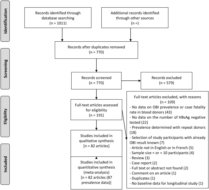
The database search yielded 1011 articles (Fig 1). Subsequently, 241 duplicates were eliminated and from the remaining 770, 579 were excluded due to irrelevant titles and abstracts. A total of 191 eligible articles were therefore fully reviewed with109 excluded with individual reasons given in S4 Appendix. A total of 82 articles were included in the study meeting the defined eligibility criteria, corresponding to 87 blood donor prevalence data (S5 Appendix).

Fig 1. PRISMA flow-chart of studies selected for the meta-analysis.

Fig 1

3.2. Study characteristics

The aggregated and individual characteristics of the included studies are reported in S6 and S7 Appendices. In most of the studies, the age range of the participants was not reported or not clearly defined (n = 64 or 73.6%), but mainly comprised of adults for the studies that reported the information. The most represented UNSD region was South Asia (n = 27 or 31.0%), closely followed by East Asia (n = 19 or 21.8%); while the most represented WHO regions were the West Pacific (n = 23 or 26.4%) and East Mediterranean (n = 22 or 25.3%). In terms of countries, China was the most represented (n = 15 or 17.2%), followed by India (n = 13 or 14.9%), and Iran (n = 10 or 11.5%). Upper middle income countries were the most represented in the various data (n = 43 or 49.4%). Most of the prevalence data came from cross-sectional studies (n = 86 or 98.9%). Most of the data came from non-probability sampling (n = 71 or 81.6%), with prospective data collection and analysis (n = 67 or 77.0%). The bulk of the data were from a single site (n = 67, 77.0%) and were obtained at the hospital (n = 86, 98.9%). The inclusion period for participants was 1991 to 2019. All data were obtained from a blood sample (n = 87, 100%). The most commonly used diagnostic method was real-time PCR (n = 59, 67.8%). Of the 87-prevalence data studied, more were at low risk of bias (n = 49 or 56.3%) (S8 Appendix).

3.3. Occult hepatitis B virus prevalence in HBsAg negative and Anti-HBc positive blood donors

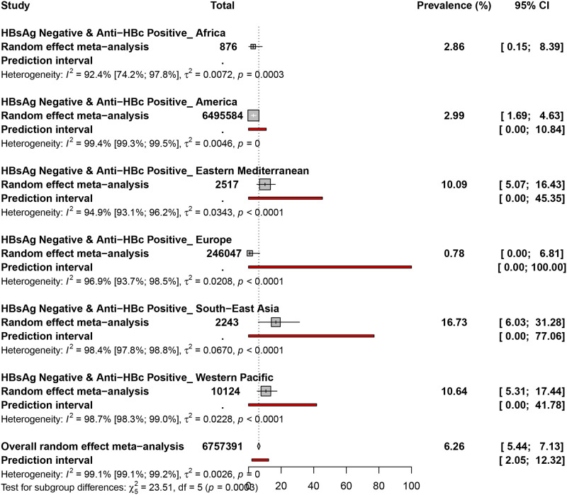
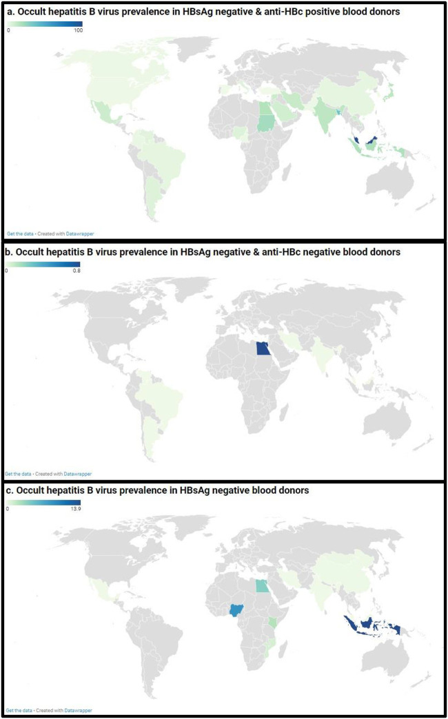
The prevalence of OBI in HBsAg negative and Anti-HBc positive blood donors was assessed in 54 prevalence data from 27 countries (Fig 2 and S9 Appendix). The overall prevalence of OBI in HBsAg negative and Anti-HBc positive blood donors was 6.2% (95% CI: 5.4–7.1). The prevalence of OBI ranged from 0.7% (Europe) to 16.7% (South-east Asia). Occult hepatitis B virus prevalence in HBsAg negative and Anti-HBc positive blood donors was 16.7% (95% CI: 6.0–31.2) in South-east Asia, 10.6% (95% CI: 5.3–17.4) in the Western Pacific, 10.0% (95% CI: 5.0–16.4) in the Eastern Mediterranean, 2.9% (95% CI: 1.6–4.6) in America, 2.8% (95% CI: 0.1–8.3) in Africa, and 0.7% (95% CI: 0.0–6.8) in Europe.

Fig 2. The pooled global prevalence of occult hepatitis B infection in HBsAg negative and anti-HBc positive blood donors.

Fig 2

3.4. Occult hepatitis B virus prevalence in HBsAg negative and Anti-HBc negative blood donors

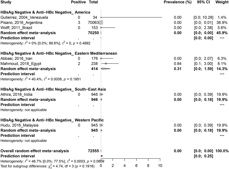
The prevalence of OBI in HBsAg negative and Anti-HBc negative blood donors was assessed in 6 prevalence data from 6 countries (Fig 3). Only two studies in Argentina (3/70063) and Egypt (2/238) reported positive OBI cases in HBsAg negative and anti-Hbc negative blood donors.

Fig 3. The pooled global prevalence of occult hepatitis B infection in HBsAg negative and anti-HBc negative blood donors.

Fig 3

3.5. Occult hepatitis B virus prevalence in HBsAg negative blood donors

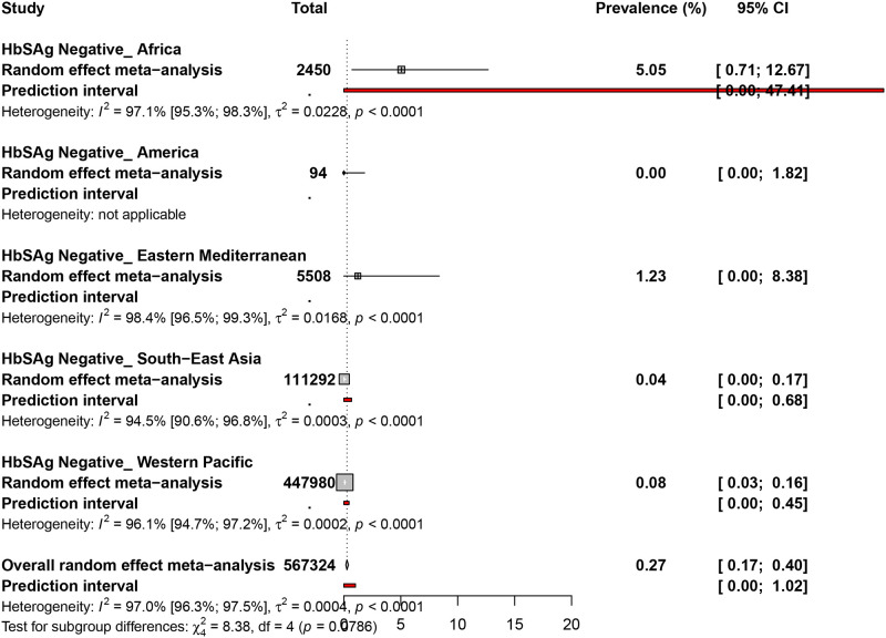
The prevalence of OBI in HBsAg negative blood donors was assessed in 27 prevalence data from 11 countries (Fig 4 and S10 Appendix). The overall prevalence of OBI in HBsAg negative blood donors was 0.2% (95% CI: 0.1–0.4). Occult hepatitis B virus prevalence in HBsAg negative blood donors was 5.0% (95% CI: 0.7–12.6) in Africa, 1.2% (95% CI: 0.0–8.3) in the Eastern Mediterranean, 0.0% (95% CI: 0.0–0.1) in the Western Pacific, 0.0% (95% CI: 0.0–0.1) in South-East Asia, and 0.0% (95% CI: 0.0–3.8) in the Americas.

Fig 4. The pooled global prevalence of hepatitis B virus infection in HBsAg negative blood donors.

Fig 4

3.6. Heterogeneity and publication bias

The degree of heterogeneity and publication bias within the prevalence data is presented in Table 1 and S11S13 Appendices. In HBsAg negative and Anti-HBc positive blood donors, the estimation of prevalence data was associated with significant heterogeneity and the presence of publication bias. The publication bias results obtained by Egger’s test were confirmed by the funnel plot (S11 Appendix). In HBsAg negative and Anti-HBc negative blood donors, the estimation of prevalence data was associated with no significant heterogeneity and publication bias. The publication bias results obtained by Egger’s test were confirmed by the funnel plot (S12 Appendix). In HBsAg negative blood donors, the estimation of prevalence data was associated with significant heterogeneity (H˃1 and I2 ˃50%) and the presence of publication bias (P < 0.05 for Egger’s test). The publication bias results obtained by Egger’s test were confirmed by the funnel plot (S13 Appendix).

Table 1. Summary of meta-analysis results for the prevalence of occult hepatitis B in blood donors.

Prevalence. % (95%CI) 95% Prediction interval N Studies N Participants H (95%CI) §I2 (95%CI) P heterogeneity
Occult hepatitis B virus prevalence in HBsAg negative & anti-HBc positive blood donors
Overall 6.6 [5.7–7.5] [2.2–12.8] 53 6756446 10.9 [10.5–11.5] 99.2 [99.1–99.2] <0.001
Cross-sectional 6.6 [5.7–7.5] [2.2–12.8] 53 6756446 10.9 [10.5–11.5] 99.2 [99.1–99.2] <0.001
Low risk of bias 9.9 [7.7–12.3] [1.1–25.2] 30 6503856 12.6 [11.9–13.3] 99.4 [99.3–99.4] <0.001
Occult hepatitis B virus prevalence in HBsAg negative & anti-HBc negative blood donors
Overall 0 [0–0] [0–0.2] 7 72555 1.4 [1–2.1] 46.7 [0–77.5] 0.081
Cross-sectional 0 [0–0] [0–0.2] 7 72555 1.4 [1–2.1] 46.7 [0–77.5] 0.081
Low risk of bias 0 [0–0] [0–0] 4 72107 1 [1–2.6] 0 [0–84.7] 0.731
Occult hepatitis B virus prevalence in HBsAg negative blood donors
Overall 0.3 [0.2–0.4] [0–1] 27 567324 5.8 [5.2–6.4] 97 [96.3–97.5] <0.001
Cross-sectional 0.2 [0.1–0.3] [0–0.9] 26 566816 5.7 [5.1–6.3] 96.9 [96.2–97.5] <0.001
Low risk of bias 0.4 [0.2–0.5] [0–1.2] 15 467345 6.7 [5.9–7.5] 97.7 [97.1–98.2] <0.001

CI: confidence interval; N: Number; 95% CI: 95% Confidence Interval; NA: not applicable.

H is a measure of the extent of heterogeneity, a value of H = 1 indicates homogeneity of effects and a value of H >1indicates a potential heterogeneity of effects.

§: I2 describes the proportion of total variation in study estimates that is due to heterogeneity, a value > 50% indicates presence of heterogeneity

3.7. Subgroup analyses

Among OBI in blood donors, subgroup analysis showed that the overall prevalence was significantly different according to sampling approach (p = 0.003, higher prevalence in probabilistic sampling), countries (p < 0.001, higher prevalence in Malaysia), WHO region (p < 0.001, higher prevalence in Eastern Mediterranean, South-East Asia, and Africa), UNSD region (p < 0.001, higher prevalence in Southeastern Asia and Northern Africa), country income level (p < 0.001, higher prevalence in Low-income economies followed by Lower-middle income economies, Upper-middle-income economies, and High-income economies), OBI categories (p <0.001, higher prevalence in HBsAg negative and Anti-HBc positive blood donors), and OBI diagnostic method (p < 0.001, higher prevalence with loop mediated isothermal amplification) (Fig 5, S13 Appendix).

Fig 5. Global prevalence estimate of occult hepatitis B virus in blood donors.

Fig 5

The letters (a, b, and c) denote blood donors with HBsAg negative and anti-HBc positive, HBsAg negative and anti-HBc negative, and HBsAg negative, respectively. Map source: https://www.datawrapper.de/.

4. Discussion

To date, the transmission of HBV through blood donation remains crucial. The pretransfusion test which relies on the detection of HBsAg in serum commonly used mainly in low-resource countries does not rule out subjects in the seronegative window and those with OBI. This study aimed to assess the prevalence of OBI in blood donors. The overall prevalence of OBI in HBsAg negative and Anti-HBc positive blood donors was 6.2%. Only 5 sporadic cases of OBI in HBsAg negative and anti-HBc negative blood donors was obtained. The overall prevalence of OBI in HBsAg negative blood donors was 0.2%. The highest prevalence of OBI was recorded in low-resource countries and in the WHO regions of Eastern Mediterranean, South-east Asia, and Africa. Only two systematic reviews previously reported data on the national prevalence of OBI in blood donors [31, 32]. The first study reported a prevalence of 16.4% in 4 studies of blood donors in Sudan [31]. The second study through a Bayesian meta-analysis determined a prevalence of 9.4% among blood donors in China [31]. Of course, our global study cannot be compared to these national systematic reviews. In addition, these two previous systematic reviews did not differentiate the subject with OBI from those in the negative window period [33]. The pooled prevalence of OBI in blood donors was highest in the regions of Eastern Mediterranean, South-East Asia, and Africa. These regions are mainly represented by developing countries which are also areas with high endemicity of HBV. These results are similar to Liu et al., study who reported a higher prevalence of OBI in low-income regions of China [32]. The prevalence of OBI shows great variability in different regions of the world depending on the study population, study design, HBV endemicity, and several factors [12, 34, 35]. Among these factors are the diagnostic approaches used for the detection of anti-HBc, anti-HBs, HBsAg and DNA which was not fully explored in this study. Although studies which explicitly stated that some participants had repeat donations were excluded, this phenomenon is common in most blood banks and therefore cannot guarantee that a residue of this limit did not influence the estimates in this review. Replacement and paid donors are also factors that affect the possibility of having repeated donors in the estimates. Although our review was limited to English or French, this study is the first to report the prevalence of OBI and the negative window period in blood donors with representativeness from all WHO regions of the world.

Molecular techniques made a significant contribution in reducing the risk of transmission of HBV through blood transfusion. The continuous improvement in the sensitivity of molecular tests further reduced the seronegative window period. The high cost, facilities, qualified personnel represent the major obstacles which slow down the adoption of molecular techniques in low-resource settings which paradoxically show the highest rates of HBV and OBI infections [36]. The introduction of molecular techniques into pretransfusion algorithms across all regions of the world and more particularly low-resource areas should be initiated to ensure safer transfusion [37]. The major considerations for the introduction of molecular testing in low-income regions include the consideration of additional number of units of discarded blood with its impact on the shortage of blood units. Also, the development of more sensitive and specific molecular tests is necessary for continuous improvement. In this sense, studies that aim to evaluate the newly developed tests should be implemented. Moreso, it would be important to adopt the vaccination of HBV negative donors without anti-HBs and apply vaccine policies that consider all population categories, is order to achieve the goal of eradicating HBV infection by 2030.

5. Conclusion

This review shows a high prevalence of OBI and / or HBV in the negative window period with the highest frequencies recorded in low-resource areas.

Supporting information

S1 Appendix. Preferred reporting items for systematic reviews and meta-analyses checklist.

(PDF)

S2 Appendix. Search strategy in PubMed.

(PDF)

S3 Appendix. Items for risk of bias assessment.

(PDF)

S4 Appendix. Main reasons of exclusion of eligible studies.

(PDF)

S5 Appendix. Reference list of included studies on global prevalence of occult hepatitis B infection in blood donors.

(PDF)

S6 Appendix. Characteristics of included studies.

(PDF)

S7 Appendix. Individual characteristics of included studies.

(PDF)

S8 Appendix. Risk of bias assessment.

(PDF)

S9 Appendix. The pooled global prevalence of hepatitis B virus infection in HBsAg negative and anti-HBc positive blood donors.

(PDF)

S10 Appendix. The pooled global prevalence of hepatitis B virus infection in HBsAg negative blood donors.

(PDF)

S11 Appendix. Funnel chart for publications of the occult hepatitis B virus prevalence in HBsAg negative and anti-HBc positive blood donors.

(PDF)

S12 Appendix. Funnel chart for publications of the occult hepatitis B virus prevalence in HBsAg negative and anti-HBc negative blood donors.

(PDF)

S13 Appendix. Funnel chart for publications of the occult hepatitis B virus prevalence in HBsAg negative blood donors.

(PDF)

S14 Appendix. Subgroup analyses of global prevalence of occult hepatitis B virus in blood donors.

(PDF)

S1 Checklist. Preferred reporting items for systematic reviews and meta-analyses checklist.

(PDF)

Abbreviations

HBsAg

Hepatitis B surface antigen

HBV

Hepatitis B virus

OBI

Occult hepatitis B infection

Data Availability

All relevant data are within the paper and its Supporting information files.

Funding Statement

This project is part of the EDCTP2 programme supported by the European Union under grant agreement TMA2019PF-2705. The funders had no role in study design, data collection and analysis, decision to publish, or preparation of the manuscript.

References

  • 1.Allain JP. Occult hepatitis B virus infection: implications in transfusion. Vox Sang. 2004;86(2):83–91. Epub 2004/03/17. doi: 10.1111/j.0042-9007.2004.00406.x . [DOI] [PubMed] [Google Scholar]
  • 2.Raimondo G, Allain JP, Brunetto MR, Buendia MA, Chen DS, Colombo M, et al. Statements from the Taormina expert meeting on occult hepatitis B virus infection. J Hepatol. 2008;49(4):652–7. Epub 2008/08/22. doi: 10.1016/j.jhep.2008.07.014 . [DOI] [PubMed] [Google Scholar]
  • 3.Raimondo G, Locarnini S, Pollicino T, Levrero M, Zoulim F, Lok AS. Update of the statements on biology and clinical impact of occult hepatitis B virus infection. J Hepatol. 2019;71(2):397–408. Epub 2019/04/21. doi: 10.1016/j.jhep.2019.03.034 . [DOI] [PubMed] [Google Scholar]
  • 4.Torbenson M, Thomas DL. Occult hepatitis B. The Lancet Infectious diseases. 2002;2(8):479–86. Epub 2002/08/02. doi: 10.1016/s1473-3099(02)00345-6 . [DOI] [PubMed] [Google Scholar]
  • 5.Thiers V, Nakajima E, Kremsdorf D, Mack D, Schellekens H, Driss F, et al. Transmission of hepatitis B from hepatitis-B-seronegative subjects. Lancet (London, England). 1988;2(8623):1273–6. Epub 1988/12/03. doi: 10.1016/s0140-6736(88)92891-7 . [DOI] [PubMed] [Google Scholar]
  • 6.Jayaraman S, Chalabi Z, Perel P, Guerriero C, Roberts I. The risk of transfusion-transmitted infections in sub-Saharan Africa. Transfusion. 2010;50(2):433–42. Epub 2009/10/22. doi: 10.1111/j.1537-2995.2009.002402.x . [DOI] [PubMed] [Google Scholar]
  • 7.Cacciola I, Pollicino T, Squadrito G, Cerenzia G, Orlando ME, Raimondo G. Occult hepatitis B virus infection in patients with chronic hepatitis C liver disease. The New England journal of medicine. 1999;341(1):22–6. Epub 1999/07/01. doi: 10.1056/NEJM199907013410104 . [DOI] [PubMed] [Google Scholar]
  • 8.Covolo L, Pollicino T, Raimondo G, Donato F. Occult hepatitis B virus and the risk for chronic liver disease: a meta-analysis. Digestive and liver disease: official journal of the Italian Society of Gastroenterology and the Italian Association for the Study of the Liver. 2013;45(3):238–44. Epub 2012/11/14. doi: 10.1016/j.dld.2012.09.021 . [DOI] [PubMed] [Google Scholar]
  • 9.Huang X, Hollinger FB. Occult hepatitis B virus infection and hepatocellular carcinoma: a systematic review. Journal of viral hepatitis. 2014;21(3):153–62. Epub 2014/01/21. doi: 10.1111/jvh.12222 . [DOI] [PubMed] [Google Scholar]
  • 10.Carman WF, Zanetti AR, Karayiannis P, Waters J, Manzillo G, Tanzi E, et al. Vaccine-induced escape mutant of hepatitis B virus. Lancet (London, England). 1990;336(8711):325–9. Epub 1990/08/11. doi: 10.1016/0140-6736(90)91874-a [DOI] [PubMed] [Google Scholar]
  • 11.Carman WF, Korula J, Wallace L, MacPhee R, Mimms L, Decker R. Fulminant reactivation of hepatitis B due to envelope protein mutant that escaped detection by monoclonal HBsAg ELISA. Lancet (London, England). 1995;345(8962):1406–7. Epub 1995/06/03. doi: 10.1016/s0140-6736(95)92599-6 . [DOI] [PubMed] [Google Scholar]
  • 12.Raimondo G, Caccamo G, Filomia R, Pollicino T. Occult HBV infection. Seminars in immunopathology. 2013;35(1):39–52. Epub 2012/07/26. doi: 10.1007/s00281-012-0327-7 [DOI] [PMC free article] [PubMed] [Google Scholar]
  • 13.Candotti D, Boizeau L, Laperche S. Occult hepatitis B infection and transfusion-transmission risk. Transfusion clinique et biologique: journal de la Societe francaise de transfusion sanguine. 2017;24(3):189–95. Epub 2017/07/05. doi: 10.1016/j.tracli.2017.06.014 . [DOI] [PubMed] [Google Scholar]
  • 14.Yu DM, Li XH, Mom V, Lu ZH, Liao XW, Han Y, et al. N-glycosylation mutations within hepatitis B virus surface major hydrophilic region contribute mostly to immune escape. J Hepatol. 2014;60(3):515–22. Epub 2013/11/19. doi: 10.1016/j.jhep.2013.11.004 . [DOI] [PubMed] [Google Scholar]
  • 15.Panhotra BR, Bahrani A, Joshi CS, ul Hassan Z. Occult hepatitis B virus infection among anti-HBc positive blood donors: Necessitates substitution of screening by HBV NAT. J Infect. 2005;51(3):263. rayyan-698181859. doi: 10.1016/j.jinf.2005.07.023 [DOI] [PubMed] [Google Scholar]
  • 16.Allain JP, Candotti D. Diagnostic algorithm for HBV safe transfusion. Blood Transfus. 2009;7(3):174–82. Epub 2009/08/07. doi: 10.2450/2008.0062-08 [DOI] [PMC free article] [PubMed] [Google Scholar]
  • 17.Urbani S, Fagnoni F, Missale G, Franchini M. The role of anti-core antibody response in the detection of occult hepatitis B virus infection. Clinical chemistry and laboratory medicine. 2010;48(1):23–9. Epub 2009/11/19. doi: 10.1515/CCLM.2010.002 . [DOI] [PubMed] [Google Scholar]
  • 18.Allain JP, Cox L. Challenges in hepatitis B detection among blood donors. Current opinion in hematology. 2011;18(6):461–6. Epub 2011/09/14. doi: 10.1097/MOH.0b013e32834bac10 . [DOI] [PubMed] [Google Scholar]
  • 19.Candotti D, Allain JP. Transfusion-transmitted hepatitis B virus infection. J Hepatol. 2009;51(4):798–809. Epub 2009/07/21. doi: 10.1016/j.jhep.2009.05.020 . [DOI] [PubMed] [Google Scholar]
  • 20.Bernvil SS, Andrews V, Kuhns MC, McNamara AL. Hepatitis B core antigen antibody as an indicator of a low grade carrier state for hepatitis B virus in a Saudi Arabian blood donor population. Transfusion science. 1997;18(1):49–53. Epub 1997/02/06. doi: 10.1016/s0955-3886(96)00076-8 . [DOI] [PubMed] [Google Scholar]
  • 21.Levicnik-Stezinar S, Rahne-Potokar U, Candotti D, Lelie N, Allain JP. Anti-HBs positive occult hepatitis B virus carrier blood infectious in two transfusion recipients. J Hepatol. 2008;48(6):1022–5. Epub 2008/04/26. doi: 10.1016/j.jhep.2008.02.016 . [DOI] [PubMed] [Google Scholar]
  • 22.Norder H, Hammas B, Larsen J, Skaug K, Magnius LO. Detection of HBV DNA by PCR in serum from an HBsAg negative blood donor implicated in cases of post-transfusion hepatitis B. Archives of virology Supplementum. 1992;4:116–8. Epub 1992/01/01. doi: 10.1007/978-3-7091-5633-9_24 . [DOI] [PubMed] [Google Scholar]
  • 23.Saraswat S, Banerjee K, Chaudhury N, Mahant T, Khandekar P, Gupta RK, et al. Post-transfusion hepatitis type B following multiple transfusions of HBsAg-negative blood. J Hepatol. 1996;25(5):639–43. Epub 1996/11/01. doi: 10.1016/s0168-8278(96)80232-7 . [DOI] [PubMed] [Google Scholar]
  • 24.Moher D, Liberati A, Tetzlaff J, Altman DG, Group P. Preferred reporting items for systematic reviews and meta-analyses: the PRISMA statement. PLoS Med. 2009;6(7):e1000097. Epub 2009/07/22. doi: 10.1371/journal.pmed.1000097 [DOI] [PMC free article] [PubMed] [Google Scholar]
  • 25.Manzini P, Abate ML, Valpreda C, Milanesi P, Curti F, Rizzetto M, et al. Evidence of acute primary occult hepatitis B virus infection in an Italian repeat blood donor. Transfusion. 2009;49(4):757–64. Epub 2009/01/28. doi: 10.1111/j.1537-2995.2008.02041.x . [DOI] [PubMed] [Google Scholar]
  • 26.Hoy D, Brooks P, Woolf A, Blyth F, March L, Bain C, et al. Assessing risk of bias in prevalence studies: modification of an existing tool and evidence of interrater agreement. J Clin Epidemiol. 2012;65(9):934–9. Epub 2012/06/30. doi: 10.1016/j.jclinepi.2011.11.014 . [DOI] [PubMed] [Google Scholar]
  • 27.Higgins JPT. Measuring inconsistency in meta-analyses. BMJ. 2003;327(7414):557–60. doi: 10.1136/bmj.327.7414.557 [DOI] [PMC free article] [PubMed] [Google Scholar]
  • 28.Egger M, Davey Smith G, Schneider M, Minder C. Bias in meta-analysis detected by a simple, graphical test. BMJ. 1997;315(7109):629–34. doi: 10.1136/bmj.315.7109.629 [DOI] [PMC free article] [PubMed] [Google Scholar]
  • 29.Borenstein M, Hedges L, Higgins J, Rothstein H. Introduction to Meta‐Analysis. 1 ed: John Wiley & Sons, Ltd; 2009 2009.
  • 30.Schwarzer G. meta: An R Package for Meta-Analysis. 2007;7:7. [Google Scholar]
  • 31.Eltom K, Albeely A, El Hussein ARM, Elkhidir IM, Enan K. Occult hepatitis B virus infection in Sudan: A systematic review and meta-analysis. JGH open: an open access journal of gastroenterology and hepatology. 2020;4(5):800–7. Epub 2020/10/27. doi: 10.1002/jgh3.12411 [DOI] [PMC free article] [PubMed] [Google Scholar]
  • 32.Liu GC, Sui GY, Liu GY, Zheng Y, Deng Y, Gao YY, et al. A Bayesian meta-analysis on prevalence of hepatitis B virus infection among Chinese volunteer blood donors. PLoS One. 2013;8(11):e79203. Epub 2013/11/16. doi: 10.1371/journal.pone.0079203 [DOI] [PMC free article] [PubMed] [Google Scholar]
  • 33.Yoshikawa A, Gotanda Y, Minegishi K, Taira R, Hino S, Tadokoro K, et al. Lengths of hepatitis B viremia and antigenemia in blood donors: preliminary evidence of occult (hepatitis B surface antigen-negative) infection in the acute stage. Transfusion. 2007;47(7):1162–71. Epub 2007/06/22. doi: 10.1111/j.1537-2995.2007.01234.x . [DOI] [PubMed] [Google Scholar]
  • 34.Makroo RN, Choudhury N, Jagannathan L, Parihar-Malhotra M, Raina V, Chaudhary RK, et al. Multicenter evaluation of individual donor nucleic acid testing (NAT) for simultaneous detection of human immunodeficiency virus -1 & hepatitis B & C viruses in Indian blood donors. The Indian journal of medical research. 2008;127(2):140–7. Epub 2008/04/12. . [PubMed] [Google Scholar]
  • 35.Romanò L, Velati C, Baruffi L, Fomiatti L, Colucci G, Zanetti AR. Multicenter evaluation of a semiautomated, standardized assay for detection of hepatitis B virus DNA in blood donations. J Clin Microbiol. 2005;43(6):2991–3. Epub 2005/06/16. doi: 10.1128/JCM.43.6.2991-2993.2005 [DOI] [PMC free article] [PubMed] [Google Scholar]
  • 36.Hans R, Marwaha N. Nucleic acid testing-benefits and constraints. Asian J Transfus Sci. 2014;8(1):2–3. Epub 2014/03/29. doi: 10.4103/0973-6247.126679 [DOI] [PMC free article] [PubMed] [Google Scholar]
  • 37.Candotti D, Laperche S. Hepatitis B Virus Blood Screening: Need for Reappraisal of Blood Safety Measures? Frontiers in medicine. 2018;5:29. Epub 2018/03/09. doi: 10.3389/fmed.2018.00029 [DOI] [PMC free article] [PubMed] [Google Scholar]

Decision Letter 0

Jason T Blackard

16 Mar 2022

PONE-D-22-03546Global epidemiology of occult hepatitis B virus infections in blood donors, a systematic review and meta-analysisPLOS ONE

Dear Dr. Kenmoe,

Thank you for submitting your manuscript to PLOS ONE. After careful consideration, we feel that it has merit but does not fully meet PLOS ONE’s publication criteria as it currently stands. Therefore, we invite you to submit a revised version of the manuscript that addresses the points raised during the review process.

Please submit your revised manuscript by Apr 30 2022 11:59PM. If you will need more time than this to complete your revisions, please reply to this message or contact the journal office at plosone@plos.org. When you're ready to submit your revision, log on to https://www.editorialmanager.com/pone/ and select the 'Submissions Needing Revision' folder to locate your manuscript file.

Please include the following items when submitting your revised manuscript:

  • A rebuttal letter that responds to each point raised by the academic editor and reviewer(s). You should upload this letter as a separate file labeled 'Response to Reviewers'.

  • A marked-up copy of your manuscript that highlights changes made to the original version. You should upload this as a separate file labeled 'Revised Manuscript with Track Changes'.

  • An unmarked version of your revised paper without tracked changes. You should upload this as a separate file labeled 'Manuscript'.

If you would like to make changes to your financial disclosure, please include your updated statement in your cover letter. Guidelines for resubmitting your figure files are available below the reviewer comments at the end of this letter.

If applicable, we recommend that you deposit your laboratory protocols in protocols.io to enhance the reproducibility of your results. Protocols.io assigns your protocol its own identifier (DOI) so that it can be cited independently in the future. For instructions see: https://journals.plos.org/plosone/s/submission-guidelines#loc-laboratory-protocols. Additionally, PLOS ONE offers an option for publishing peer-reviewed Lab Protocol articles, which describe protocols hosted on protocols.io. Read more information on sharing protocols at https://plos.org/protocols?utm_medium=editorial-email&utm_source=authorletters&utm_campaign=protocols.

We look forward to receiving your revised manuscript.

Kind regards,

Jason T. Blackard, PhD

Academic Editor

PLOS ONE

Journal requirements:

When submitting your revision, we need you to address these additional requirements.

1. Please ensure that your manuscript meets PLOS ONE's style requirements, including those for file naming. The PLOS ONE style templates can be found at

https://journals.plos.org/plosone/s/file?id=wjVg/PLOSOne_formatting_sample_main_body.pdf and

https://journals.plos.org/plosone/s/file?id=ba62/PLOSOne_formatting_sample_title_authors_affiliations.pdf.

2. Thank you for stating the following in the Acknowledgments Section of your manuscript:

“This project is part of the EDCTP2 programme supported by the European Union.”

We note that you have provided funding information that is not currently declared in your Funding Statement. However, funding information should not appear in the Acknowledgments section or other areas of your manuscript. We will only publish funding information present in the Funding Statement section of the online submission form.

Please remove any funding-related text from the manuscript and let us know how you would like to update your Funding Statement. Currently, your Funding Statement reads as follows:

“This project is part of the EDCTP2 programme supported by the European Union under grant agreement TMA2019PF-2705. The funders had no role in study design, data collection and analysis, decision to publish, or preparation of the manuscript.”

Please include your amended statements within your cover letter; we will change the online submission form on your behalf.

3. We note that [Figure 4] in your submission contain [map/satellite] images which may be copyrighted. All PLOS content is published under the Creative Commons Attribution License (CC BY 4.0), which means that the manuscript, images, and Supporting Information files will be freely available online, and any third party is permitted to access, download, copy, distribute, and use these materials in any way, even commercially, with proper attribution. For these reasons, we cannot publish previously copyrighted maps or satellite images created using proprietary data, such as Google software (Google Maps, Street View, and Earth). For more information, see our copyright guidelines: http://journals.plos.org/plosone/s/licenses-and-copyright.

We require you to either (1) present written permission from the copyright holder to publish these figures specifically under the CC BY 4.0 license, or (2) remove the figures from your submission:

a. You may seek permission from the original copyright holder of [Figure 4] to publish the content specifically under the CC BY 4.0 license. 

We recommend that you contact the original copyright holder with the Content Permission Form (http://journals.plos.org/plosone/s/file?id=7c09/content-permission-form.pdf) and the following text:

“I request permission for the open-access journal PLOS ONE to publish XXX under the Creative Commons Attribution License (CCAL) CC BY 4.0 (http://creativecommons.org/licenses/by/4.0/). Please be aware that this license allows unrestricted use and distribution, even commercially, by third parties. Please reply and provide explicit written permission to publish XXX under a CC BY license and complete the attached form.”

Please upload the completed Content Permission Form or other proof of granted permissions as an ""Other"" file with your submission.

In the figure caption of the copyrighted figure, please include the following text: “Reprinted from [ref] under a CC BY license, with permission from [name of publisher], original copyright [original copyright year].”

b. If you are unable to obtain permission from the original copyright holder to publish these figures under the CC BY 4.0 license or if the copyright holder’s requirements are incompatible with the CC BY 4.0 license, please either i) remove the figure or ii) supply a replacement figure that complies with the CC BY 4.0 license. Please check copyright information on all replacement figures and update the figure caption with source information. If applicable, please specify in the figure caption text when a figure is similar but not identical to the original image and is therefore for illustrative purposes only.

The following resources for replacing copyrighted map figures may be helpful:

USGS National Map Viewer (public domain): http://viewer.nationalmap.gov/viewer/

The Gateway to Astronaut Photography of Earth (public domain): http://eol.jsc.nasa.gov/sseop/clickmap/

Maps at the CIA (public domain): https://www.cia.gov/library/publications/the-world-factbook/index.html and https://www.cia.gov/library/publications/cia-maps-publications/index.html

NASA Earth Observatory (public domain): http://earthobservatory.nasa.gov/

Landsat: http://landsat.visibleearth.nasa.gov/

USGS EROS (Earth Resources Observatory and Science (EROS) Center) (public domain): http://eros.usgs.gov/#

Natural Earth (public domain): http://www.naturalearthdata.com/.

Additional Editor Comments:

This is a meta-analysis of occult HBV in blood donors.

There are multiple awkward phrases, and the manuscript should be revised carefully by a native English speaker and/or a professional editing service.

The introduction includes several unsubstantiated sentences and requires additional references and/or restructuring.  For example:

·      These two factors contribute to making HBV the virus whose transmission during blood transfusion is the most frequent [6]. . . . confusing wording / should be reworded

·      Occult HBV infections are explained by several mechanisms including reduction . . . what does “reduction” mean here?

·      In high-income countries, weakly endemic for HBV,  . . . “weakly endemic” is not the correct term

·      Screening for anti-HBc is generally associated with insufficient blood in areas highly endemic for HBV . . . does “insufficient blood” mean insufficient volume of blood for testing or something else?

·      Although OBI is not always transmissible through blood transfusion, several studies have reported cases of post-transfusion infections with OBI and during the seronegative window period [5, 21-23]. . . . there are plenty of studies suggesting that OBI can be transferred to another individual who then develops OBI or chronic HBV, so then what is the meaning of this sentence?

·      The risk of transmission is also very high in immunocompromised recipients or multiple transfused subjects [1]. . . . very high compared to what?

The authors are confusing the seronegative window period with occult HBV infection.  These are not the same thing and should not be combined into one analysis.  This is a major limitation and must be addressed prior to publication.

What does UNSD region stand for?  The full name is never provided.

What does “low risk of bias” mean for some studies?

In figure 1, the N for the “additional records identified through other sources” is absent.

In figure 2, change the prevalence scale from 0 to 100 to 0 to ~20 would be more helpful to visualize the results.

Appendix 4 should be updated with consistent formatting – capitalization of the author’s name, no use of all caps, etc.

[Note: HTML markup is below. Please do not edit.]

Reviewers' comments:

Reviewer's Responses to Questions

Comments to the Author

1. Is the manuscript technically sound, and do the data support the conclusions?

The manuscript must describe a technically sound piece of scientific research with data that supports the conclusions. Experiments must have been conducted rigorously, with appropriate controls, replication, and sample sizes. The conclusions must be drawn appropriately based on the data presented.

Reviewer #1: Yes

Reviewer #2: Partly

**********

2. Has the statistical analysis been performed appropriately and rigorously?

Reviewer #1: Yes

Reviewer #2: Yes

**********

3. Have the authors made all data underlying the findings in their manuscript fully available?

The PLOS Data policy requires authors to make all data underlying the findings described in their manuscript fully available without restriction, with rare exception (please refer to the Data Availability Statement in the manuscript PDF file). The data should be provided as part of the manuscript or its supporting information, or deposited to a public repository. For example, in addition to summary statistics, the data points behind means, medians and variance measures should be available. If there are restrictions on publicly sharing data—e.g. participant privacy or use of data from a third party—those must be specified.

Reviewer #1: Yes

Reviewer #2: Yes

**********

4. Is the manuscript presented in an intelligible fashion and written in standard English?

PLOS ONE does not copyedit accepted manuscripts, so the language in submitted articles must be clear, correct, and unambiguous. Any typographical or grammatical errors should be corrected at revision, so please note any specific errors here.

Reviewer #1: Yes

Reviewer #2: No

**********

5. Review Comments to the Author

Please use the space provided to explain your answers to the questions above. You may also include additional comments for the author, including concerns about dual publication, research ethics, or publication ethics. (Please upload your review as an attachment if it exceeds 20,000 characters)

Reviewer #1: This is a well-conducted analysis that would be a useful addition to the literature. I suggest the authors add further context for the observed heterogeneity. While the I2 values are very large (meaning that the vast majority of variation is due to differences among studies, rather than imprecision within individual studies), this is not surprising because many of the studies are quite large. As the sample sizes become large within-study variation shrinks toward zero, so I2 must increase even if the absolute degree of among-study variation is negligible. In some cases the individual estimates appear similar but I2 is very large. The authors should comment on whether the large "relative" variation is accompanied by enough "absolute" variation to be meaningful (worth attempting to explain by examining factors that differed among studies).

Reviewer #2: This manuscript began with the laudable objective of determined OBI prevalence worldwide among blood donors. It may be a question of semantics, but as the article search was limited to English or French languages, it is difficult to assume that global prevalence of occult HBV could be accurately estimated. There were multiple other weaknesses that dampened my enthusiasm. For example, testing of donor pool blood samples for HBV DNA is the most critical piece of OBI detection; yet the discussion states that this was not a point of examination for the current manuscript. I think it is difficult to draw meaningful conclusions without a good assessment of the diagnostic methods used for HBV DNA at the least (not to mention the serologies). Moreover, I had a great deal of difficulty reading the article. The term “prevalence” was used to describe numbers of articles from different countries; that is an incorrect use of the term, and was quite confusing. Similarly, there are contradictory statements made: e.g., most of the data came from cross-sectional studies, and most of the data came from prospective sampling (which would not constitute a cross-sectional study). I did not see an assessment of study quality, which is a key factor when conducting a meta-analysis. The fact that many studies did not report age certainly renders quality suspect. Net: it is not clear that the conclusions in this manuscript are supported by the data.

**********

6. PLOS authors have the option to publish the peer review history of their article (what does this mean?). If published, this will include your full peer review and any attached files.

If you choose “no”, your identity will remain anonymous but your review may still be made public.

Do you want your identity to be public for this peer review? For information about this choice, including consent withdrawal, please see our Privacy Policy.

Reviewer #1: No

Reviewer #2: No

[NOTE: If reviewer comments were submitted as an attachment file, they will be attached to this email and accessible via the submission site. Please log into your account, locate the manuscript record, and check for the action link "View Attachments". If this link does not appear, there are no attachment files.]

While revising your submission, please upload your figure files to the Preflight Analysis and Conversion Engine (PACE) digital diagnostic tool, https://pacev2.apexcovantage.com/. PACE helps ensure that figures meet PLOS requirements. To use PACE, you must first register as a user. Registration is free. Then, login and navigate to the UPLOAD tab, where you will find detailed instructions on how to use the tool. If you encounter any issues or have any questions when using PACE, please email PLOS at figures@plos.org. Please note that Supporting Information files do not need this step.

PLoS One. 2022 Aug 22;17(8):e0272920. doi: 10.1371/journal.pone.0272920.r002

Author response to Decision Letter 0


14 Apr 2022

Review Comments to the Author

Please remove any funding-related text from the manuscript and let us know how you would like to update your Funding Statement. Currently, your Funding Statement reads as follows:

“This project is part of the EDCTP2 programme supported by the European Union under grant agreementTMA2019PF-2705. The funders had no role in study design, data collection and analysis, decision to publish, or preparation of the manuscript.”

Please include your amended statements within your cover letter; we will change the online submission form on your behalf.

Authors: corrected, thank you. Our funding statement is as above. “This project is part of the EDCTP2 programme supported by the European Union under grant agreementTMA2019PF-2705. The funders had no role in study design, data collection and analysis, decision to publish, or preparation of the manuscript.”

We note that [Figure 4] in your submission contain [map/satellite] images which may be copyrighted. All PLOS content is published under the Creative Commons Attribution License (CC BY 4.0), which means that the manuscript, images, and Supporting Information files will be freely available online, and any third party is permitted to access, download, copy, distribute, and use these materials in any way, even commercially, with proper attribution. For these reasons, we cannot publish previously copyrighted maps or satellite images created using proprietary data, such as Google software (Google Maps, Street View, and Earth). For more information, see our copyright guidelines: http://journals.plos.org/plosone/s/licenses-and-copyright.

We require you to either (1) present written permission from the copyright holder to publish these figures specifically under the CC BY 4.0 license, or (2) remove the figures from your submission:

a. You may seek permission from the original copyright holder of [Figure 4] to publish the content specifically under the CC BY 4.0 license.

We recommend that you contact the original copyright holder with the Content Permission Form(http://journals.plos.org/plosone/s/file?id=7c09/content-permission-form.pdf) and the

following text:

“I request permission for the open-access journal PLOS ONE to publish XXX under the Creative Commons Attribution License (CCAL) CC BY 4.0 (http://creativecommons.org/licenses/by/4.0/). Please be aware that this license allows unrestricted use and distribution, even commercially, by third parties. Please reply and provide explicit written permission to publish XXX under a CC BY license and complete the attached form.”

Please upload the completed Content Permission Form or other proof of granted permissions as an ""Other"" file with your submission.

In the figure caption of the copyrighted figure, please include the following text: “Reprinted from [ref] under a CC BY license, with permission from [name of publisher], original copyright [original copyright year].”

b. If you are unable to obtain permission from the original copyright holder to publish these figures under the CC BY4.0 license or

if the copyright holder’s requirements are incompatible with the CC BY 4.0 license, please either i) remove the figure or ii) supply a replacement figure that complies with the CC BY 4.0 license. Please check copyright information on all replacement figures and update the figure caption with source information. If applicable, please specify in the figure caption text when a figure is similar but not identical to the original image and is therefore for illustrative purposes only.

The following resources for replacing copyrighted map figures may be helpful:

USGS National Map Viewer (public domain): http://viewer.nationalmap.gov/viewer/

The Gateway to Astronaut Photography of Earth (public domain): http://eol.jsc.nasa.gov/sseop/clickmap/

Maps at the CIA (public domain): https://www.cia.gov/library/publications/the-world-factbook/index.html andhttps://www.cia.gov/library/publications/cia-maps-publications/index.html

NASA Earth Observatory

(public domain): http://earthobservatory.nasa.gov/

Landsat: http://landsat.visibleearth.nasa.gov/

USGS EROS (Earth Resources Observatory and Science (EROS) Center) (public domain): http://eros.usgs.gov/#

Natural Earth (public domain): http://www.naturalearthdata.com/.

Authors: We checked the license terms of Datawrapper (https://www.datawrapper.de/: the site we created the figure with), and it looks like they request the label "Created with Datawrapper" that you find below our Fig. 4 to be kept on the figure. We therefore assure you that we meet the CC BY 4.0 license requirements for our fig. 4 for this article.

This is a meta-analysis of occult HBV in blood donors. There are multiple awkward phrases, and the manuscript should be revised carefully by a native English speaker and/or a professional editing service.

The introduction includes several unsubstantiated sentences and requires additional references and/or restructuring. For example:

· These two factors contribute to making HBV the virus whose transmission during blood transfusion is the most frequent [6]. . . . confusing wording / should be reworded

· Occult HBV infections are explained by several mechanisms including reduction . . . what does “reduction” mean here?

· In high-income countries, weakly endemic for HBV, . . . “weakly endemic” is not the correct term

· Screening for anti-HBc is generally associated with insufficient blood in areas highly endemic for HBV . . . does “insufficient blood” mean insufficient volume of blood for testing or something else?

· Although OBI is not always transmissible through blood transfusion, several studies have reported cases of post-transfusion infections with OBI and during the seronegative window period [5, 21-23]. . . . there are plenty of studies suggesting that OBI can be transferred to another individual who then develops OBI or

chronic HBV, so then what is the meaning of this sentence?

· The risk of transmission is also very high in immunocompromised recipients or multiple transfused subjects [1]. .. . very high compared to what?

Authors: We thank the reviewer for all the comments. The manuscript has been improved

according to the comments and suggestions.

The authors are confusing the seronegative window period with occult HBV infection. These are not the same thing and should not be combined into one analysis. This is a major limitation and must be addressed prior to publication.

Authors: Thank you for the comment. We reread all included articles and extracted data when possible, distinguishing studies with positive (occult hepatitis B virus) and negative (window period) anti-HBc. We also repeated all the analyses.

What does UNSD region stand for? The full name is never provided.

Authors: Thank you corrected, United Nations Statistics Division.

What does “low risk of bias” mean for some studies?

Authors: We used the scale proposed by Hoy et al (doi: 10.1016/j.jclinepi.2011.11.014) to assess the risk of bias in prevalence studies. Each study is evaluated on 10 questions (10 marks) which makes it possible to categorize all the included studies in low, moderate or high risk of bias. We also conducted a sensitivity analysis including only studies with a low risk of bias.

In figure 1, the N for the “additional records identified through other sources” is absent.

Authors: corrected as suggested, thank you.

In figure 2, change the prevalence scale from 0 to 100 to 0 to ~20 would be more helpful to visualize the results.

Authors: Fig. 2 is the simplified version of the Appendix S9. We have provided this simplified version since the full version is larger than the A4 printable format. The figure actually hides multiple underlying information such as the prevalence of individual studies which are above 20 and which prevent us from being able to reduce the scale. Thanks for the suggestion.

Appendix 4 should be updated with consistent formatting – capitalization of the author’s name, no use of all caps, etc.

Authors: corrected as suggested, thank you.

Reviewer #1: This is a well-conducted analysis that would be a useful addition to the literature.

Authors: Thank you for this appreciation.

I suggest the authors add further context for the observed heterogeneity. While the I2 values are very large (meaning that the vast majority of variation is due to differences among studies, rather than imprecision within individual studies), this is not surprising because many of the studies are quite large. As the sample sizes become large within-study variation shrinks toward zero, so I2 must increase even if the absolute degree of among-study variation is negligible. In some cases, the individual estimates appear similar but I2 is very large. The authors should comment on whether the large "relative" variation is accompanied by enough "absolute" variation to be meaningful (worth attempting to explain by examining factors that differed among studies).

Authors: We thank the Reviewer for this relevant comment. Heterogeneity is known to be inherent in meta-analyses of observational studies. We also thank the reviewer for pointing out that most of the variability is due to differences between studies and not differences within individual studies. To reduce the influence of sources of heterogeneity on our estimates, we distinguished, when possible, between negative and positive anti-HBc donors. In discussion section, we strengthened the explanation of potential sources of heterogeneity previously mentioned such as the population studied with parameters such as age, type of donors (voluntary, replacement, remunerated, etc.), the possibility of having repeat donors, endemicity of HBV, study design and diagnostic approach to HBV markers (anti-HBc, anti-HBs, HBsAg, and DNA).

Reviewer #2: This manuscript began with the laudable objective of determined OBI prevalence worldwide among blood donors. It may be a question of semantics, but as the article search was limited to English or French languages, it is difficult to assume that global prevalence of occult HBV could be accurately estimated.

Authors: Our review is influenced by multiple factors including the language restriction which we reported in the discussion section, thank you.

There were multiple other weaknesses that dampened my enthusiasm. For example, testing of donor pool blood samples for HBV DNA is the most critical piece of OBI detection; yet the discussion states that this was not a point of examination for the current manuscript.

Authors: For studies that tested pooled blood samples, we included only those that resolved positive pools by further analysis of individual samples as reported in the methodology, thank you.

I think it is difficult to draw meaningful conclusions without a good assessment of the diagnostic methods used for HBV DNA at the least (not to mention the serologies).

Authors: The OBI considers only HBsAg negative subjects with positive and/or negative results for anti-HBc and anti-HBs. We agree on the great variability of sensitivity or specificity of HBV serological tests. This suggests the possibility of having HBsAg false-negative subjects recruited in some included studies. We mentioned these aspects as the limitations of the study which are responsible for the observed heterogeneity. We conducted a subgroup analysis according to the DNA detection test which unfortunately cannot allow us to draw a conclusion although the difference according to the techniques was statistically significant. The assay with the highest prevalence, LAMP, being represented by a single prevalence data.

Moreover, I had a great deal of difficulty reading the article. The term “prevalence” was used to describe numbers of articles from different countries; that is an incorrect use of the term, and was quite confusing. Similarly, there are contradictory statements made: e.g., most of the data came from cross-sectional studies, and most of the data came from prospective sampling (which would not constitute a cross-sectional study).

Authors: A study could present several prevalence data according to the number of population categories. We clarified this aspect in the methodology, thank you.

I did not see an assessment of study quality, which is a key factor when conducting a meta-analysis.

Authors: We presented the results of the risk of bias assessment at the end of the study characteristics (results section) and in Appendix S8

The fact that many studies did not report age certainly renders quality suspect. Net: it is not clear that the conclusions in this manuscript are supported by the data.

Authors: Although very few of the included studies reported donor age information, most blood transfusion policy worldwide relies on adult donors. We have now mentioned that this factor could be a potential source of heterogeneity in our estimates, thank you.

Decision Letter 1

Jason T Blackard

29 Jul 2022

Global epidemiology of occult hepatitis B virus infections in blood donors, a systematic review and meta-analysis

PONE-D-22-03546R1

Dear Dr. Kenmoe,

We’re pleased to inform you that your manuscript has been judged scientifically suitable for publication and will be formally accepted for publication once it meets all outstanding technical requirements.

Within one week, you’ll receive an e-mail detailing the required amendments. When these have been addressed, you’ll receive a formal acceptance letter and your manuscript will be scheduled for publication.

An invoice for payment will follow shortly after the formal acceptance. To ensure an efficient process, please log into Editorial Manager at http://www.editorialmanager.com/pone/, click the 'Update My Information' link at the top of the page, and double check that your user information is up-to-date. If you have any billing related questions, please contact our Author Billing department directly at authorbilling@plos.org.

If your institution or institutions have a press office, please notify them about your upcoming paper to help maximize its impact. If they’ll be preparing press materials, please inform our press team as soon as possible -- no later than 48 hours after receiving the formal acceptance. Your manuscript will remain under strict press embargo until 2 pm Eastern Time on the date of publication. For more information, please contact onepress@plos.org.

Kind regards,

Jason T. Blackard, PhD

Academic Editor

PLOS ONE

Additional Editor Comments (optional):

None

Reviewers' comments:

Reviewer's Responses to Questions

Comments to the Author

1. If the authors have adequately addressed your comments raised in a previous round of review and you feel that this manuscript is now acceptable for publication, you may indicate that here to bypass the “Comments to the Author” section, enter your conflict of interest statement in the “Confidential to Editor” section, and submit your "Accept" recommendation.

Reviewer #1: All comments have been addressed

**********

2. Is the manuscript technically sound, and do the data support the conclusions?

The manuscript must describe a technically sound piece of scientific research with data that supports the conclusions. Experiments must have been conducted rigorously, with appropriate controls, replication, and sample sizes. The conclusions must be drawn appropriately based on the data presented.

Reviewer #1: (No Response)

**********

3. Has the statistical analysis been performed appropriately and rigorously?

Reviewer #1: (No Response)

**********

4. Have the authors made all data underlying the findings in their manuscript fully available?

The PLOS Data policy requires authors to make all data underlying the findings described in their manuscript fully available without restriction, with rare exception (please refer to the Data Availability Statement in the manuscript PDF file). The data should be provided as part of the manuscript or its supporting information, or deposited to a public repository. For example, in addition to summary statistics, the data points behind means, medians and variance measures should be available. If there are restrictions on publicly sharing data—e.g. participant privacy or use of data from a third party—those must be specified.

Reviewer #1: (No Response)

**********

5. Is the manuscript presented in an intelligible fashion and written in standard English?

PLOS ONE does not copyedit accepted manuscripts, so the language in submitted articles must be clear, correct, and unambiguous. Any typographical or grammatical errors should be corrected at revision, so please note any specific errors here.

Reviewer #1: (No Response)

**********

6. Review Comments to the Author

Please use the space provided to explain your answers to the questions above. You may also include additional comments for the author, including concerns about dual publication, research ethics, or publication ethics. (Please upload your review as an attachment if it exceeds 20,000 characters)

Reviewer #1: (No Response)

**********

7. PLOS authors have the option to publish the peer review history of their article (what does this mean?). If published, this will include your full peer review and any attached files.

If you choose “no”, your identity will remain anonymous but your review may still be made public.

Do you want your identity to be public for this peer review? For information about this choice, including consent withdrawal, please see our Privacy Policy.

Reviewer #1: No

**********

Acceptance letter

Jason T Blackard

12 Aug 2022

PONE-D-22-03546R1

Global epidemiology of occult hepatitis B virus infections in blood donors, a systematic review and meta-analysis

Dear Dr. Kenmoe:

I'm pleased to inform you that your manuscript has been deemed suitable for publication in PLOS ONE. Congratulations! Your manuscript is now with our production department.

If your institution or institutions have a press office, please let them know about your upcoming paper now to help maximize its impact. If they'll be preparing press materials, please inform our press team within the next 48 hours. Your manuscript will remain under strict press embargo until 2 pm Eastern Time on the date of publication. For more information please contact onepress@plos.org.

If we can help with anything else, please email us at plosone@plos.org.

Thank you for submitting your work to PLOS ONE and supporting open access.

Kind regards,

PLOS ONE Editorial Office Staff

on behalf of

Dr. Jason T. Blackard

Academic Editor

PLOS ONE

Associated Data

    This section collects any data citations, data availability statements, or supplementary materials included in this article.

    Supplementary Materials

    S1 Appendix. Preferred reporting items for systematic reviews and meta-analyses checklist.

    (PDF)

    S2 Appendix. Search strategy in PubMed.

    (PDF)

    S3 Appendix. Items for risk of bias assessment.

    (PDF)

    S4 Appendix. Main reasons of exclusion of eligible studies.

    (PDF)

    S5 Appendix. Reference list of included studies on global prevalence of occult hepatitis B infection in blood donors.

    (PDF)

    S6 Appendix. Characteristics of included studies.

    (PDF)

    S7 Appendix. Individual characteristics of included studies.

    (PDF)

    S8 Appendix. Risk of bias assessment.

    (PDF)

    S9 Appendix. The pooled global prevalence of hepatitis B virus infection in HBsAg negative and anti-HBc positive blood donors.

    (PDF)

    S10 Appendix. The pooled global prevalence of hepatitis B virus infection in HBsAg negative blood donors.

    (PDF)

    S11 Appendix. Funnel chart for publications of the occult hepatitis B virus prevalence in HBsAg negative and anti-HBc positive blood donors.

    (PDF)

    S12 Appendix. Funnel chart for publications of the occult hepatitis B virus prevalence in HBsAg negative and anti-HBc negative blood donors.

    (PDF)

    S13 Appendix. Funnel chart for publications of the occult hepatitis B virus prevalence in HBsAg negative blood donors.

    (PDF)

    S14 Appendix. Subgroup analyses of global prevalence of occult hepatitis B virus in blood donors.

    (PDF)

    S1 Checklist. Preferred reporting items for systematic reviews and meta-analyses checklist.

    (PDF)

    Data Availability Statement

    All relevant data are within the paper and its Supporting information files.


    Articles from PLoS ONE are provided here courtesy of PLOS

    RESOURCES