Abstract
Purpose:
To assess the response to pexidartinib treatment in six cohorts of adult patients with advanced, incurable solid tumors associated with colony-stimulating factor 1 receptor (CSF1R) and/or KIT proto-oncogene receptor tyrosine kinase activity.
Patients and Methods:
From this two-part phase I, multicenter study, pexidartinib, a small-molecule tyrosine kinase inhibitor that targets CSF1R, KIT, and FMS-like tyrosine kinase 3 (FLT3), was evaluated in six adult patient cohorts (part 2, extension) with advanced solid tumors associated with dysregulated CSF1R. Adverse events, pharmacokinetics, and tumor responses were assessed for all patients; patients with tenosynovial giant cell tumor (TGCT) were also evaluated for tumor volume score (TVS) and patient-reported outcomes (PRO). CSF1 transcripts and gene expression were explored in TGCT biopsies.
Results:
Ninety-one patients were treated: TGCT patients (n = 39) had a median treatment duration of 511 days, while other solid tumor patients (n = 52) had a median treatment duration of 56 days. TGCT patients had response rates of 62% (RECIST 1.1) and 56% (TVS) for the full analysis set. PRO assessments for pain showed improvement in patient symptoms, and 76% (19/25) of TGCT tissue biopsy specimens showed evidence of abnormal CSF1 transcripts. Pexidartinib treatment of TGCT resulted in tumor regression and symptomatic benefit in most patients. Pexidartinib toxicity was manageable over the entire study.
Conclusions:
These results offer insight into outcome patterns in cancers whose biology suggests use of a CSF1R inhibitor. Pexidartinib results in tumor regression in TGCT patients, providing prolonged control with an acceptable safety profile.
Translational Relevance.
The results in this study extend previously reported observations from phase I clinical trial that treatment of tenosynovial giant cell tumors (TGCT) with pexidartinib resulted in sustained tumor regression in most patients with TGCT. Treatment did not translate into the same level of efficacy in the other cohorts that were selected based on presumptive biology targeted by pexidartinib, demonstrating the complexity of these malignancies as compared with TGCT, a neoplasm almost completely dependent on CSF1R signaling. In addition, the molecular data demonstrated how the alterations may provide a mechanism of sustained CSF1R production in TGCT and an explanation as to why inhibition of CSF1R is an effective therapeutic intervention. Future studies would need to be conducted in order to assess if there is a direct correlation between specific patient tissue sample expressing high levels of CSF1 and efficacy.
Introduction
Tenosynovial giant cell tumor (TGCT), formerly known as pigmented villonodular synovitis (PVNS) or giant cell tumor of tendon sheath (GCT-TS), is a rare and locally aggressive neoplasm that affects joints, tendon sheaths, and bursae and is characterized by synovial proliferation and tumors with multinucleated giant cells (1). Intra-articular cellular overexpression of colony-stimulating factor 1 (CSF1) in TGCT plays an important role in the pathogenesis and propagation of TGCT (1, 2). Inhibitors of the CSF1 receptor (CSF1R) have shown compelling antitumor activity in patients with TGCT (3–8). Pexidartinib is a novel oral small-molecule inhibitor that selectively targets CSF1R, as well as the KIT receptor tyrosine kinase (KIT) and FMS-like tyrosine kinase 3 (FLT3) harboring an internal tandem duplication mutation (8–11). In the phase III ENLIVEN trial, pexidartinib was associated with a robust tumor response and improvements in symptoms and functionality among adult patients with severe symptomatic TGCT (4). Based on these results, pexidartinib was the first approved treatment for adult patients with symptomatic TGCT associated with severe morbidity or functional limitations not amenable to improvement with surgery (9, 12, 13). Pexidartinib is available in the United States only through a restricted program under a Risk Evaluation and Mitigation Strategy (REMS) because of the risk of hepatotoxicity (9). Prior to the ENLIVEN study, pexidartinib was evaluated in a two-part phase I study. Results from the part 1 dose-escalation portion have been previously reported (8). In the part 2 extension, six cohorts were enrolled comprising patients with: (i) mucoepidermal carcinoma (MEC) of the salivary gland, (ii) TGCT, (iii) gastrointestinal stromal tumor (GIST), (iv) anaplastic thyroid carcinoma (ATC), (v) solid tumors with documented malignant pleural or peritoneal effusions, and (vi) miscellaneous tumor types with scientific evidence supporting the involvement of CSF1R/KIT signaling in tumorigenesis.
The present analysis reports the results for 91 patients from all six extension cohorts, with emphasis on evaluating long-term efficacy and safety in the TGCT patient cohort. Preliminary results from the first 23 patients in the TGCT cohort were previously reported (8). Owing to those encouraging results as well as the lack of nonsurgical therapy options for patients with advanced diffuse TGCT, this cohort was expanded, and novel tools that might better capture TGCT-specific treatment effect, disease status, and patient-reported outcomes (PRO) were incorporated. These tools included the TVS to measure disease burden more accurately than Response Evaluation Criteria in Solid Tumors (RECIST; refs. 8, 14) and PRO (15, 16) questions to measure symptom improvement; these measures were customized to capture unique aspects of TGCT. Also reported is a post hoc analysis on the CSF1 transcript alterations in patients in the TGCT cohort.
Patients and Methods
Study design, patients, and procedures
This is the part 2 extension of a phase I, multicenter, open-label, uncontrolled, two-part study (Supplementary Appendix). Patients with advanced, incurable solid tumors were enrolled in six extension cohorts and treated with the recommended phase II dose (RP2D) of 1,000 mg/day (taken as 600 mg in the morning and 400 mg in the evening), identified in the part 1 dose-escalation study. Patients continued treatment in part 2 until disease progression or unacceptable toxicity occurred. The primary objective of the extension was to evaluate the potential clinical benefit of single-agent pexidartinib in patients with these specific neoplasms. Pharmacokinetics and safety were also assessed; area under the concentration–time curves over time interval 0 to 4 hours (AUC0–4 h) were estimated from data collection at five time points (nominal time 0, 1, 2, 4, and 6 hours). Adult patients (18 years or older) were enrolled at 12 centers in the United States. The first patient was enrolled on November 16, 2011, and as of March 22, 2021, the enrollment was complete. Briefly, eligibility criteria included: (i) for advanced or recurrent MEC of the salivary gland, patients could not be candidates for curative surgery or radiotherapy; (ii) for TGCT, patients had to have a histologically confirmed diagnosis of inoperable progressive or relapsing TGCT; (iii) for GIST, patients had to have progressed on previous therapy with imatinib and sunitinib; (iv) for ATC, patients had to have histologically or cytologically diagnosed advanced ATC; (v) patients must not have been receiving specific therapy for the effusion or have an indwelling drain; and (vi) other solid tumor types could be included in the miscellaneous cohort. Details of patient eligibility, study design, and procedures have been previously described (8), and key eligibility criteria are listed in the Supplementary Appendix. Patients provided written informed consent. Dose adjustments, usually in increments of 200 mg daily, were allowed, as were temporary drug holds for toxicity or other reasons. This interim analysis presents cumulative data for exposure and response to pexidartinib up to the data cutoff of January 31, 2018 (March 3, 2017, for pharmacokinetic analysis).
Efficacy was assessed locally by imaging at baseline and every 2 months using RECIST version 1.1 [primary outcome, best overall tumor response (i.e., complete response, partial response)]. For patients with TGCT, tumor response was also assessed centrally using a TVS specifically developed for this disease (8, 14). For patients with TGCT enrolled after protocol amendment 8 (August 2013), we used five numeric rating scale (NRS) questions targeting symptoms that are relevant for patients with TGCT (i.e., worst pain, stiffness, limited motion, swelling, and instability; ref. 15). Procedures for assessment of TVS, PRO (for pain), and safety are described in the Supplementary Appendix.
The study protocol was approved by the Institutional Review Board at each study center. All patients provided written informed consent before study eligibility screening. This study was conducted in accordance with Good Clinical Practice guidelines, as provided by the International Conference on Harmonisation and principles of the Declaration of Helsinki, and all applicable regulatory and ethical requirements. This trial is registered with ClinicalTrials.gov, number NCT01004861.
TGCT-targeted RNA sequencing (RNA-seq)
As part of the protocol, archival TGCT tissue biopsies were collected when possible to study the molecular characteristics of this disease. Samples were collected between June 9, 2015 and April 12, 2017. RNA isolation from formalin-fixed paraffin-embedded (FFPE) TGCT specimens was performed at AltheaDx Inc. using the Qiagen FFPE RNeasy kit. An Anchored Multiplex PCR (AMP) RNA panel consisting of 33 gene-specific primer sets was specifically designed to target CSF1, including at least one primer set to detect each exon/exon boundary as well as primer sets to tile the 3′-untranslated regions (3′UTR). Libraries were prepared from ∼200 ng RNA and sequenced on an Illumina MiSeq at a depth greater than 1 million reads per sample. Sequencing data were analyzed with Archer Analysis software (Archer DX, Inc.) to identify chimeric alignments. Potential rearrangements were manually reviewed with Integrative Genomics Viewer (17) and prioritized based on the number of unique reads supporting each rearrangement. CSF1 gene expression was estimated by counting unique CSF1 reads and normalizing to a set of housekeeping genes.
Statistical analysis
For the five non-TGCT disease-specific extension cohorts, a sample size of 10 was considered the minimum number of patients required to test the hypothesis that single-agent pexidartinib could provide a clinical benefit in the individual patient population. Additional statistical analysis details are provided in the Supplementary Appendix.
Data availability statement
Deidentified individual participant data and applicable supporting clinical trial documents may be available upon request at https://www.clinicalstudydatarequest.com. In cases where clinical trial data and supporting documents are provided pursuant to our company policies and procedures, Daiichi Sankyo, Inc., will continue to protect the privacy of our clinical trial participants. Details on data sharing criteria and the procedure for requesting access can be found at https://www.clinicalstudydatarequest.com/Study-Sponsors/Study-Sponsors-DS.aspx.
Results
Patient characteristics and drug exposure
A total of 91 patients in six cohorts were enrolled in the extension study, including 39 patients with advanced TGCT and 52 patients with other tumor types. Patient demographic and baseline characteristics are provided in Table 1. Mean (± standard deviation) patient age was 51 years (±15.5), with 45 years (±14) for the TGCT cohort and 56 years (±14.9) for the non-TGCT cohorts.
Table 1.
Demographics and baseline characteristics of patients in the extension cohorts—safety population.
| Extension cohorts (1,000 mg/day) | |||
|---|---|---|---|
| Patients with TGCT | Non-TGCT patientsa | Total | |
| (n = 39) | (n = 52) | (n = 91) | |
| Age, mean (Standard Deviation) (y) | 45.1 (13.99) | 56.2 (14.92) | 51.4 (15.46) |
| Sex, n (%) | |||
| Male | 17 (44%) | 31 (60%) | 48 (53%) |
| Female | 22 (56%) | 21 (40%) | 43 (47%) |
| Race, n (%) | |||
| White | 33 (85%) | 48 (92%) | 81 (89%) |
| Black | 3 (8%) | 1 (2%) | 4 (4%) |
| Asian | 3 (8%) | 1 (2%) | 4 (4%) |
| Native Hawaiian or other Pacific Islander | 0 | 1 (2%) | 1 (1%) |
| Multiple races | 0 | 1 (2%) | 1 (1%) |
| BMI, mean (Standard Deviation) (kg/m2) | 28.03 (5.866) | 26.90 (6.508) | 27.40 (6.225) |
| Tumor location, n | |||
| Knee | 21 | — | — |
| Foot/ankle | 7 | — | — |
| Hip/thigh | 7 | — | — |
| Forearm/wrist | 2 | — | — |
| Elbow | 1 | — | — |
| Gastroc muscle | 1 | — | — |
| Upper extremity | 1 | — | — |
| Previous treatment, n (%) | |||
| Surgery | 31 (80%) | — | — |
| Radiation | 3 (8%) | — | — |
| TKI | 4 (10%) | — | — |
| Other systemic treatment (denosumab or sirolimus) | 2 (5%) | — | — |
Abbreviations: ATC, anaplastic thyroid carcinoma; BMI, body mass index; GIST, gastrointestinal stromal tumor; MEC, mucoepidermal carcinoma; TGCT, tenosynovial giant cell tumor; TKI, tyrosine kinase inhibitor.
aNon-TGCT patients: The 5 non-TGCT cohorts include the following tumor types: ATC (n = 9), GIST (n = 11), malignant effusion (n = 8), MEC (n = 4), and other tumor types (n = 20). Malignant effusions included mesothelioma (2), colon cancer (2), ovarian adenocarcinoma (1), unknown primary with lung and liver metastases (1), breast cancer (1), and non–small cell lung cancer (1). The category of “other tumor types” included mesothelioma (7), malignant peripheral nerve sheath tumor (3), Erdheim–Chester disease (2), neurofibromatosis (2), leiomyosarcoma (1), adenoid cystic carcinoma (1), pancreatic neuroendocrine tumor (1), acinic cell carcinoma of the parotid (1), familial schwannomatosis (1), and desmoplastic small round cell tumor (1).
Among the 39 patients with TGCT, 31 had undergone previous surgery, 3 had received radiation, 4 had received prior treatment with a kinase inhibitor (imatinib or nilotinib), and 2 had received other systemic treatment (denosumab or sirolimus). Two patients had metastatic disease. The most common tumor location was in the knee (21 patients).
The median duration of treatment for all 91 patients was 111 days (4 months, range, 1–1,814 days), with a longer median duration in the TGCT cohort of 511 days (17 months, range, 15–1,814 days) than in the non-TGCT cohorts [56 days (2 months) range, 1–494 days]. Seven non-TGCT patients (13%) received treatment for more than 6 months. Twenty-three patients with TGCT (59%) were treated for 1 year, 17 (44%) of these patients received treatment for 2 years, 14 (36%) patients for 3 years, and 11 (28%) and 5 (13%) for 4 and 5 years, respectively (as of May 31, 2019).
Of the 91 patients, 30 (33%) required dose reduction and 40 (44%) experienced a drug holiday. Of the 39 patients with TGCT, 24 (62%) required dose reduction and 27 (69%) had a drug holiday. The most common reason for dose interruption among the patients with TGCT was missed doses. Missed doses, dose reductions, and drug holidays were often associated with an adverse event (AE).
Safety
All 91 patients were included in the safety analysis. Treatment-related treatment-emergent AEs (TEAE; ≥10%) are summarized in Supplementary Table S1, and TEAEs (≥20%) are provided in Supplementary Table S2. All 39 patients with TGCT reported at least 1 AE. For patients with TGCT, the most frequently reported treatment-related AEs were fatigue (74%), hair color changes (72%), nausea (56%), periorbital edema (39%), dysgeusia (36%), and pruritus (31%). The most common grade ≥ 3 treatment-related AEs observed in patients with TGCT were hypophosphatemia (10%), alanine aminotransferase increase (10%), and aspartate aminotransferase (AST) increase (8%); all were reversible and well managed with dose interruption or reduction. Two patients with TGCT developed treatment-related serious adverse events (SAE), consisting of hyponatremia and elevated transaminases.
For non-TGCT patients, the most frequently reported TEAEs were fatigue (40%), decreased appetite (39%), nausea (33%), and hair color changes (31%). The most common treatment-related AE of grade ≥ 3 in these non-TGCT patients included fatigue (6%), AST increase (4%), and hypophosphatemia (4%). Four non-TGCT patients developed treatment-related SAEs that led to permanent drug withdrawal. Both patients with GIST withdrew: 1 due to grade 4 liver hemorrhage (a metastatic liver lesion with possible hemorrhage present at baseline) and 1 due to grade 3 hypoxia. The third treatment-related SAE was febrile neutropenia that resolved with medication in a mesothelioma patient. The fourth was a fatal cerebrovascular accident (CVA) occurring in an Erdheim–Chester disease patient with a history of CVA and intraparenchymal hemorrhage who had been on study for more than a year. The patient died 10 days after the onset of the event. Six other patients (1 MEC, 3 GIST, 1 ATC, and 1 maligant effusion patient) died due to disease progression during the study.
Of the 91 patients, 6 had grade 3 elevations in aminotransferase levels; for 5 of these patients, the increases resolved after temporary drug withdrawals or dose reductions. One patient continued to have intermittent liver transaminase elevations more than 1 year after permanent drug withdrawal and was eventually diagnosed with primary biliary cirrhosis considered unrelated to the study drug. Two clinically distinct types of hepatotoxocity have previously been observed—aminotransferase elevations and mixed or cholestatic hepatotoxicity (4, 18, 19). In all cases of drug-related increases in aminotransferase levels in the current study, total bilirubin levels were normal, and there were no cases that met criteria for Hy's law.
Decreases in platelets, hemoglobin, and white blood cells—specifically, neutrophils and lymphocytes—were also observed. These effects were generally grade 2 or less. No patient had clinically significant electrocardiogram abnormalities.
Pharmacokinetics
The AUC0–4 h was estimated for 78 patients (36 patients with TGCT and 42 non-TGCT patients). There were no significant diffferences between the disease-specific cohorts in geometric mean AUC0–4 h (Supplementary Fig. S1). The geometric mean AUC0–4h averaged over 78 patients was 26,052 ng·h/mL and for the 36 patients with TGCT was 23,581 ng·h/mL.
Efficacy
Response by tumor assessment
In the non-TGCT populaiton (n = 52), 1 (Erdheim–Chester disease) was treated for 494 days and had a partial response (PR). Fifteen non-TGCT patients (GIST, n = 4; malignant effusion, n = 3; mesothelioma, n = 2; neurofibromatosis, n = 2; pancreatic neuroendocrine tumor, familial schwannomatosis, MEC, adenoid cystic carcinoma, n = 1 each) had stable disease (SD) as best response (Table 2), 6 of them had prolonged (>6 months) SD while on study drug (Supplementary Table S3). The median progression-free survival (PFS) for patients with TGCT could not be determined [95% confidence interval (CI), 667 days–not applicable; Supplementary Fig. S2], while that for the non-TGCT patients was 56 days (95% CI, 53–161 days; Supplementary Fig. S3).
Table 2.
Treatment duration and best response of SD or PR in non-TGCT patients.
| Tumor | Total treatment duration (days) | Best response |
|---|---|---|
| GIST | 80 | SD |
| GIST | 111 | SD |
| GIST | 169 | SD |
| GIST | 345 | SD |
| MEC | 350 | SD |
| Malignant effusiona | 55 | SD |
| Malignant effusionb | 263 | SD |
| Malignant effusionc | 56 | SD |
| Familial schwannomatosis | 187 | SD |
| Neurofibromatosis | 199 | SD |
| Neurofibromatosis | 113 | SD |
| ACC | 57 | SD |
| Mesothelioma | 150 | SD |
| Pancreatic neuroendocrine tumor | 413 | SD |
| Erdheim–Chester disease | 494 | PR |
| Mesothelioma | 55 | SD |
Abbreviations: ACC, adenoid cystic carcinoma; GIST, gastrointestinal stromal tumor; MEC, mucoepidermal carcinoma; SD, stable disease; PR, partial response; TGCT, tenosynovial giant cell tumor.
aNon–small cell lung cancer.
bMesothelioma.
cColon cancer.
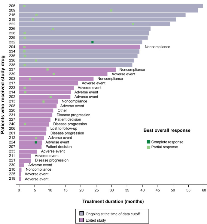
Of the 39 TGCT full analysis set (FAS) patients, 37 met the criteria of the efficacy-evaluable subset (i.e., had a baseline and at least 1 post-baseline radiographic scan). As assessed by RECIST 1.1 per local reading, 24 patients with TGCT achieved a response [2 complete response (CR) and 22 PR], for an objective response rate (ORR) of 62% (95% CI, 45%–77%) and 65% (95% CI, 47%–80%) for the FAS (n = 39) and efficacy-evaluable populations (n = 37), respectively (Fig. 1). The 25th percentile for duration of response (DOR) was 943 days (95% CI, 169 days–not applicable). In addition to those that responded, 8 patients had SD that lasted for at least 6 months, for a disease control rate (DCR = CR + PR + SD) of 82% (95% CI, 66%–92%) and 86% (95% CI, 71%–95%) for the FAS patients (n = 39) and efficacy-evaluable patients (n = 37), respectively. Fifteen patients experienced at least a 50% reduction in sum of longest tumor diameter (Supplementary Fig. S4). Many of these patients who continued on treatment for more than a year demonstrated prolonged tumor response by pexidartinib (18). Thirteen patients remained on pexidartinib treatment as of the January 31, 2018, cutoff, all of whom had been receiving treatment for more than 3 years. As of February 2021, the median DOR and PFS for these 13 subjects that remained on pexidartinib treatment was 1,529 and 1,764 days, respectively.
Figure 1.
Tumor response assessed by local RECIST, treatment duration, and disposition for patients with TGCT. Data cutoff: January 31, 2018. Each line represents 1 patient in the study. Patients received an initial dose of 1,000 mg/day. Treatment duration in months is calculated as treatment duration in days divided by 30.4167. RECIST, Response Evaluation Criteria in Solid Tumors; TGCT, tenosynovial giant cell tumor.
Response by tumor volume score (TVS)
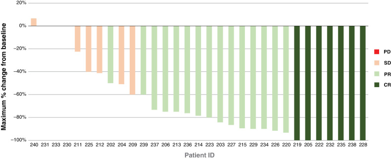
Of the 39 patients with TGCT, 31 were evaluable radiologically by MRI. Similar to RECIST, the TVS assessment showed that the majority of patients experienced a significant decrease in tumor burden (Fig. 2). Of the 31 efficacy-evaluable patients, 7 achieved a CR, and 15 achieved a PR, giving a TVS ORR of 56% (95% CI, 40%–72%) and 71% (95% CI, 52%–86%) for the FAS (n = 39) and efficacy-evaluable (n = 31) patients, respectively. Nine patients had SD that lasted for at least 6 months, giving a DCR of 79% (95% CI, 64%–91%) and 100% (95% CI, 89%–100%) for FAS and efficacy-evaluable patients, respectively.
Figure 2.
Maximum percentage change from baseline in TVS for patients with TGCT. The graph shows the maximum percentage change in TVS score from baseline by individual patient for the 31 MRI-evaluable patients with TGCT. Data cutoff: January 31, 2018. CR, complete response; PD, progressive disease; PR, partial response; RECIST, Response Evaluation Criteria in Solid Tumors; SD, stable disease; TGCT, tenosynovial giant cell tumor; TVS, tumor volume score.
PROs
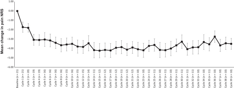
TGCT-related symptoms reported by patients using the NRS [range from 0 (normal) to 10 (extreme)] for TGCT symptoms of worst pain, stiffness, limited motion, swelling, and instability were assessed in 22 patients from baseline and monthly on treatment. All symptoms assessed showed a consistent trend of decreased scores (i.e., symptom improvement) over the course of pexidartinib treatment. Pain has been shown to be one of the most common symptoms in TGCT (15), and the measurement of pain NRS as a tool has been validated in many neoplastic diseases (20). The mean change in the pain NRS from baseline through cycle 39 in the FAS population showed a decrease starting at approximately cycle 1 that was sustained over time, with sustained improvement from cycle 3 through cycle 39 (Fig. 3).
Figure 3.
Mean change from baseline: pain by NRS for patients with TGCT. Data cutoff: January 31, 2018. n drops below 10 after cycle 39, and the results are not shown. NRS, numeric rating scale; TGCT, tenosynovial giant cell tumor.
An assessment of the relationship between TVS and PRO (Brief Pain Inventory NRS Worst Pain item) showed that changes in TVS corresponded with changes in pain in the same direction (Supplementary Fig. S5). Although 31 patients were evaluable by TVS, in this assessment, n = 13, as PRO measurements were assessed toward the end of patient enrollment. Some patients were excluded from TVS of imaging assessment because metal-induced artifacts on MRI.
Targeted RNA-seq identified abnormal CSF1 transcripts and suggested high expression of CSF1 in TGCT biopsy
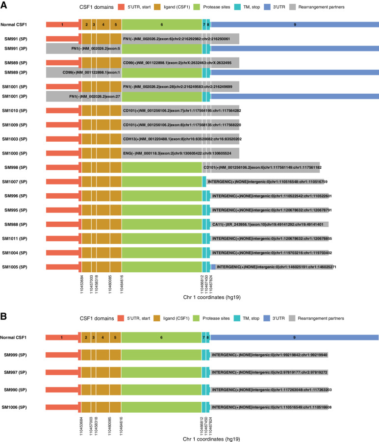
RNA from 25 TGCT samples was successfully isolated and sequenced, of which 19 (76%) showed evidence of abnormal CSF1 transcripts (Fig. 4A and B). Nine samples showed strong evidence of gene rearrangements at the junctions of CSF1 exon 5 (n = 7), exon 6 (n = 1), or exon 7 (n = 1); 8 of those were predicted to be in frame. Three of the 7 exon 5 fusion events were supported by evidence of the reciprocal fusion event (FN1:CSF1 and CD99:CSF1), no COL6A3-CSF1 fusions were observed. Ten specimens showed evidence of alteration at exon 8 or within the 3′UTR, with 6 supported by strong-evidence events and 4 with low confidence.
Figure 4.
TGCT biopsy-targeted RNA-seq identified abnormal CSF1 transcripts. Ten specimens showed evidence of alteration at exon 8 or within the 3′UTR, with 6 strong-evidence events (A) and 4 low-confidence events (B). CA11, carbonic anhydrase 11; CD101, CD101 molecule; CD99, CD99 molecule (Xg blood group); CDH13, cadherin 13; Chr, chromosome; CSF1, colony-stimulating factor 1; ENG, endoglin; FN1, fibronectin 1; SM, sample; TGCT, tenosynovial giant cell tumor; TM, transmembrane; UTR, untranslated region.
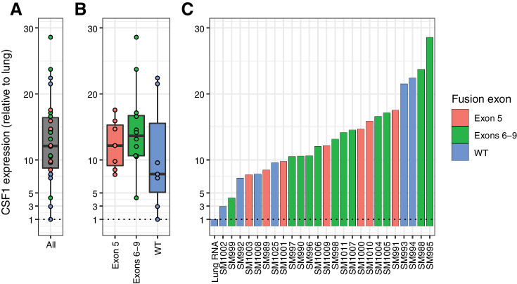
There was no obvious expression difference between the distinct fusion categories that occurred at or near the exon 5 or exon 6–9 junction of CSF1 (Fig. 5A and B). Regardless of CSF1 fusion status, all 25 TGCT libraries exceeded 2× CSF1 expression relative to the lung RNA control library (Fig. 5C).
Figure 5.
CSF1 RNA expression in TGCT relative to lung. Relative to the lung RNA control library, all 25 TGCT libraries had higher CSF1 expression; all 25 exceeded 2× lung CSF1 expression (A–C). There was no obvious expression difference between the distinct fusion categories. CSF1, colony-stimulating factor 1; SM, sample; TGCT, tenosynovial giant cell tumor; WT, wild-type.
Discussion
TGCT is an uncommon neoplasm that can confer significant morbidity, especially the diffuse form of the disease. Though rarely life-threatening or metastatic, TGCT can be a locally invasive, debilitating disease that causes significant suffering and physical impairment. In recent years, systemic therapies, particularly agents that target the CSF1/CSF1R signaling pathways, have shown promising results as novel treatment options for patients with TGCT (3–8, 21–26). Based on the phase III ENLIVEN study, pexidartinib is now approved in the United States for the treatment of adult patients with symptomatic TGCT associated with severe morbidity or functional limitations with surgery at 800-mg total daily dose, reflecting equivalent efficacy and less toxicity (9, 12, 13). Preliminary data from this phase I study enabled the design and conduct of the ENLIVEN study (8). More specifically, to better evaluate the effect of pexidartinib in patients with TGCT, we implemented novel quantitative endpoints, including TVS, a new quantitative radiographical scoring system to fully measure tumor burden, and TGCT-specific PRO questions to understand whether and how a patient's tumor response might correlate with decreased symptoms and improved quality of life (15). These tools were used in ENLIVEN as key secondary endpoints. Overall, the development of these tools represents a step forward for understanding the efficacy and outcomes that are unique to patients with TGCT as they appropriately and longitudinally monitor the effect of a treatment in clinical development.
In this phase I extension study, response to pexidartinib treatment was assessed in six cohorts of adult patients with advanced, incurable solid tumors associated with CSF1R and/or KIT activity. Efficacy was most notable in patients with TGCT, where a considerable proportion of patients experienced substantial and prolonged tumor reduction, in addition to improvements in pain-related PRO. There was no substantial evidence of responses among the non-TGCT patients in this study. The safety profile was similar among all six cohorts, and no late-emerging safety signals were observed in the TGCT cohort. All TGCT patient samples assessed showed elevated expression of CSF1. A significant proportion (15/25, 60%) of these samples also had sequence alterations that could alter CSF1 gene expression or result in the fusion of CSF1 to another protein.
Here, we evaluated response in our TGCT cohort in several ways. First, we used the standard radiologic response criteria, RECIST 1.1, which showed an ORR of over 60% for both the FAS and efficacy-evaluable populations. Many patients who responded by RECIST continued on treatment for more than 3 years, indicating that pexidartinib may provide prolonged control of TGCT. Of note, 3 of the 4 patients (75%) who had previously received other tyrosine kinase inhibitor (TKI) treatment achieved a RECIST response on pexidartinib (8).
Second, we evaluated TGCT response by TVS, which showed an ORR of 56% and 71% for the FAS and efficacy-evaluable populations, respectively. These volumetric measurements could potentially be more sensitive in capturing the full magnitude of response in diffuse TGCT as opposed to the unidimensional measurement of RECIST 1.1.
In addition, we evaluated symptomatic improvement using PRO questions that were created specifically for patients with TGCT. The present study demonstrates a strong relationship between the radiologic outcome (tumor size change) and the patient-reported pain and stiffness outcomes, which highlights both the strength of the relationship between these outcomes and the importance of each of these measures as unique indicators of treatment benefit.
Importantly, our study also gives insight into the long-term application of pexidartinib in patients with TGCT. With pexidartinib, patients usually achieve a radiologic response within 4 months, and the tumor response can be sustained while remaining on drug (4, 13). Additional analysis of tumor response assessment based on modified tumor response criteria showed that only 3 patients progressed while on therapy: 1 with a locally aggressive forearm lesion that never responded and 2 with metastatic TGCT who had either prolonged SD or PR before relapsing. One question to explore is whether tumor reduction and functional improvement would persist after chronic treatment (beyond 1 year) followed by a drug holiday, a dose reduction, or a simple surgery. Currently, a phase IV study is assessing discontinuation and retreatment with pexidartinib in patients with TGCT from multiple studies (NCT02371369, NCT02734433, and NCT03291288), including patients with TGCT from this phase I extension cohort (NCT01004861). The dose used in this phase I extension was calibrated primarily for patients with lethal, end-stage cancers. Based on findings from this study and the ENLIVEN phase III study, the final approved label indication dose was determined to be 800 mg total daily dose, reflecting equivalent efficacy and less toxicity (9).
Overall, short-term safety between patients with TGCT and non-TGCT patients was similar. However, in the TGCT group, dose reductions and holidays were made on a more liberal basis in consideration of the non–life-threatening nature of this disease, the dramatic initial responses, and the longer cumulative duration of therapy. Long-term treatment with pexidartinib demonstrated a tolerable safety profile, with no late-emerging toxicity. Reported TEAEs were mostly low grade, the most frequent being hair color change (18).
Long-term treatment with pexidartinib has been reported to have a predictable effect on hepatic aminotransferases and an unpredictable risk of serious cholestatic or mixed liver injury (19). Prolonged pexidartinib treatment may be associated with hepatic laboratory abnormalities, including hepatic adverse reactions (AR). The vast majority of patients who experienced hepatic ARs had 1 of 2 clinically distinct types. The first type was isolated aminotransferase elevations (> 90%), which were frequent, reversible with dose interruption, and dose dependent. The second type of hepatic AR was mixed or cholestatic hepatotoxicity (< 5%), which, in clinically significant cases, presented as an increase in alkaline phosphatase and total bilirubin with aminotransferase elevations. Notably, all cases of serious liver toxicity observed presented in the first 8 weeks of treatment, and all resolved in patients with TGCT (4, 18, 19).
This study included an in-depth analysis of the CSF1 gene in TGCT tissues. Previous findings identified gross chromosomal aberrations involving the CSF1 locus using break-apart FISH probes (27). In another study, two cohorts of patients with TGCT were investigated for CSF1 rearrangements using FISH and either RNA-seq or DNA-seq with Sanger validation (28). CSF1 rearrangements were identified by FISH in 30/39 cases, and sequencing confirmed CSF1 breakpoints in 28 cases. Patients with TGCT, in their large cohort, were characterized by variable alterations, all of which led to truncation of the 3′ end of CSF1, instead of the COL6A3–CSF1 fusions previously reported in some TGCTs. Another report suggested the importance of CSF1 exon 9 deletion in the molecular pathogenesis of TGCT (29). This analysis and review of the sequencing data revealed two general patterns of CSF1 rearrangement in the TGCT tissues. The most common pattern was a rearrangement near the exon 8/9 junction or within exon 9 (3′UTR) of CSF1, which typically partnered with intergenic sequences, often downstream of CSF1 on chromosome 1. The other common pattern was gene fusion of CSF1 exon 5/6 junction with exons of various other genes. Both patterns of CSF1 alteration would eliminate the 3′UTR microRNA regulatory sites (30), suggesting a loss of negative regulation of CSF1 gene expression.
Analysis of the RNA-seq data also suggested that relative to a lung RNA control library, TGCTs have high expression of CSF1. Together, these results suggest that somatic CSF1 alterations may provide a mechanism of sustained CSF1 production in TGCT and an explanation as to why inhibition of CSF1R is an effective therapeutic intervention.
We note several limitations of this study. First, the study design is open-label and uncontrolled; this especially affects interpretation of the PRO findings for pain. Second, dose reductions were made based on the clinical judgment of the investigators, which led to substantial dose variation. Third, the RECIST results were based on local readings not blinded to visit order. In contrast, assessments by TVS were based on centralized, time-blinded reading, but as a post hoc analysis. Last, for the RNA-seq study, lung RNA is not the most appropriate control. However, lung samples have the fifth highest median CSF1 expression of all tissues in the public GTEx database (> 50 tissues; ref. 31).
Supplementary appendix
Additional data and information about this study are provided in the Supplementary Appendix.
Authors' Disclosures
W.D. Tap reports grants from Plexxikon during the conduct of the study, as well as personal fees from Eli Lilly, EMD-Serono, Mundipharma, C4 Therapeutics, Daiichi Sankyo, Blueprint, Agios Pharmaceuticals, NanoCarrier, Deciphera, Adcendo, Ayala, Kowa, Servier, Bayer, Epizyme, Cogent, Medpactor, and Foghorn outside the submitted work. In addition, W.D. Tap has a patent for Companion Diagnostic for CDK4 inhibitors 14/854,329 pending to MSK/SKI and a patent for Enigma and CDH18 as companion diagnostics for CDK4 inhibition SKI2016-021-03 pending to MSK/SKI; reports scientific advisory board membership for Certis Oncology Solutions and Innova Therapeutics; reports stock ownership from Certis Oncology Solutions and Atropos Therapeutics; and is co-founder of Atropos Therapeutics. A.S. Singh reports grants and personal fees from Daiichi Sankyo during the conduct of the study. A.S. Singh also reports personal fees from Eli Lilly, OncLive, and Expert Perspectives; grants and personal fees from Deciphera, Eisai, Roche, and Blueprint Medicines; grants from NanoCarrier; and other support from Certis Oncology Solutions outside the submitted work. S.P. Anthony reports grants from Plexxikon during the conduct of the study. M. Sterba reports personal fees from Plexxikon Inc. outside the submitted work. J.H. Healey reports personal fees from Daiichi Sankyo during the conduct of the study, as well as personal fees from Daiichi Sankyo outside the submitted work. B. Chmielowski reports grants from Daiichi Sankyo during the conduct of the study, as well as personal fees from Iovance Biotherapeutics, Sanofi Genzyme, OncoSec, IDEAYA Biosciences, Epizyme, Nektar, Regeneron, Biothera, Novartis, Genentech, and Deciphera outside the submitted work. G.I. Shapiro reports other support from Plexxikon during the conduct of the study. G.I. Shapiro also reports grants and personal fees from Eli Lilly, Merck KGaA/EMD-Serono, Sierra Oncology, and Pfizer; personal fees from G1 Therapeutics, Roche, Bicycle Therapeutics, Fusion Pharmaceuticals, Cybrexa Therapeutics, Astex, Daiichi Sankyo, Seattle Genetics, Boehringer Ingelheim, ImmunoMet, Almac, Ipsen, Bayer, Angiex, Asana, Artios, Atrin, Concarlo Holdings, Syros, Zentalis, CytomX Therapeutics, Blueprint Medicines, and Kymera; and grants from Merck & Co outside the submitted work. In addition, G.I. Shapiro has a patent for dosage regimen for sapacitabine and seliciclib issued to Cyclacel Pharmaceuticals and Geoffrey Shapiro and a patent for compositions and methods for predicting response and resistance to CDK4/6 inhibition pending to Liam Cornell and Geoffrey Shapiro. V.L. Keedy reports grants from Plexxikon during the conduct of the study. V.L. Keedy also reports grants from Medpacto, Lilly, Immune Design, GSK, Tracon, Advenchen, Deciphera, and Springworks; grants and personal fees from Daiichi Sankyo; and personal fees from Karyopharm outside the submitted work. Z.A. Wainberg reports grants from AbbVie; Z.A. Wainberg also reports personal fees from Amgen, Bayer, AstraZeneca, Roche, Merck, Incyte, BMS, Seagen, and Genentech, as well as grants and personal fees from Daiichi, Novartis, Ipsen, and Plexxikon outside the submitted work. I. Puzanov reports personal fees from Merck, Nouscom, Oncorus, Amgen, Nektar, and Iovance outside the submitted work. G.M. Cote reports other support from Daiichi Sankyo during the conduct of the study. G.M. Cote also reports other support from Agios, Eisai, Macrogenics, Merck KGaA/EMD-Serono Research and Development Institute, SpringWorks Therapeutics, Bayer, and Repare; personal fees and other support from Epizyme, Foghorn, and PharmaMar; and personal fees from Ikena and C4 Therapeutics outside the submitted work. A.J. Wagner reports grants from Plexxikon and Daiichi Sankyo during the conduct of the study. A.J. Wagner also reports grants and personal fees from Aadi Bioscience, Deciphera, Eli Lilly, Five Prime, and Foghorn; personal fees from Mundipharma, Boehringer Ingelheim, Cogent Biosciences, and NanoCarrier; and grants from Karyopharm outside the submitted work. F. Braiteh reports personal fees from Lilly, AstraZeneca, BMS, Incyte, Deciphera, Eisai, Seagen, Daiichi Sankyo, Boehringer Ingelheim, Pfizer, Amgen, Genentech, and Regeneron outside the submitted work. E. Sherman reports grants and non-financial support from Daiichi Sankyo during the conduct of the study. E. Sherman also reports grants and personal fees from Regeneron; personal fees from Eisai and Exelixis; and grants, personal fees, and non-financial support from Eli Lilly/Loxo and Roche outside the submitted work. H.H. Hsu reports personal fees from Plexxikon during the conduct of the study. C. Peterfy reports personal fees from Daiichi Sankyo during the conduct of the study, as well as personal fees from Five Prime, Deciphera, AmMax, and SynOx outside the submitted work. H.L. Gelhorn reports other support from DSI during the conduct of the study. X. Ye reports other support from Daiichi Sankyo, Inc. during the conduct of the study, as well as other support from Daiichi Sankyo, Inc. outside the submitted work. P.S. Lin reports other support from Plexxikon Inc. during the conduct of the study, as well as a patent for US 9358235 B2 issued and a patent for US 9730918 B2 issued. No disclosures were reported by the other authors.
Supplementary Material
Acknowledgments
We thank the patients who volunteered to participate in this study, their family members and caregivers, and the study center staff members who cared for the patients. Medical writing assistance was provided by Phillip Giannopoulos and Shruthi Satish of SciStrategy Communications and funded by Daiichi Sankyo, Inc. We also thank Karen Getz for her support with writing the manuscript. Research sponsored by Daiichi Sankyo, Inc. and Plexxikon Inc., a subsidiary of Daiichi Sankyo, Inc.; ClinicalTrials.gov number: NCT01004861. W.D. Tap and J.H. Healey acknowledge that all research funding for Memorial Sloan Kettering is supported in part by a grant from the NIH/NCI (#P30 CA008748). Manuscript writing support was sponsored by Daiichi Sankyo, Inc.
The publication costs of this article were defrayed in part by the payment of publication fees. Therefore, and solely to indicate this fact, this article is hereby marked “advertisement” in accordance with 18 USC section 1734.
Footnotes
Note: Supplementary data for this article are available at Clinical Cancer Research Online (http://clincancerres.aacrjournals.org/).
Authors' Contributions
W.D. Tap: Conceptualization, resources, supervision, investigation, visualization, methodology, writing–original draft, project administration, writing–review and editing. A.S. Singh: Investigation, writing–review and editing. S.P. Anthony: Resources, writing–review and editing. M. Sterba: Data curation, supervision, project administration, writing–review and editing. C. Zhang: Conceptualization, formal analysis, supervision, investigation, writing–review and editing. J.H. Healey: Conceptualization, investigation, writing–review and editing. B. Chmielowski: Formal analysis, supervision, validation, investigation, writing–review and editing. A.L. Cohn: Data curation, supervision, investigation, writing–review and editing. G.I. Shapiro: Resources, supervision, investigation, writing–review and editing. V.L. Keedy: Resources, investigation, writing–review and editing. Z.A. Wainberg: Investigation, writing–review and editing. I. Puzanov: Resources, validation, investigation, writing–review and editing. G.M. Cote: Investigation, writing–review and editing. A.J. Wagner: Resources, investigation, writing–review and editing. F. Braiteh: Resources, data curation, investigation, writing–review and editing. E. Sherman: Resources, investigation, writing–review and editing. H.H. Hsu: Conceptualization, supervision, investigation, methodology, project administration, writing–review and editing. C. Peterfy: Resources, formal analysis, writing–review and editing. H.L. Gelhorn: Investigation, methodology, writing–review and editing. X. Ye: Conceptualization, visualization, methodology, project administration, writing–review and editing. P. Severson: Data curation, formal analysis, writing–review and editing. B.L. West: Conceptualization, data curation, supervision, investigation, writing–review and editing. P.S. Lin: Conceptualization, supervision, writing–review and editing. S. Tong-Starksen: Conceptualization, formal analysis, investigation, project administration, writing–review and editing.
References
- 1. Cassier PA, Italiano A, Gomez-Roca CA, Le Tourneau C, Toulmonde M, Cannarile MA, et al. CSF1R inhibition with emactuzumab in locally advanced diffuse-type tenosynovial giant cell tumours of the soft tissue: a dose-escalation and dose-expansion phase 1 study. Lancet Oncol 2015;16:949–56. [DOI] [PubMed] [Google Scholar]
- 2. Gelderblom H, Cropet C, Chevreau C, Boyle R, Tattersall M, Stacchiotti S, et al. Nilotinib in locally advanced pigmented villonodular synovitis: a multicentre, open-label, single-arm, phase 2 trial. Lancet Oncol 2018;19:639–48. [DOI] [PubMed] [Google Scholar]
- 3. Turalio (Pexidartinib) Capsules, for Oral Use [prescribing information]. Basking Ridge, NJ: Daiichi Sankyo, Inc; 2020. [Google Scholar]
- 4. Gounder MM, Thomas DM, Tap WD. Locally aggressive connective tissue tumors. J Clin Oncol 2018;36:202–9. [DOI] [PMC free article] [PubMed] [Google Scholar]
- 5. Staals EL, Ferrari S, Donati DM, Palmerini E. Diffuse-type tenosynovial giant cell tumour: current treatment concepts and future perspectives. Eur J Cancer 2016;63:34–40. [DOI] [PubMed] [Google Scholar]
- 6. Tap WD, Gelderblom H, Palmerini E, Desai J, Bauer S, Blay JY, et al. Pexidartinib versus placebo for advanced tenosynovial giant cell tumour (ENLIVEN): a randomised phase 3 trial. Lancet 2019;394:478–87. [DOI] [PMC free article] [PubMed] [Google Scholar]
- 7. Tap WD, Wainberg ZA, Anthony SP, Ibrahim PN, Zhang C, Healey JH, et al. Structure-guided blockade of CSF1R kinase in tenosynovial giant-cell tumor. N Engl J Med 2015;373:428–37. [DOI] [PubMed] [Google Scholar]
- 8. Verspoor FGM, Mastboom MJL, Hannink G, Maki RG, Wagner A, Bompas E, et al. Long-term efficacy of imatinib mesylate in patients with advanced tenosynovial giant cell tumor. Sci Rep 2019;9:14551. [DOI] [PMC free article] [PubMed] [Google Scholar]
- 9. Benner B, Good L, Quiroga D, Schultz TE, Kassem M, Carson WE, et al. Pexidartinib, a novel small molecule CSF-1R inhibitor in use for tenosynovial giant cell tumor: a systematic review of pre-clinical and clinical development. Drug Des Devel Ther 2020;14:1693–704. [DOI] [PMC free article] [PubMed] [Google Scholar]
- 10. Monestime S, Lazaridis D. Pexidartinib (TURALIO): the first FDA-indicated systemic treatment for tenosynovial giant cell tumor. Drugs R D 2020;20:189–95. [DOI] [PMC free article] [PubMed] [Google Scholar]
- 11. Smith CC, Zhang C, Lin KC, Lasater EA, Zhang Y, Massi E, et al. Characterizing and overriding the structural mechanism of the quizartinib-resistant FLT3 “Gatekeeper” F691L mutation with PLX3397. Cancer Discov 2015;5:668–79. [DOI] [PMC free article] [PubMed] [Google Scholar]
- 12. FDA Briefing Document. Oncologic Drugs Advisory Committee Meeting. NDA 211810 Pexidartinib 2019.Available from:https://www.fda.gov/drugs/resources-information-approved-drugs/fda-approves-pexidartinib-tenosynovial-giant-cell-tumor. FDA approves pexidartinib for tenosynovial giant cell tumor.
- 13. Gelderblom H, van de Sande M. Pexidartinib: first approved systemic therapy for patients with tenosynovial giant cell tumor. Future Oncol 2020;16:2345–56. [DOI] [PubMed] [Google Scholar]
- 14. Peterfy C, Ye X, Gelhorn HL, Speck RM, Countryman PJ, Keedy VL, et al. Tumor volume score (TVS), modified recist, and tissue damage score (TDS) as novel methods for assessing response in tenosynovial giant cell tumors (TGCT) treated with pexidartinib: relationship with patient-reported outcomes (PROs). J Clin Oncol 2017;35:11048. [Google Scholar]
- 15. Gelhorn HL, Tong S, McQuarrie K, Vernon C, Hanlon J, Maclaine G, et al. Patient-reported symptoms of tenosynovial giant cell tumors. Clin Ther 2016;38:778–93. [DOI] [PMC free article] [PubMed] [Google Scholar]
- 16. Gelhorn HL, Ye X, Speck RM, Tong S, Healey JH, Bukata SV, et al. The measurement of physical functioning among patients with tenosynovial giant cell tumor (TGCT) using the patient-reported outcomes measurement information system (PROMIS). J Patient Rep Outcomes 2019;3:6. [DOI] [PMC free article] [PubMed] [Google Scholar]
- 17. Robinson JT, Thorvaldsdottir H, Winckler W, Guttman M, Lander ES, Getz G, et al. Integrative genomics viewer. Nat Biotechnol 2011;29:24–6. [DOI] [PMC free article] [PubMed] [Google Scholar]
- 18. Gelderblom H, Wagner AJ, Tap WD, Palmerini E, Wainberg ZA, Desai J, et al. Long-term outcomes of pexidartinib in tenosynovial giant cell tumors. Cancer 2021;127:884–93. [DOI] [PMC free article] [PubMed] [Google Scholar]
- 19. Lewis JH, Gelderblom H, van de Sande M, Stacchiotti S, Healey JH, Tap WD, et al. Pexidartinib long-term hepatic safety profile in patients with tenosynovial giant cell tumors. Oncologist 2021;26:e863–73. [DOI] [PMC free article] [PubMed] [Google Scholar]
- 20. Cleeland CS, Ryan KM. Pain assessment: global use of the Brief Pain Inventory. Ann Acad Med Singap 1994;23:129–38. [PubMed] [Google Scholar]
- 21. Brahmi M, Vinceneux A, Cassier PA. Current systemic treatment options for tenosynovial giant cell tumor/pigmented villonodular synovitis: targeting the CSF1/CSF1R axis. Curr Treat Options Oncol 2016;17:10. [DOI] [PubMed] [Google Scholar]
- 22. Nakayama R, Jagannathan JP, Ramaiya N, Ferrone ML, Raut CP, Ready JE, et al. Clinical characteristics and treatment outcomes in six cases of malignant tenosynovial giant cell tumor: initial experience of molecularly targeted therapy. BMC Cancer 2018;18:1296. [DOI] [PMC free article] [PubMed] [Google Scholar]
- 23. Palmerini E, Staals EL, Maki RG, Pengo S, Cioffi A, Gambarotti M, et al. Tenosynovial giant cell tumour/pigmented villonodular synovitis: outcome of 294 patients before the era of kinase inhibitors. Eur J Cancer 2015;51:210–7. [DOI] [PubMed] [Google Scholar]
- 24. Ravi V, Wang WL, Lewis VO. Treatment of tenosynovial giant cell tumor and pigmented villonodular synovitis. Curr Opin Oncol 2011;23:361–6. [DOI] [PubMed] [Google Scholar]
- 25. Rebuzzi SE, Grassi M, Catalano F, Buscaglia M, Bertulli R, Satragno C, et al. Multiple systemic treatment options in a patient with malignant tenosynovial giant cell tumour. Anticancer Drugs 2020;31:80–4. [DOI] [PMC free article] [PubMed] [Google Scholar]
- 26. Temple HT. Pigmented villonodular synovitis therapy with MSCF-1 inhibitors. Curr Opin Oncol 2012;24:404–8. [DOI] [PubMed] [Google Scholar]
- 27. West RB, Rubin BP, Miller MA, Subramanian S, Kaygusuz G, Montgomery K, et al. A landscape effect in tenosynovial giant-cell tumor from activation of CSF1 expression by a translocation in a minority of tumor cells. Proc Natl Acad Sci U S A 2006;103:690–5. [DOI] [PMC free article] [PubMed] [Google Scholar]
- 28. Ho J, Peters T, Dickson BC, Swanson D, Fernandez A, Frova-Seguin A, et al. Detection of CSF1 rearrangements deleting the 3' UTR in tenosynovial giant cell tumors. Genes Chromosomes Cancer 2020;59:96–105. [DOI] [PubMed] [Google Scholar]
- 29. Tsuda Y, Hirata M, Katayama K, Motoi T, Matsubara D, Oda Y, et al. Massively parallel sequencing of tenosynovial giant cell tumors reveals novel CSF1 fusion transcripts and novel somatic CBL mutations. Int J Cancer 2019;145:3276–84. [DOI] [PubMed] [Google Scholar]
- 30. Woo HH, Laszlo CF, Greco S, Chambers SK. Regulation of colony stimulating factor-1 expression and ovarian cancer cell behavior in vitro by miR-128 and miR-152. Mol Cancer 2012;11:58. [DOI] [PMC free article] [PubMed] [Google Scholar]
- 31. Gene expression for CSF1. GTExPortal website. GTEx Analysis Release V8 (dbGaP Accession phs000424.v8.p2). [cited May 24, 2021]. Available from: https://gtexportal.org/home/gene/CSF1.
Associated Data
This section collects any data citations, data availability statements, or supplementary materials included in this article.
Supplementary Materials
Data Availability Statement
Deidentified individual participant data and applicable supporting clinical trial documents may be available upon request at https://www.clinicalstudydatarequest.com. In cases where clinical trial data and supporting documents are provided pursuant to our company policies and procedures, Daiichi Sankyo, Inc., will continue to protect the privacy of our clinical trial participants. Details on data sharing criteria and the procedure for requesting access can be found at https://www.clinicalstudydatarequest.com/Study-Sponsors/Study-Sponsors-DS.aspx.


