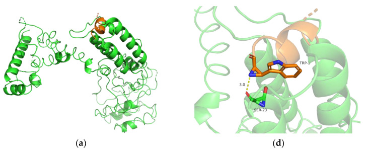
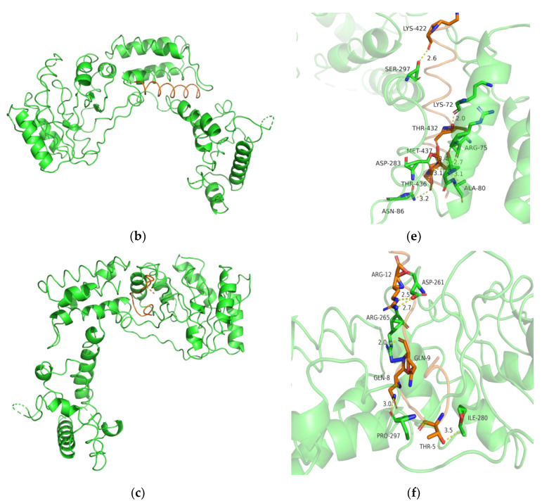
Figure 9.
Molecular docking of different signal peptides with Escherichia coli SRP. SRP and signal peptides are shown as cartoons colored in green and orange, respectively, and the related residues are shown as stick models. The spatial structures of the Escherichia coli DsbA signal peptide (a), the nattokinase signal peptide (b), and the nattokinase signal peptide containing the upstream sequence of peT28a(+) (c) docking with SRP are shown in the left picture; the interactions between the Escherichia coli DsbA signal peptide (d), the nattokinase signal peptide (e), the nattokinase signal peptide containing the upstream sequence of peT28a(+) (f) and SRP are shown in the picture on the right.


