Abstract
Purposes: To quantitatively predict the risk of neck lymph node metastasis for unilateral and bilateral papillary thyroid microcarcinomas (PTMC) that may guide individual treatment strategies for the neck region. Methods: A total of 717 PTMC patients from three medical centers were enrolled for analysis. Results: Bilateral PTMCs were demonstrated to be more aggressive with a much higher cervical lymph node metastasis rate including for both central (CLNM) and lateral lymph node metastasis (LLNM) when being compared to unilateral PTMCs. In unilateral PTMC, five (age < 55 years old, male, maximum tumor diameter (MTD) ≥ 0.5 cm, and the presence of thyroid capsular invasion (TCI) and multifocality) and three (maximum diameter of positive CLN (MDCLN) > 0.5 cm, the presence of multifocality and nodular goiter, iNG) factors were identified as independent risk factors for CLNM and LLNM, respectively. In bilateral PTMC, three (age < 55 and presence of TCI and multifocality in at least one side of thyroid lobe) and two (MDCLN > 0.5 cm and presence of nodular goiter (iNG)) factors were identified as independent factors for CLNM and LLNM, respectively. Predictive models of CLNM and LLNM for patients with unilateral disease and of CLNM for patients with the bilateral disease were established based on the described risk factors. Bilateral patients with positive CLNM were also stratified into different subgroups according to the presence and absence of independent risk factors. Conclusion: An evaluation system based on independent factors of CLNM and LLNM for PTMC patients with bilateral and unilateral disease was established. Our newly established evaluation system can efficaciously quantify risks of CLNM and LLNM for PTMC patients with bilateral and unilateral disease and may guide individual treatment strategy including both surgical and postoperative adjuvant treatment of the neck region for these patients.
Keywords: papillary thyroid microcarcinoma, central lymph node metastasis, lateral lymph node metastasis, bilateral disease, unilateral disease
1. Introduction
Papillary thyroid carcinoma (PTC) is the most prevalent type of thyroid cancer and has seen a continued increase in incidence over the recent decades globally [1,2]. PTC that has a maximum tumor diameter of 1.0 cm or less is defined as papillary thyroid microcarcinoma (PTMC), accounting for over half of all newly diagnosed thyroid cancer cases [3,4]. Although the nature of PTMC is generally indolent and the long-term prognosis is satisfactory, cervical lymph node involvement remains a concern [5], especially for those with bilateral lesions where lymph node metastasis rate may reach up to 60% [6]. Considering that the 2015 American Thyroid Association (ATA) places all intrathyroidal PTMCs, whether unifocal or multifocal, in the low-risk category, it is important to clarify the risk of lymph node metastasis involvement in PTMC [7]. It has also been reported that the occurrence of lymph node involvement and locoregional recurrence are both significantly more frequent in PTC patients with bilateral lesions than those with unilateral lesions [8]. In addition, the prevalence of BRAF V600E mutation in patients with bilateral PTMC was also significantly higher than in those with unilateral disease [9], indicating that marked differences exist between these two different entities.
Whether prophylactic central lymph node dissection (CLND) should be conducted for PTMC patients with no clinically detected central lymph node metastasis (CLNM) is still controversial. Although the occult CLNM rate is not negligible with an incidence rate over 60% in some series [10,11], the relatively higher incidence of postoperative complications caused by CLND put into question the routine conduction of prophylactic surgery involving this region [12]. Developing an effective method to accurately predict cervical metastasis is thus crucial for the surgical management strategy of PTMC patients. Previous studies have revealed significantly different histopathological findings including lymph node metastasis between patients with bilateral and unilateral PTMC, however, none of these studies have quantitatively summarized the risk of cervical lymph node metastasis including both CLNM and lateral lymph node metastasis (LLNM) in PTMC patients with bilateral and unilateral diseases. Here in our current research, a comprehensive and meticulous evaluating system that can efficaciously quantify risks of CLNM and LLNM for bilateral and unilateral PTMC was established.
2. Materials and methods
2.1. Study Population
Between 2018 and 2020, 1477 patients with PTC received initial surgery at three clinical centers: Department of Otorhinolaryngology, Head and Neck Surgery at the Eye, Ear, Nose, and Throat Hospital of Fudan University, Department of General Surgery at Ruijin Hospital of Shanghai Jiao Tong University School of Medicine; and Department of General Surgery, Civil Aviation Shanghai Hospital. Among them, 751 patients were diagnosed with PTMC by postoperative pathology. Patients meeting any of the following conditions were excluded from our study: (1) having received thyroid-related surgery previously (n = 22); (2) history or coexistence of other primary tumors (n = 12). As a result, a total of 717 patients were enrolled for further analysis. This study was approved by the Institutional Ethics Committee of the Eye and ENT Hospital of Fudan University and the Ruijin Hospital of Shanghai Jiao Tong University School of Medicine.
2.2. Surgical Management and Clinicopathological Features
Clinical and pathological features were retrospectively collected and analyzed. Preoperative fine-needle aspiration (FNA) was performed in all enrolled patients with PTMC and diagnosed by cytology. A total thyroidectomy or thyroid lobectomy was conducted for all patients in our cohort. Central lymph node dissection (CLND) was also performed for all patients considering both prophylactic and therapeutic purposes. Lateral lymph node dissection (LLND) was performed therapeutically for those with pre-operatively detected lateral lymph node metastasis (LLNM) using both preoperative ultrasonography and FNA. The LLND was performed for those with clinically detected lateral lymph nodes that were highly suspected as having tumor involvement using preoperative ultrasonography but later proven LLNM negative by FNA biopsy. Patients enrolled were treated with postoperative TSH suppression and radioactive iodine (RAI) therapy according to the 2015 American Thyroid Association Guidelines [7]. For patients receiving CLND only, if positive LLNM was found by ultrasonography and FNA within six months after initial surgery, they would be regarded as having lateral involvement at the time of operation. The thyroid glands were categorized into three equal volumes (upper portion, middle portion, and lower portion) based on the consensus of most clinical medical centers. Patients with tumors involving both sides of thyroid lobes were defined as having bilateral disease.
2.3. Statistical Analyses
The categorical and continuous variables were compared using the Pearson Chi-square test and independent t-test, respectively. Logistic univariate and multivariate regression analyses were used for screening out risk factors that were significantly correlated with LLNM by the SPSS 24.0 package (SPSS Inc., Chicago, IL, USA). A p-value of <0.05 was considered statistically significant. The prediction models for the risk of CLNM and LLNM were created based on the selected independent risk factors, respectively; and the corresponding concordance index (C-index), receiver operating characteristic (ROC) curve, and the calibration curve were constructed using R software (version 3.5.1; R Development Core Team, Bell Laboratories, Lucent Technologies, Murray Hill, NJ, USA).
3. Results
3.1. Clinicopathological Characteristics of PTMC Patients with Unilateral and Bilateral Diseases
A total of 582 (81.2%) patients were confirmed as having unilateral disease and were classified into the Uni group among all PTMC patients, while the other 135 (18.8%) patients that were diagnosed as having the bilateral disease were categorized into the Bil group. CLND was conducted routinely for all patients while LLND was only conducted for 47 (6.6%) patients with clinically detected or highly suspected LLNM. As a result, 307 (42.8%) and 41 (5.7%) patients were confirmed as having CLNM and LLNM by postoperative pathology. In addition, eight patients receiving CLND alone and detected as having lateral neck involvement within six months in post-operation follow-up were also regarded as having preoperative LLNM. In total, 49 (6.8%) patients were considered as having LLNM at the time of initial operation.
The basic clinicopathological features of patients within the Uni and Bil groups are shown and compared in Table 1. Tumor size was significantly larger in patients of the Bil group than that of the Uni group ((0.58 ± 0.23) cm vs. (0.54 ± 0.22) cm, p-value = 0.031). The presence of thyroid capsular invasion (TCI, 40.7% vs. 27.7%, p-value = 0.003), multifocality in at least one side of thyroid lobe (49.6% vs. 23.2%, p-value = 0.000), upper portion tumor of thyroid (33.3% vs. 24.4%, p-value = 0.033), and Hashimoto thyroiditis (HT, 28.1% vs. 18.4%, p-value = 0.011) were significantly more frequent in patients of the Bil group than those of the Uni group. In terms of cervical lymph node metastasis, the overall CLNM and LLNM rates were 42.8% (307 in 717) and 6.8% (49 in 717), respectively, for all patients enrolled, and the incidence of CLNM was significantly higher in patients within the Bil group than those within the Uni group (55.6% vs. 39.9%, p-value = 0.001). Detailed information on CLNM was also collected and analyzed, and the result showed that although patients with bilateral and unilateral disease exhibited comparable levels in terms of counts of positive central lymph node (CLN), patients within the Bil group showed significantly larger positive CLN sizes than those within the Uni group ((0.64 ± 0.42) cm vs. (0.49 ± 0.36) cm, p-value = 0.003). Moreover, for patients with positive CLNM, those with bilateral disease also showed a significantly higher lateral neck involvement rate (25.3% vs. 12.9%, p-value = 0.011).
Table 1.
The clinicopathological characteristics of patients with PTMC.
| All Patients | Unilateral | Bilateral | |||||
|---|---|---|---|---|---|---|---|
| n = 717 | % | n = 582 | % | n = 135 | % | p Value | |
| Age (mean ± SD) | 43.40 ± 12.31 | 43.25 ± 12.37 | 44.04 ± 12.09 | 0.502 | |||
| BMI (mean ± SD) | 23.60 ± 3.68 | 23.51 ± 3.66 | 23.98 ± 3.77 | 0.180 | |||
| Maximum tumor diameter (mean ± SD) | 0.55 ± 0.23 | 0.54 ± 0.22 | 0.58 ± 0.23 | 0.031 | |||
| Gender | 0.499 | ||||||
| Male | 230 | 32.1 | 190 | 32.6 | 40 | 29.6 | |
| Female | 487 | 67.9 | 392 | 67.4 | 95 | 70.4 | |
| Thyroid capsular invasion | 0.003 | ||||||
| No | 501 | 69.9 | 421 | 72.3 | 80 | 59.3 | |
| Yes | 216 | 30.1 | 161 | 27.7 | 55 | 40.7 | |
| Multifocality | 0.000 | ||||||
| Absent | 515 | 71.8 | 447 | 76.8 | 68 | 50.4 | |
| Present | 202 | 28.2 | 135 | 23.2 | 67 | 49.6 | |
| Tumor location | 0.033 | ||||||
| Upper portion | 187 | 26.1 | 142 | 24.4 | 45 | 33.3 | |
| Middle/Lower portion | 530 | 73.9 | 440 | 75.6 | 90 | 66.7 | |
| PTMC with Hashimoto thyroiditis | 0.011 | ||||||
| No | 572 | 79.8 | 475 | 81.6 | 97 | 71.9 | |
| Yes | 145 | 20.2 | 107 | 18.4 | 38 | 28.1 | |
| PTMC with ipsilateral nodular goiter | 0.942 | ||||||
| No | 517 | 72.1 | 420 | 72.2 | 97 | 71.9 | |
| Yes | 200 | 27.9 | 162 | 27.8 | 38 | 28.1 | |
| CLNM | 0.001 | ||||||
| No | 410 | 57.2 | 350 | 60.1 | 60 | 44.4 | |
| Yes | 307 | 42.8 | 232 | 39.9 | 75 | 55.6 | |
| Number of positive CLN | 0.621 | ||||||
| (For patients with CLNM only, n = 307) | |||||||
| 1–2 | 178 | 58.0 | 136 | 58.6 | 42 | 56.0 | |
| 3–4 | 71 | 23.1 | 55 | 23.7 | 16 | 21.3 | |
| ≥5 | 58 | 18.9 | 41 | 17.7 | 17 | 22.7 | |
| Maximum diameter of positive CLN | 0.003 | ||||||
| Mean ± SD, cm | 0.53 ± 0.38 | 0.49 ± 0.36 | 0.64 ± 0.42 | ||||
| Median (range), cm | 0.4 (0.1–2.5) | 0.4 (0.1–2.5) | 0.5 (0.1–2.0) | ||||
| 0.024 | |||||||
| ≤0.5cm | 216 | 70.4 | 171 | 73.7 | 45 | 60.0 | |
| >0.5cm | 91 | 29.6 | 61 | 26.3 | 30 | 40.0 | |
| LLNM | 0.011 | ||||||
| No | 258 | 84.0 | 202 | 87.1 | 56 | 74.7 | |
| Yes | 49 | 16.0 | 30 | 12.9 | 19 | 25.3 | |
SD, standard error; PTMC, papillary thyroid microcarcinoma; BMI, body mass index; CLNM, central lymph node metastasis; CLN, central lymph node; LLNM, lateral lymph node metastasis.
3.2. Comparisons between PTMC with or without CLNM and LLNM for Patients within Uni and Bil Groups
Further analyses were conducted between patients with or without CLNM and LLNM within Uni and Bil groups (Shown in Table 2). For patients in the Uni group, the age of patients with positive CLNM was significantly younger than those with negative CLNM (40.32 ± 11.98 years old vs. 45.20 ± 12.26 years old, p-value = 0.000). The maximum tumor diameter (MTD) was larger in patients with positive CLNM than in those with negative CLNM (p-value = 0.000). In addition, being male and the presence of TCI and ipsilateral multifocality were significantly more frequent in patients with positive CLNM (p-value = 0.000, 0.000, and 0.000, respectively). We further divided patients with positive CLNM into two subgroups according to the status of lateral lymph node involvement: patients with positive (n = 30) and negative (n = 202) LLNM. The presence of ipsilateral multifocality (70.0% vs. 35.1%) and nodular goiter (iNG, 56.7% vs. 23.8%) were significantly more common in patients with positive LLNM than those without (p-value = 0.000 and 0.000, respectively). Patients with positive LLNM also showed significantly larger sizes and higher counts of positive CLN than those with negative LLNM (p-value = 0.000 and 0.001, respectively).
Table 2.
The clinicopathological characteristics of PTMC patients with different lymph node metastasis status within Bil and Uni groups.
| Uni Group (n = 582) | Bil Group (n = 135) | |||||||||||
|---|---|---|---|---|---|---|---|---|---|---|---|---|
| All Patients (n = 582) (n (%)) | p Value | Patients with CLNM (n = 232) (n (%)) | p Value | All Patients (n = 135) (n (%)) | p Value | Patients with CLNM (n = 75) (n (%)) | p Value | |||||
| No-CLNM | CLNM | No-LLNM | LLNM | No-CLNM | CLNM | No-LLNM | LLNM | |||||
| n = 350 | n = 232 | n = 202 | n =30 | n = 60 | n = 75 | n = 56 | n = 19 | |||||
| Age (mean ± SD) | 45.20 ± 12.26 | 40.32 ± 11.98 | 0.000 | 40.78 ± 12.15 | 37.23 ± 10.43 | 0.130 | 48.82 ± 11.74 | 40.23 ± 11.03 | 0.000 | 39.82 ± 10.71 | 41.42 ± 12.16 | 0.588 |
| BMI (mean ± SD) | 23.44 ± 3.32 | 23.61 ± 4.13 | 0.588 | 23.66 ± 4.18 | 23.30 ± 3.82 | 0.658 | 23.25 ± 3.33 | 24.56 ± 4.01 | 0.045 | 24.46 ± 4.22 | 24.85 ± 3.40 | 0.720 |
| Maximum tumor diameter (mean ± SD) | 0.49 ± 0.22 | 0.61 ± 0.23 | 0.000 | 0.61 ± 0.20 | 0.63 ± 0.21 | 0.654 | 0.53 ± 0.23 | 0.63 ± 0.23 | 0.009 | 0.61 ± 0.22 | 0.68 ± 0.26 | 0.285 |
| Gender | 0.000 | 0.749 | 0.070 | 0.021 | ||||||||
| Male | 88 (25.1) | 102 (44.0) | 88 (43.6) | 14 (46.7) | 13 (21.7) | 27 (36.0) | 16 (28.6) | 11 (57.9) | ||||
| Female | 262 (74.9) | 130 (56.0) | 114 (56.4) | 16 (53.3) | 47 (78.3) | 48 (64.0) | 40 (71.4) | 8 (42.1) | ||||
| Thyroid capsular invasion | 0.000 | 0.202 | 0.009 | 0.739 | ||||||||
| No | 303 (86.6) | 118 (50.9) | 106 (52.5) | 12 (40.0) | 43 (71.7) | 37 (49.3) | 27 (48.2) | 10 (52.6) | ||||
| Yes | 47 (13.4) | 114 (49.1) | 96 (47.5) | 18 (60.0) | 17 (28.3) | 38 (50.7) | 29 (51.8) | 9 (47.4) | ||||
| Multifocality | 0.000 | 0.000 | 0.000 | 0.060 | ||||||||
| Absent | 307 (87.7) | 140 (60.3) | 131 (64.9) | 9 (30.0) | 43 (71.7) | 25 (33.3) | 22 (39.3) | 3 (15.8) | ||||
| Present | 43 (12.3) | 92 (39.7) | 71 (35.1) | 21 (70.0) | 17 (28.3) | 50 (66.7) | 34 (60.7) | 16 (84.2) | ||||
| Tumor location | 0.752 | 0.683 | 0.142 | 0.435 | ||||||||
| Upper portion | 87 (24.9) | 55 (23.7) | 47 (23.3) | 8 (26.7) | 24 (40.0) | 54 (72.0) | 17 (30.4) | 4 (21.1) | ||||
| Middle/Lower portion | 263 (75.1) | 177 (76.3) | 155 (76.7) | 22 (73.3) | 36 (60.0) | 21 (28.0) | 39 (69.6) | 15 (78.9) | ||||
| Number of positive CLN | / | 0.001 | / | 0.209 | ||||||||
| 1–2 | / | 136 (58.6) | 126 (62.4) | 10 (33.3) | / | 42 (56.0) | 34 (60.7) | 8 (42.1) | ||||
| 3–4 | / | 55 (23.7) | 47 (23.3) | 8 (26.7) | / | 16 (21.3) | 12 (21.4) | 4 (21.1) | ||||
| ≥5 | / | 41 (17.7) | 29 (14.4) | 12 (40.0) | / | 17 (22.7) | 10 (17.9) | 7 (36.8) | ||||
| Maximum diameter of positive CLN | / | 0.000 | / | 0.000 | ||||||||
| ≤0.5cm | / | 171 (73.7) | 169 (83.7) | 2 (6.7) | / | 45 (60.0) | 42 (75.0) | 3 (15.8) | ||||
| >0.5cm | / | 61 (26.3) | 33 (16.3) | 28 (93.3) | / | 30 (40.0) | 14 (25.0) | 16 (84.2) | ||||
| PTMC with Hashimoto thyroiditis | 0.309 | 0.964 | 0.467 | 0.634 | ||||||||
| No | 281 (80.3) | 194 (83.6) | 169 (83.7) | 25 (83.3) | 45 (75.0) | 52 (69.3) | 38 (67.9) | 14 (73.7) | ||||
| Yes | 69 (19.7) | 38 (16.4) | 33 (16.3) | 5 (16.7) | 15 (25.0) | 23 (30.7) | 18 (32.1) | 5 (26.3) | ||||
| PTMC with ipsilateral nodular goiter | 0.936 | 0.000 | 0.966 | 0.006 | ||||||||
| No | 253 (72.3) | 167 (72.0) | 154 (76.2) | 13 (43.3) | 43 (71.7) | 54 (72.0) | 45 (80.4) | 9 (47.4) | ||||
| Yes | 97 (27.7) | 65 (28.0) | 48 (23.8) | 17 (56.7) | 17 (28.3) | 21 (28.0) | 11 (19.6) | 10 (52.6) | ||||
PTMC, papillary thyroid microcarcinoma; BMI, body mass index; CLNM, central lymph node metastasis; CLN, central lymph node; LLNM, lateral lymph node metastasis.
For patients in the Bil group, younger age, larger MTD, being male, and a more common presence of TCI and multifocality in at least one side of the thyroid lobe were also found in patients with positive CLNM compared with those with negative CLNM (Shown in Table 2). However, the presence of iNG, which was proven to be significantly more frequent in patients with positive CLNM among all patients within the Uni group, showed no difference in patients with positive CLNM among those within the Bil group (28.0% vs. 28.3%, p-value = 0.966). For patients with positive CLNM within the Bil group, the percentages of factors including male patients and iNG were significantly higher in patients with positive LLNM (p-value = 0.021 and 0.006, respectively). Additionally, patients with positive LLNM also showed a larger size of positive CLN than those with negative LLNM (p-value = 0.000).
3.3. Creation of Risk Prediction Model for Cervical Lymph Node Metastasis of PTMC Patients within Uni Group
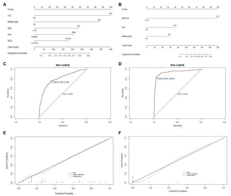
The result of the univariate and multivariate regression analyses showed that five factors (age less than 55 years old, male, MTD ≥ 0.5cm, and the presence of TCI and multifocality) were proven to be independent risk factors of CLNM for PTMC patients within the Uni group (Shown in Table 3). Meanwhile, for patients with positive CLNM, the result of multivariate analysis exhibited that three factors (maximum diameter of positive CLN (MDCLN) > 0.5cm, the presence of multifocality, and iNG) were screened out as independent risk factors of LLNM. The prediction models for quantitatively assessing the risk of CLNM and LLNM for all patients within the Uni group and unilateral patients with positive CLNM respectively were then created based on the independent risk factors described (Shown in Figure 1A,B). To validate the accuracy of the newly created nomogram, an internal validation by 1000 bootstrap resamples was performed and assessed in terms of the C-index. Validation results returned a C-index of 0.806 (95% CI, 0.769–0.843), and 0.803 (95% CI, 0.790–0.816) after bootstrapping, demonstrating our nomogram’s excellent accuracy in CLNM risk prediction. The ROC curve and the calibration plot are shown in Figure 1C,E, both exhibiting satisfactory agreement between the actual and predicted probability of CLNM for patients in the Uni group. For the nomogram used to assess the risk of LLNM in unilateral patients with positive CLNM, a C-index of 0.938 (95% CI, 0.881–0.994) was yielded, and 0.931 (95% CI, 0.913–0.949) after bootstrapping. The ROC curve and the calibration plot (Shown in Figure 1D,F) also confirmed high efficiency and accuracy for predicting LLNM.
Table 3.
Univariate and multivariate analyses for PTMC patients with unilateral disease.
| Univariate Analysis | Multivariate Analysis | Univariate Analysis | Multivariate Analysis | ||||||
|---|---|---|---|---|---|---|---|---|---|
| Hazard Ratio (95% CI) | p Value | Hazard Ratio (95% CI) | p Value | Hazard Ratio (95% CI) | p Value | Hazard Ratio (95% CI) | p Value | ||
| Factors selected | Factors selected | ||||||||
| Age | 0.000 | 0.000 | Age | 0.261 | |||||
| ≥55 vs. <55 | 0.426 (0.273–0.666) | 0.360 (0.213–0.609) | ≥55 vs. <55 | 0.426 (0.096–1.886) | |||||
| BMI | 0.436 | BMI | 0.596 | ||||||
| >23 vs. ≤23 | 0.876 (0.629–1.222) | >23 vs. ≥23 | 0.812 (0.375–1.758) | ||||||
| Gender | 0.000 | 0.000 | Gender | 0.749 | |||||
| Male vs. Female | 2.336 (1.639–3.329) | 2.505 (1.643–3.818) | Male vs. Female | 1.134 (0.525–2.446) | |||||
| TCI | 0.000 | 0.000 | TCI | 0.205 | |||||
| Yes vs. No | 6.228 (4.171–9.299) | 5.894 (3.736–9.299) | Yes vs. No | 1.656 (0.759–3.616) | |||||
| Maximum tumor diameter | 0.000 | 0.000 | Maximum tumor diameter | 0.228 | |||||
| >0.5 cm vs. ≤0.5 cm | 2.788 (1.978–3.929) | 2.181 (1.456–3.269) | >0.5cm vs. ≤0.5cm | 1.694 (0.719–3.993) | |||||
| Tumor location | 0.752 | Tumor location | 0.683 | ||||||
| Upper vs. Middle/Lower | 0.939 (0.637–1.384) | Upper vs. Middle/Lower | 1.199 (0.501–2.870) | ||||||
| Multifocality | 0.000 | 0.000 | Multifocality | 0.001 | 0.010 | ||||
| Yes vs. No | 4.692 (3.103–7.095) | 4.514 (2.831–7.196) | Yes vs. No | 4.305 (1.872–9.899) | 4.439 (1.423–13.847) | ||||
| PTMC with ipsilateral nodular goiter | 0.936 | PTMC with ipsilateral nodular goiter | 0.000 | 0.003 | |||||
| Yes vs. No | 1.015 (0.701–1.470) | Yes vs. No | 4.196 (1.901–9.258) | 6.311 (1.883–21.159) | |||||
| PTMC with Hashimoto thyroiditis | 0.310 | PTMC with Hashimoto thyroiditis | 0.964 | ||||||
| Yes vs. No | 0.798 (0.516–1.234) | Yes vs. No | 1.024 (0.366–2.869) | ||||||
| Maximum diameter of positive CLN | 0.000 | 0.000 | |||||||
| >0.5cm vs. ≤0.5cm | 71.697 (16.284–315.670) | 107.399 (20.011–576.400) | |||||||
| Number of positive CLN | 0.001 | 0.826 | |||||||
| ≥3 vs. <3 | 2.282 (1.429–3.644) | 0.924 (0.458–1.866) | |||||||
CI, confidence interval; PTMC, papillary thyroid microcarcinoma; TCI, thyroid capsular invasion; BMI, body mass index; CLN, central lymph node.
Figure 1.
Construction, assessment, and validation of the predictive model of CLNM and LLNM. (A,B) The nomograms for predicting CLNM and LLNM risk in PTMC patients within the Uni group, respectively; (C,D) the ROC curve and AUC of the nomograms for predicting CLNM and LLNM risk in PTMC patients within the Uni group, respectively; (E,F) the calibration curves of the nomogram for predicting CLNM and LLNM risk in PTMC patients within the Uni group, respectively. Actual probability is plotted on the y-axis, and nomogram predicted probability on the x-axis. PTMC, papillary thyroid microcarcinoma; CLNM, central lymph node metastasis; LLNM, lateral lymph node metastases; TCI, thyroid capsular invasion; MTD, maximum tumor diameter; MDCLN, the maximum diameter of positive central lymph node; iNG, ipsilateral nodular goiter; ROC, receiver operating characteristics.
3.4. Creation of Risk Prediction Model for Cervical Lymph Node Metastasis of PTMC Patients within the Bil Group
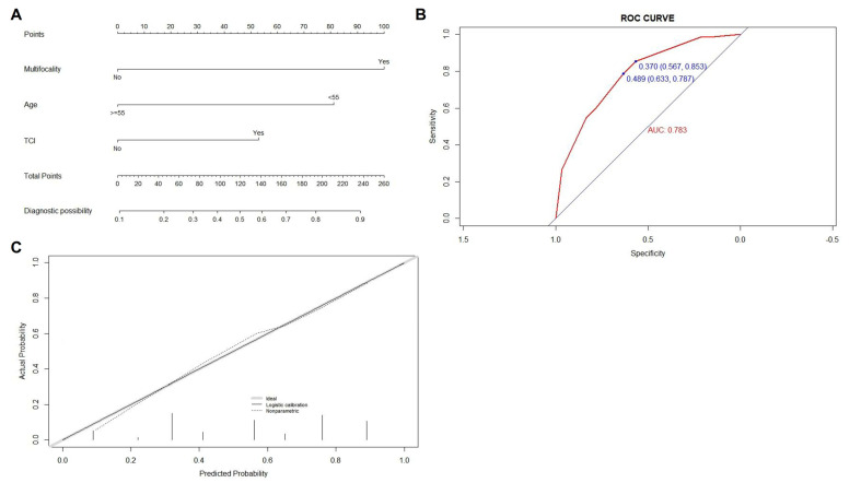
Three factors including age less than 55 and the presence of TCI and multifocality in at least one side of the thyroid lobe were confirmed as independent risk factors of CLNM for patients within the Bil group by multivariate analysis (shown in Table 4). Similarly, the prediction model for quantitatively assessing the risk of CLNM for these patients was established (shown in Figure 2A), and the ROC and the calibration curves were plotted and shown in Figure 2B,C, both indicating a high degree of accuracy of our newly created model (with C-index of 0.783 (95% CI, 0.706–0.860) and 0.776 (95% CI, 0.754–0.798) for training group and after bootstrapping respectively).
Table 4.
Univariate and multivariate analyses for PTMC patients with bilateral disease.
| Univariate Analysis | Multivariate Analysis | Univariate Analysis | Multivariate Analysis | ||||||
|---|---|---|---|---|---|---|---|---|---|
| Hazard Ratio (95% CI) | p Value | Hazard Ratio (95% CI) | p Value | Hazard Ratio (95% CI) | p Value | Hazard Ratio (95% CI) | p Value | ||
| Factors selected | Factors selected | ||||||||
| Age | 0.007 | 0.004 | Age | 0.716 | |||||
| ≥55 vs. <55 | 0.308 (0.131–0.724) | 0.224 (0.080–0.626) | ≥55 vs. <55 | 1.312 (0.303–5.683) | |||||
| BMI | 0.609 | BMI | 0.255 | ||||||
| >23 vs. ≤23 | 1.199 (0.599–2.401) | >23 vs. ≤23 | 1.952 (0.617–6.173) | ||||||
| Gender | 0.072 | Gender | 0.025 | 0.293 | |||||
| Male vs. Female | 2.034 (0.938–4.411) | Male vs. Female | 3.437 (1.168–10.118 ) | 2.052 (0.538–7.824) | |||||
| TCI | 0.009 | 0.020 | TCI | 0.739 | |||||
| Yes vs. No | 2.598 (1.263–5.344) | 2.624 (1.161–5.929) | Yes vs. No | 0.838 (0.296–2.375) | |||||
| Maximum tumor diameter | 0.038 | 0.133 | Maximum tumor diameter | 0.850 | |||||
| >0.5 cm vs. ≤0.5 cm | 2.074 (1.040–4.137) | 1.836 (0.832–4.055) | >0.5 cm vs. ≤0.5 cm | 1.109 (0.378–3.251) | |||||
| Tumor location | 0.143 | Tumor location | 0.438 | ||||||
| Upper vs. Middle/Lower | 0.583 (0.283–1.200) | Upper vs. Middle/Lower | 0.612 (0.177–2.117) | ||||||
| Multifocality | 0.000 | 0.000 | Multifocality | 0.071 | |||||
| Yes vs. No | 5.059 (2.417–10.590) | 6.278 (2.728–14.451) | Yes vs. No | 3.451 (0.899–13.241) | |||||
| PTMC with ipsilateral nodular goiter | 0.966 | PTMC with ipsilateral nodular goiter | 0.008 | 0.038 | |||||
| Yes vs. No | 0.984 (0.463–2.092) | Yes vs. No | 4.545 (1.489–13.876) | 4.375 (1.083–17.670) | |||||
| PTMC with Hashimoto thyroiditis | 0.468 | PTMC with Hashimoto thyroiditis | 0.635 | ||||||
| Yes vs. No | 1.327 (0.619–2.846) | Yes vs. No | 0.754 (0.235–2.417) | ||||||
| Maximum diameter of positive CLN | 0.000 | 0.000 | |||||||
| >0.5 cm vs. ≤0.5 cm | 16.000 (4.052–63.185) | 13.868 (3.226–59.610) | |||||||
| Number of positive CLN | 0.090 | ||||||||
| ≥3 vs. <3 | 1.707 (0.919–3.170) | ||||||||
Figure 2.
Construction, assessment, and validation of the predictive model of CLNM for PTMC patients within the Bil group. (A) The nomograms for predicting CLNM risk in PTMC patients within the Bil group; (B) the ROC curve and AUC of the nomogram for predicting CLNM risk in PTMC patients within the Bil group; (C) the calibration curve of the nomogram for predicting CLNM risk in PTMC patients within the Bil group. Actual probability is plotted on the y-axis, and nomogram predicted probability on the x-axis. PTMC, papillary thyroid microcarcinoma; CLNM, central lymph node metastasis; TCI, thyroid capsular invasion; ROC, receiver operating characteristics.
For bilateral PTMC patients with CLNM, two factors including MDCLN > 0.5 cm and the presence of iNG were screened out as independent factors of LLNM. These patients were then divided into four subgroups according to the presence of the two factors: patients with none of the two risk factors, patients with iNG only, patients with MDCLN > 0.5 cm only, and patients with both of the two risk factors. The incidences of LLNM for patients within different subgroups were shown and compared in Table 5. Patients exhibiting both of the two risk factors (8 in 11, 72.7%) showed a much higher LLNM rate than those within the other three subgroups, while only 1 (2.9%) in 35 patients with none of the two factors was proven to have lateral neck involvement in our cohort.
Table 5.
Risk stratification of CLNM and LLNM for PTMC patients within Uni and Bil groups.
| Low Risk (TS ≤ 100) | Moderate Risk (100 < TS < 180) | High Risk (TS ≥ 180) | ||||
|---|---|---|---|---|---|---|
| (n = 197, %) | (n = 212, %) | (n = 173, %) | p Value | |||
| Uni group | ALL patients (n = 582) | Negative CLNM | 173 (87.8) | 144 (67.9) | 33 (19.1) | 0.000 |
| Positive CLNM | 24 (12.2) | 68 (32.1) | 140 (80.9) | |||
| Low risk (TS ≤ 100) | High risk (TS > 100) | |||||
| (n = 196, %) | (n = 36, %) | p value | ||||
| Patients with positive CLNM (n = 232) | Negative LLNM | 192 (98.0) | 10 (27.8) | 0.000 | ||
| Positive LLNM | 4 (2.0) | 26 (72.2) | ||||
| Low risk (TS < 80) | Moderate risk (80 ≤ TS < 100) | High risk (TS ≥ 100) | ||||
| (n = 14, %) | (n = 31, %) | (n = 90, %) | p value | |||
| Bil group | All patients (n = 135) | Negative LLNM | 14 (100.0) | 22 (71.0) | 24 (26.7) | 0.000 |
| Positive LLNM | 0 (0.0) | 9 (29.0) | 66 (73.3) | |||
| No risk factor | iNG only MDCLN > 0.5 cm only | Both two risk factors | ||||
| (n = 35, %) | (n = 10, %) (n = 19, %) | (n = 11, %) | p value | |||
| Patients with positive CLNM (n = 75) | Negative LLNM | 34 (97.1) | 8 (80.0) 11 (57.9) | 3 (27.3) | 0.000 | |
| Positive LLNM | 1 (2.9) | 2 (20.0) 8 (42.1) | 8 (72.7) | |||
PTMC, papillary thyroid microcarcinoma; CLNM, central lymph node metastasis; LLNM, lateral lymph node metastasis; iNG, ipsilateral nodular goiter; MDCLN, maximum diameter of positive central lymph node.
3.5. Risk Stratification and Cervical Lymph Node Metastasis Risk Assessment Flow Chart for PTMC Patients
Each factor enrolled for the construction of the nomogram has its own risk points. Patients within different groups would gain a total risk score by summing up the risk scores of each factor based on their own nomogram. According to the distribution of the total risk scores, all patients within the Uni group, unilateral patients with positive CLNM, and patients within the Bil group were separately classified into different subgroups with significantly distinct CLNM or LLNM risks (shown in Table 5, p-value = 0.000, 0.000, and 0.000, respectively) by different cutoff values.
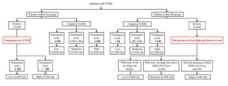
The risk stratification according to the nomogram score was shown in Table 6. The aforementioned three nomograms and the risk stratification strategy of LLNM for bilateral patients with positive CLNM were further integrated and were presented as a comprehensive flow diagram for quantitatively evaluating the risk of cervical lymph node involvement for all patients with PTMC (exhibited in Figure 3).
Table 6.
Risk stratification of Uni and Bil group of PTC patients based on the model database.
| Uni Group CLNM Risk | Uni Group LLNM Risk | Bil Group CLNM Risk | ||||||
|---|---|---|---|---|---|---|---|---|
| Low | Moderate | High | Low | High | Low | Moderate | High | |
| Nomogramscore | 0–100 | 100–180 | >180 | <100 | >100 | <80 | 80–100 | >100 |
| Value | 24/197 | 68/212 | 140/173 | 4/196 | 26/36 | 0/14 | 9/31 | 66/90 |
| % | 12.2 | 32.1 | 80.9 | 2 | 72.2 | 0 | 29 | 73.3 |
Figure 3.
A flow chart of cervical lymph node metastasis risk including both CLNM and LLNM for PTMC patients with unilateral and bilateral diseases. CLNM, central lymph node metastasis; LLNM, lateral lymph node metastases; PTMC, papillary thyroid microcarcinoma; iNG, ipsilateral nodular goiter.
4. Discussion
Previous studies have demonstrated that bilateral PTCs usually present more aggressively, and more advanced, as well as having poorer recurrence and survival outcomes [13]. Bilateral disease was also reported to be significantly associated with clinicopathological features including both primary tumor sites such as larger tumor size and thyroid capsular invasion, and cervical lymph node metastasis [14]. Furthermore, here in our study, compared with the previous research, more detailed factors have been enrolled and analyzed. As a result, the presence of multifocality in at least one side of the thyroid lobe, upper portion tumor of the thyroid, and iNG were significantly more frequent in patients with bilateral disease. In terms of further information regarding cervical lymph node metastasis, central lymph node metastasis (CLNM) was significantly more common in patients within the Bil group than those within the Uni group, where higher counts and larger-sized central lymph nodes were found in patients within the Bil group. For patients with positive CLNM, those within the Bil group also showed a significantly higher incidence of LLNM. All these differences further indicate the extremely more invasive nature of bilateral tumors. Wang et al. [15] proved that bilateral tumors within one patient share the same clone source, and this result also suggests that bilateral disease may be a more aggressive status of intra-thyroid metastasis from the primary tumor at the contralateral lobe, which further validates our conclusions. The aforementioned significant difference between PTMC patients with unilateral and bilateral diseases further indicates the significant implications of our study to discuss patients within these two groups separately.
Several studies have revealed that male gender, larger tumor size, multifocality, younger age, and the presence of TCI are independent risk factors of CLNM for patients with PTMC [16,17,18], however, little research has focused on the central neck involvement for PTMC patients with unilateral and bilateral diseases respectively. Here in our research, factors including age < 55 years old, male, MTD ≥ 0.5cm, and the presence of TCI and multifocality were identified as independent factors of CLNM for PTMC patients with unilateral disease, while these factors including age < 55 years old, and the presence of TCI and multifocality for those with bilateral disease. Two prediction models for quantitatively assessing the risk of CLNM for PTMC patients with unilateral and bilateral diseases were established based on their respective independent risk factors. Then, patients in each group were stratified into three subgroups with significantly different CLNM risks according to the distribution of the total score received from their own prediction models. For PTMC patients with unilateral disease, who are traditionally considered as having a low risk of CLNM among all PTC patients, a subgroup of patients were screened out and proven to have a much higher risk of CLNM (140 in 173, 80.9%). However, for PTMC patients with bilateral disease, who displayed a significantly higher incidence of CLNM than those with unilateral disease, a small subgroup of patients with no incidence of CLNM has also been selected (0 in 14, 0.0%), showing much lower CLNM risk compared to those classified as a high-risk subgroup (66 in 90, 73.3%). For patients with PTMC, aside from resection of the primary tumor site, a “wait and see” strategy for clinical negative central neck region is recommended by many guidelines including the 2015 American Thyroid Association (ATA) guideline to avoid unnecessary surgery-related complications. However, several research works have revealed that the incidence of central lymph node involvement for patients with PTMC is not that low, with reported CLNM ranging from 27.4% to 53.9% [19,20,21], and that cervical lymph node involvement is likely associated with an elevated incidence of loco-regional tumor recurrence [22]. Considering this, an effective method that can accurately assess the risk of CLNM for PTMC patients is necessary. According to our results, a more cautious examination of central neck regions, as well as a more frequent postoperative follow-up, should be conducted for unilateral and bilateral patients with a total score of no less than 180 and 90, according to their respective nomograms. Prophylactic CLND could also be considered as a second choice given the extremely high CLNM risk. However, for those within the low CLNM risk subgroup, resection of the primary tumor site is enough, and intervention of the central neck region is unnecessary.
Existing literature showed that factors such as upper portion tumor and a high count of positive central lymph nodes are independent risk factors of LLNM for PTMC patients [23,24]. Here in our research, the risk of LLNM was also quantitatively analyzed for PTMC patients within the Uni group. The LLNM risk for patients in low LLNM risk subgroup was only 2.0% yet could reach up to 72.2% for those in the high LLNM risk subgroup. For those in the Bil group, only two factors including the presence of iNG and the maximum diameter of positive lymph nodes in the central compartment >0.5 cm were confirmed as independent factors, and those with none of these two factors showed an extremely low LLNM risk. Given that those exhibiting both of these two risk factors showed a high LLNM risk of 72.7%, the rationality and validity of our classification are confirmed. For unilateral and bilateral patients within high LLNM risk groups according to their respective prediction models, a more frequent postoperative follow-up schedule is necessary. For those defined as having low LLNM risk, therapeutic CLND is enough and no additional intervention involving the lateral neck is needed.
5. Conclusion
An evaluation system based on independent factors of CLNM and LLNM for PTMC patients with bilateral and unilateral disease was established. Our newly established evaluating system can efficaciously quantify risks of CLNM and LLNM for PTMC patients with bilateral and unilateral disease and may guide individual treatment strategy including both surgical and postoperative adjuvant treatment of neck region for these patients.
Author Contributions
All authors contributed to the study conception and design. Material preparation, data collection and analysis were performed by Z.Y., Y.H. and L.T. The first draft of the manuscript was written by Z.Y. and Y.H., W.C. and W.Q. revised the article, and all authors commented on previous versions of the manuscript. All authors have read and agreed to the published version of the manuscript.
Institutional Review Board Statement
This study was performed in line with the principles of the Declaration of Helsinki. Approval was granted by the Chinese Clinical Trial (ChiCTR2100043353).
Informed Consent Statement
Informed consent was obtained from all individual participants included in the study.
Data Availability Statement
The datasets generated and analyzed during the current study are available from the corresponding author on reasonable request.
Conflicts of Interest
The authors have no relevant financial or non-financial interests to disclose.
Funding Statement
This research was supported by the National Natural Science Foundation of China under Grant [81772878, 82072948]; the Shanghai Jiao Tong University [YG2019ZDA15].
Footnotes
Publisher’s Note: MDPI stays neutral with regard to jurisdictional claims in published maps and institutional affiliations.
References
- 1.Pellegriti G., Frasca F., Regalbuto C., Squatrito S., Vigneri R. Worldwide increasing incidence of thyroid cancer: Update on epidemiology and risk factors. J. Cancer Epidemiol. 2013;2013:965212. doi: 10.1155/2013/965212. [DOI] [PMC free article] [PubMed] [Google Scholar]
- 2.Hughes D.T., Haymart M.R., Miller B.S., Gauger P.G., Doherty G.M. The most commonly occurring papillary thyroid cancer in the United States is now a microcarcinoma in a patient older than 45 years. Thyroid. 2011;21:231–236. doi: 10.1089/thy.2010.0137. [DOI] [PubMed] [Google Scholar]
- 3.Siegel R.L., Miller K.D., Jemal A. Cancer statistics, 2018. CA Cancer J. Clin. 2018;68:7–30. doi: 10.3322/caac.21442. [DOI] [PubMed] [Google Scholar]
- 4.Lim H., Devesa S.S., Sosa J.A., Check D., Kitahara C.M. Trends in Thyroid Cancer Incidence and Mortality in the United States, 1974–2013. J. Am. Med. Assoc. 2017;317:1338–1348. doi: 10.1001/jama.2017.2719. [DOI] [PMC free article] [PubMed] [Google Scholar]
- 5.Mehanna H., Al-Maqbili T., Carter B., Martin E., Campain N., Watkinson J., McCabe C., Boelaert K., Franklyn J.A. Differences in the recurrence and mortality outcomes rates of incidental and nonincidental papillary thyroid microcarcinoma: A systematic review and meta-analysis of 21 329 person-years of follow-up. J. Clin. Endocrinol. Metab. 2014;99:2834–2843. doi: 10.1210/jc.2013-2118. [DOI] [PubMed] [Google Scholar]
- 6.Wada N., Duh Q.Y., Sugino K., Iwasaki H., Kameyama K., Mimura T., Ito K., Takami H., Takanashi Y. Lymph node metastasis from 259 papillary thyroid microcarcinomas: Frequency, pattern of occurrence and recurrence, and optimal strategy for neck dissection. Ann. Surg. 2003;237:399–407. doi: 10.1097/01.SLA.0000055273.58908.19. [DOI] [PMC free article] [PubMed] [Google Scholar]
- 7.Haugen B.R., Alexander E.K., Bible K.C., Doherty G.M., Mandel S.J., Nikiforov Y.E., Pacini F., Randolph G.W., Sawka A.M., Schlumberger M., et al. 2015 American Thyroid Association Management Guidelines for Adult Patients with Thyroid Nodules and Differentiated Thyroid Cancer: The American Thyroid Association Guidelines Task Force on Thyroid Nodules and Differentiated Thyroid Cancer. Thyroid. 2016;26:1–133. doi: 10.1089/thy.2015.0020. [DOI] [PMC free article] [PubMed] [Google Scholar]
- 8.Wang W., Su X., He K., Wang Y., Wang H., Wang H., Zhao Y., Zhao W., Zarnegar R., Fahey T.J., III, et al. Comparison of the clinicopathologic features and prognosis of bilateral versus unilateral multifocal papillary thyroid cancer: An updated study with more than 2000 consecutive patients. Cancer. 2016;122:198–206. doi: 10.1002/cncr.29689. [DOI] [PubMed] [Google Scholar]
- 9.Liu Z., Lv T., Xie C., Di Z. BRAF V600E Gene Mutation Is Associated with Bilateral Malignancy of Papillary Thyroid Cancer. Am. J. Med. Sci. 2018;356:130–134. doi: 10.1016/j.amjms.2018.04.012. [DOI] [PubMed] [Google Scholar]
- 10.Roh J.L., Kim J.M., Park C.I. Central lymph node metastasis of unilateral papillary thyroid carcinoma: Patterns and factors predictive of nodal metastasis, morbidity, and recurrence. Ann. Surg. Oncol. 2011;18:2245–2250. doi: 10.1245/s10434-011-1600-z. [DOI] [PubMed] [Google Scholar]
- 11.So Y.K., Son Y.I., Hong S.D., Seo M.Y., Baek C.H., Jeong H.S., Chung M.K. Subclinical lymph node metastasis in papillary thyroid microcarcinoma: A study of 551 resections. Surgery. 2010;148:526–531. doi: 10.1016/j.surg.2010.01.003. [DOI] [PubMed] [Google Scholar]
- 12.Viola D., Materazzi G., Valerio L., Molinaro E., Agate L., Faviana P., Seccia V., Sensi E., Romei C., Piaggi P., et al. Prophylactic central compartment lymph node dissection in papillary thyroid carcinoma: Clinical implications derived from the first prospective randomized controlled single institution study. J. Clin. Endocrinol. Metab. 2015;100:1316–1324. doi: 10.1210/jc.2014-3825. [DOI] [PubMed] [Google Scholar]
- 13.Wang W., Zhao W., Wang H., Teng X., Wang H., Chen X., Li Z., Yu X., Fahey T.J., 3rd, Teng L. Poorer prognosis and higher prevalence of BRAF (V600E) mutation in synchronous bilateral papillary thyroid carcinoma. Ann. Surg. Oncol. 2012;19:31–36. doi: 10.1245/s10434-011-2096-2. [DOI] [PubMed] [Google Scholar]
- 14.Karatzas T., Vasileiadis I., Charitoudis G., Karakostas E., Tseleni-Balafouta S., Kouraklis G. Bilateral versus unilateral papillary thyroid microcarcinoma: Predictive factors and associated histopathological findings following total thyroidectomy. Hormones. 2013;12:529–536. doi: 10.14310/horm.2002.1441. [DOI] [PubMed] [Google Scholar]
- 15.Wang W., Wang H., Teng X., Wang H., Mao C., Teng R., Zhao W., Cao J., Fahey T.J., III, Teng L. Clonal analysis of bilateral, recurrent, and metastatic papillary thyroid carcinomas. Hum. Pathol. 2010;41:1299–1309. doi: 10.1016/j.humpath.2010.02.008. [DOI] [PubMed] [Google Scholar]
- 16.Zhao J., Zhao Y., Ling Y., Kang H. Risk Factors of Central Lymph Node Metastasis in Papillary Thyroid Microcarcinoma and the Value of Sentinel Lymph Node Biopsy. Front. Surg. 2021;8:680493. doi: 10.3389/fsurg.2021.680493. [DOI] [PMC free article] [PubMed] [Google Scholar]
- 17.Gui C.Y., Qiu S.L., Peng Z.H., Wang M. Clinical and pathologic predictors of central lymph node metastasis in papillary thyroid microcarcinoma: A retrospective cohort study. J. Endocrinol. Investig. 2018;41:403–409. doi: 10.1007/s40618-017-0759-y. [DOI] [PubMed] [Google Scholar]
- 18.Liviu H., Paul-Andrei Ș., Doina P. Total Tumor Diameter and Unilateral Multifocality as Independent Predictor Factors for Metastatic Papillary Thyroid Microcarcinoma. J. Clin. Med. 2021;10:3707. doi: 10.3390/jcm10163707. [DOI] [PMC free article] [PubMed] [Google Scholar]
- 19.Wu X., Li B., Zheng C., He X. Risk factors for central lymph node metastases in patients with papillary thyroid microcarcinoma. Endocr. Pract. 2018;24:1057–1062. doi: 10.4158/EP-2018-0305. [DOI] [PubMed] [Google Scholar]
- 20.Wang Y., Guan Q., Xiang J. Nomogram for predicting central lymph node metastasis in papillary thyroid microcarcinoma: A retrospective cohort study of 8668 patients. Int. J. Surg. 2018;55:98–102. doi: 10.1016/j.ijsu.2018.05.023. [DOI] [PubMed] [Google Scholar]
- 21.Wei X., Min Y., Feng Y., He D., Zeng X., Huang Y., Fan S., Chen H., Chen J., Xiang K., et al. Development and validation of an individualized nomogram for predicting the high-volume (>5) central lymph node metastasis in papillary thyroid microcarcinoma. J. Endocrinol. Investig. 2022;45:507–515. doi: 10.1007/s40618-021-01675-5. [DOI] [PubMed] [Google Scholar]
- 22.Jianyong L., Jinjing Z., Zhihui L., Tao W., Rixiang G., Jingqiang Z. A Nomogram Based on the Characteristics of Metastatic Lymph Nodes to Predict Papillary Thyroid Carcinoma Recurrence. Thyroid. 2018;28:301–310. doi: 10.1089/thy.2017.0422. [DOI] [PubMed] [Google Scholar]
- 23.Zeng R.C., Zhang W., Gao E.L., Cheng P., Huang G.L., Zhang X.H., Li Q. Number of central lymph node metastasis for predicting lateral lymph node metastasis in papillary thyroid microcarcinoma. Head Neck. 2014;36:101–106. doi: 10.1002/hed.23270. [DOI] [PubMed] [Google Scholar]
- 24.Zhang L., Wei W.J., Ji Q.H., Zhu Y.X., Wang Z.Y., Wang Y., Huang C.P., Shen Q., Li D.S., Wu Y. Risk factors for neck nodal metastasis in papillary thyroid microcarcinoma: A study of 1066 patients. J. Clin. Endocrinol. Metab. 2012;97:1250–1257. doi: 10.1210/jc.2011-1546. [DOI] [PubMed] [Google Scholar]
Associated Data
This section collects any data citations, data availability statements, or supplementary materials included in this article.
Data Availability Statement
The datasets generated and analyzed during the current study are available from the corresponding author on reasonable request.


