

Abstract
In present world of contemporary techniques of microscopic ear surgery and single handed endoscopic ear surgery, we propose the technique of two handed endoscopic tympanoplasty using endoscope holders. The aim of the study is to evaluate the functional and anatomical results of our technique of endoscopic type 3 cartilage tympanoplasty using endoscope holder. It is a Retrospective Non Randomized Clinical Study. A total of 67 endoscope holder assisted exclusively two handed endoscopic type 3 cartilage tympanoplasties performed from December 2014 to March 2017 with our technique were included in the study. Patients with pars tensa retractions and perforations with absent incus were included in the study. Those with cholesteatoma were excluded from the study. Full thickness tragal cartilage disc of 3 × 3 mm dimensions with a circular slot of 1 mm to fit onto the head of the stapes was used for reconstruction. Tympanic membrane reconstruction was done along with attic reconstruction, using sliced tragal cartilage of 0.5 mm thickness. Patients were assessed at 6, 12 and 24 months for graft status. In early follow up period ranging from 24 to 52 months, the graft take up was seen in 64 ears with three perforations giving a success rate of 95.52%. The pre-operative air-bone gap was 42.6 ± 3.26 dB and the post-operative air-bone gap at 6 months, 1 and 2 years was 18.36 ± 3.46 dB, 19.42 ± 4.32 dB and 19.53 ± 4.33 dB respectively. The study reports good air-bone closure to 20 dB postoperatively following type 3 endoscopic tympanoplasty using endoscope holder. Slotted cartilage graft is definitely an excellent option for ossiculoplasty in cases of absent incus providing a stable assembly.
Level of evidence: Level 4.
Keywords: Endoscope holder, Two handed technique, Type 3 endoscopic cartilage Tympanoplasty, Ossiculoplasty, Eroded incus
Introduction
Discontinuity of the ossicles due to erosion of the incudostapedial joint is one of the commonest defects in chronic otitis media. Various techniques for reconstruction in cases of erosion of the incus are described in the literature. The earliest attempt at ossiculoplasty [1] was made in 1901. Since then, many techniques and materials have been proposed for ossiculoplasty with varying degree of success. The goal of ossiculoplasty is to repair the conductive hearing loss giving a long term stability. Type 3 tympanoplasty is the surgery of choice for middle ear reconstruction in ossicular reconstruction with an integral and functional stapes suprastructure and mobile footplate. The most commonly used autograft material is the incus body, which is fashioned to fit between the manubrium of the malleus and the stapes head. Various grafts for reconstruction of the ossicular defect include bone cement, cartilage, hydroxyapatite, glass ionomers etc. PORP (partial ossicular prostheses) is one of the options which requires surgical expertise and experience [2]. Inspite of all the advancements in the biotechnical and biomedical methods of ossicular reconstruction, there is no dramatic increase in the success results. We have been using tragal cartilage since 2003 for reconstructing the hearing mechanism in cases of eroded or absent incus. In this technique, cartilage disc of 3 × 3 mm dimensions with a circular slot of 0.8 mm is placed over the stapes to accommodate the head of the stapes. Till 2013, we were operating with this technique using microscope. After 2013, we have been operating with endoscopic two handed ear surgery using endoscope holders. Endoscopic ear surgery (EES) is still in its developing stage. Though appealing, single handed ear surgery has disadvantages.
Endoscopic ear surgery till now has been single handed technique with the endoscope being held in the left or the non-dominant hand and the micro ear instruments in the right or the dominant hand. The only disadvantage of endoscopic ear surgeries as compared to microscopic ear surgery is that it is one-handed surgical technique in which the non-dominant left hand of the surgeon is utilized for holding and manipulating the endoscope. The problem of one handed endoscopic ear surgery is more evident during the atticotomy while drilling (in which simultaneous suction and drilling is not possible), in cases of hemorrhage (in which simultaneous suction and instrumentation is not possible), during fogging of the endoscope (one needs to remove endoscope again and again for lens cleaning), during prosthesis fitting in ossiculoplasty (manipulation may be difficult with one hand). In these situations, one feels the immense need for the two handed technique as in the microscopic ear surgery. To incorporate the advantages of the endoscope and to retain the two handed nature of the ear surgery, we developed 2 endoscope holders [3–5]. These endoscope holders allow both hands of the surgeon to be free for surgical manipulation during endoscopic ear surgery. After development of the endoscope holders (Fig. 1), we incorporated all our techniques previously used microscopically in the two handed endoscopic ear surgery in the year 2013. The purpose of the study is to report our experience with this technique. This is the first study of endoscopic sliced cartilage type 3 tympanoplasty with the two handed technique using the endoscope holder.
Fig. 1.

Endoscope holder with endoscope
Materials and Methods
A total of 67 endoscope holder assisted type 3 cartilage tympanoplasties were performed from December 2014 to March 2017 in Sushrut ENT Hospital with a follow up period ranging from 24 to 52 months with our technique to evaluate the feasibility and results of a two handed technique. Patients with pars tensa perforations and those with pars tensa retractions with absent incus were included in the study. Those with cholesteatoma requiring atticoantrostomy were excluded from the study. The mean age of the study group was 26.2 years. The youngest patient was 10 years old and the oldest was 57 years. There were 38 males and 29 females in the study population. The preoperative air bone gap was 42.6 ± 3.26 dB. All patients were operated under general anaesthesia. Patients were assessed at 6, 12, and 24 months for graft status.
Adapting to the Two Handed Technique of Endoscopic Ear Surgery
During the early days of development of the Endoscope Holder, its application was limited to Cadaveric Temporal Bones for learning the two handed endoscopic ear surgery. Each of the authors have dissected minimum of five human cadaveric temporal bones endoscopically with endoscope holders before starting the endoscope holder assisted endoscopic ear surgery. The hand–eye-monitor coordination of the surgical steps with the suction in the left hand and the micro ear instruments in the right hand was practiced. Then it was extended to grommet insertions, small perforation tympanoplasties and later on for subtotal perforations and ossiculoplasty.
Operating Theatre Requirements
Zero degree 3 mm and 45° 4 mm Karl Storz endoscopes with triple charge coupled device Camera (Karl Storz, Germany) placed onto endoscope holder [3–5].
Preparation of the Patient
The patient is prepared and draped in the usual fashion with the ipsilateral shoulder pulled down. All patients were operated under general anaesthesia. The ear canal is infiltrated with 2 percent lidocaine with 1 in 2, 00,000 adrenaline.
Procedure of Endoscopic Type 3 Cartilage Tympanoplasty with Endoscope Holder (Figs. 2–3, video)
Fig. 2.

Sliced tragal cartilages and perichondrium for TM reconstruction
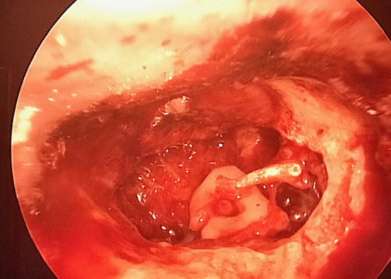
Fig. 3.

Note the coordinated action of the two instruments in two handed EES with cartilage with slot over stapes
Instruments are similar to those used in microscopic ear surgery. Transcanal incision is taken. Tympanomeatal flap is elevated to visualise the middle ear pathology. Middle ear inspection is done to detect ossicular erosion and ventilation problems. We prefer tragal cartilage as the graft of choice for tympanic membrane and ossicular reconstruction. It is harvested via the horizontal incision on the tragus. Approximately 15 × 15mm cartilage is harvested. The perichondrium is separated/ peeled off from one of the sides of the cartilage. A 4 × 4 mm unsliced full thickness cartilage devoid of perichondrium on both sides is kept aside to be fashioned as a cartilage cap over the stapes head. The remaining tragal cartilage graft is sliced [6–13] with metallic thickness plate of 0.3 mm with cartilage splitter to obtain two sliced tragal grafts, approximately 0.3 and 0.5 mm thickness (considering the tragal cartilage is 1 mm in thickness, each perichondrium 0.1 mm and after slicing with 0.3 mm we get 0.3 and 0.5 mm). A central circular slot of 0.8 mm is made onto the 4 × 4 mm disc (Fig. 2). This circular slot is fashioned as a cap so as to accommodate and fit onto the head of the stapes (Figs. 3). This not only ensures stability of the cartilage graft over the stapes head but also helps to increase the middle ear space by 1 mm. Sliced cartilage of 0.5 mm thickness kept as underlay shield graft or island graft for tympanic membrane reconstruction. Tympanomeatal flap reposed back. During the procedure the suction is held in left hand to avoid fogging and to achieve cooling of endoscope generated heat as reported in previous study [14]. Intermittent irrigation is done for cooling of endoscope as well as to achieve cleaning of endoscope lens [14]. Canalplasty was not performed in this series. The 3 mm 14 cm Karl Storz zero degree endoscope could give a panoramic view even in narrow canals. All procedures performed were recorded for documentation. The advantage of endoscope holder is that both the hands are free for intervention with the left hand holding the suction and right hand holding the microear instruments. The two handed technique is of immense benefit during fitting of the graft over the stapes head.
Modification of the Technique
In cases of presence of malleus, we use an oblong/boat shaped cartilage graft of dimensions 6 × 4 mm and make a slot of 0.8 mm at the peripheral part of the boat shaped graft (Fig. 4). The other end of the boat is placed medial to the malleus and the slot is snugly fitting onto the head of the stapes.
Fig. 4.
Modified cartilage (boat shaped) one part medial to malleus handle and the other fitted over the stapes superstructure
Results
In early follow up period ranging from 24 to 52 months, the graft take up was seen in 64 ears with three recurrent perforations giving a success rate of 95.52%. The recurrent perforations were noted at 6 months follow up and one year follow up. These perforations did not heal with conservative management. The pre-operative air-bone gap was 42.6 ± 3.26 dB and the post-operative air-bone gap at 6 months, one year and 2 years was 18.36 ± 3.46 dB, 19.42 ± 4.32 dB and 19.53 ± 4.33 dB respectively. The cartilage to stapes superstructure assembly was stable with no extrusion in our series of cases or any worsening of hearing (Fig. 5). Middle ear inflammation had no adverse effect on the cartilage and the postoperative hearing.
Fig. 5.

Post operative 1 year intact tm cartilage graft
Discussion
The reconstruction of the ossicular chain as a surgical treatment of chronic otitis media is challenging to the otologist [15]. Ossiculoplasty involves reconstruction of sound-conducting mechanism between the graft and oval window. Variety of graft materials which have been used for the ossicular reconstruction include the use of a prosthesis, autologous ossicle (most commonly the incus), bone cement, cartilage, hydroxyapatite, glass ionomers etc. [16].
The overall long-term results of ossiculoplasty have not dramatically improved in recent years, in spite of all the great advances in biomedical technologies [17]. Type 3 tympanoplasty is the surgery of choice for middle ear reconstruction in ossicular reconstruction with an integral and functional stapes suprastructure and mobile footplate. The most commonly used autograft material is the incus body, which is fashioned to fit between the manubrium of the malleus and the stapes head. In our study involving 67 patients, the post-operative air-bone gap at 6 months, one year and 2 years was 18.36 ± 3.46 dB, 19.42 ± 4.32 dB and 19.53 ± 4.33 dB respectively. The slotted cartilage to stapes superstructure assembly was stable with no extrusion in our series of cases or any worsening of hearing.
In a review of cases that were performed over a 10-year period, Slater et al. [18] noted the advantages of porous polyethylene in its ability to form a fibrous union and decreased extrusion rate. The air-bone gap was closed to within 20 dB in 81% of cases with PORPs. In a study by Dornhoffer [19], prostheses composed of hydroxyapatite were used. It required preserving of the tensor tendon. He also noted the ideal weight of a middle ear prosthesis, the need to accommodate the malleus and minimize the angle formed with the tympanic membrane. In a study by Vashishth et al. [20] ossicular reconstruction was done titanium prostheses with a success rate of 90% with pure tone air bone gap pre- and postoperatively was 32.4 and 8.8 dB, respectively and no evidence of implant extrusion. The tympanic membrane reconstruction was done with full thickness broad tragal cartilage palisades. In the study by Martin et al. [21], the average air‐bone gap (ABG) improvement was 13 dB with closure of the ABG to within 20 dB in 57% of cases. They proposed that titanium prosthesis is easy to insert, well tolerated, and has a low extrusion rate. Hearing results were better for primary versus revision cases for PORPs versus TORPs and for intact canal wall (ICW) procedures versus canal wall‐down (CWD) procedures.
The surgery performed is dependent on the ossicular deficit. In a study by Bernal-Sprekelsen et al. [22] closure of the tympanic membrane was achieved in 98.3% of patients with postoperative air-bone gap <10 dB in 29.8% and between 11 and 20 dB in 32.3% of the patients. Ossiculoplasty using three different PORP: titanium angle prosthesis, autologous incus interposition, and titanium clip prosthesis was studied by Neumann et al. Their results did not generally favour the use of any specific prosthesis, rather they suggest that the correct choice of a prosthesis should be based on the anatomic and pathophysiologic conditions found in the individual patient [23].
In a study by Merchant et al. [24], the structural features that are responsible for the large variation in postoperative hearing results after Type 3 stapes columella tympanoplasty were evaluated. Larger air-bone gaps of 30–70 dB occurred as a result of stapes fixation, non-aeration of the middle ear, or both. When the stapes was mobile and the middle ear was aerated, a fascia graft resulted in air-bone gaps of 15–30 dB. Interposing a thin disc of cartilage between the fascia graft and stapes head improved the effective vibrating graft area and gave better hearing, with air-bone gaps of 10–25 dB. The clinical Type 3 results were consistent with predictions based on experimental investigations of mechanics of the Type 3 procedure in a temporal bone model.
In a cadaveric experimental type 3 tympanoplasty [25], it was concluded that mobile stapes and aerated middle ear were essential for a successful outcome in Type 3 tympanoplasty. There was little effect of varying the mechanical properties of the tympanic membrane graft or changing the tightness of connection between the graft and stapes head. Improved results were achieved by interposing a thin cartilage disc between the graft and stapes head to increase the effective vibrating area of the graft.
The methods of tympanoplasty are aimed to reconstruct the ossicular chain and the TM and eradication of the middle ear disease. Despite its wide application, the postoperative hearing results after tympanoplasty vary widely. The reasons for poor audiological outcomes after Type 3 stapes columella tympanoplasty have been postulated as non-aeration of the middle ear cavity, stapes fixation, and poor coupling between the TM graft and stapes. It is difficult to pinpoint the cause for a poor hearing outcome in any patient without surgical re-exploration. Non-aeration of the middle ear and stapes fixation lead to large residual conductive hearing losses after Type 3 tympanoplasty [26].
Our technique of slotted cartilage over intact stapes superstructure using exclusively two handed technique of endoscopic tympanoplasty has lot of advantages. Firstly, advantages of the use of endoscopes in ear surgery. (1) It extends visualization of the operative field in transcanal procedures to structures, (2) It extends the operative field in transcanal procedures to structures usually hidden from the microscope (anterior tympanic perforation, posterior retraction pocket, facial recess, sinus tympani and hypotympanum) and (3) it visualizes structures from multiple angles as opposed to the microscope's single axis along the ear canal [27].
Advantages of two handed endoscopic type 3 cartilage tympanoplasty:
Uses of endoscope holders
Stable image on the monitor
No fatigue as the endoscope is mounted on endoscope holder
Simultaneous use of both hands as in microscopic ear surgery
Simultaneous use of drill and the suction possible
Comfort during handling and manipulation during ossiculoplasty
Precision during cartilage placement due to the two handed technique
Advantages of slotted cartilage mounted onto the stapes head
Gives better graft stability
no displacement or extrusion
Good hearing with post-operative airbone gap closure upto 20 dB.
It is reinforced by a sliced tragal cartilage for TM reconstruction.
Conclusion
Our study of type 3 cartilage tympanoplasty reports good air-bone closure to 20 dB postoperatively with a stable assembly of slotted cartilage on intact stapes superstructure. Slotted cartilage graft is definitely a good option for ossiculoplasty in cases of absent incus. The study also proves successful application of the use of the endoscope holder in two handed technique of endoscopic type 3 tympanoplasty. The endoscope holder overcomes the disadvantages of single handed surgery and is definitely a good option for those who wish to perform endoscopic ear surgery using both hands.
Supplementary Information
Below is the link to the electronic supplementary material.
Acknowledgements
We express our sincere gratitude to Dr Mrs Shirin Mubarak Khan for the technical support provided during the study. We also express our gratitude to Asim Khan for the proof reading of the manuscript.
Funding
This study was not financially supported from external sources.
Declarations
Conflict of Interest
None.
Footnotes
Publisher's Note
Springer Nature remains neutral with regard to jurisdictional claims in published maps and institutional affiliations.
Supplementary Information
The online version contains supplementary material available at 10.1007/s12070-021-02484-1.
References
- 1.Aldosari B, Thomassin JM. Audiological results of endoscopic surgical repair of the long process of incus. World J Otorhinolaryngol Head Neck Surg. 2017;3(3):148–152. doi: 10.1016/j.wjorl.2017.08.004. [DOI] [PMC free article] [PubMed] [Google Scholar]
- 2.Hildmann H, Sudhoff H. Middle ear surgery. Berlin: Springer Science & Business Media; 2006. [Google Scholar]
- 3.Khan MM, Parab SR. Endoscopic cartilage tympanoplasty: a two-handed technique using an endoscope holder. Laryngoscope. 2016;126:1893–1898. doi: 10.1002/lary.25760. [DOI] [PubMed] [Google Scholar]
- 4.Khan MM, Parab SR. Concept, design and development of innovative endoscope holder system for endoscopic otolaryngological surgeries. Indian J Otolaryngol Head Neck Surg. 2015;67(2):113–119. doi: 10.1007/s12070-014-0738-y. [DOI] [PMC free article] [PubMed] [Google Scholar]
- 5.Khan MM, Parab SR. Novel concept of attaching endoscope holder to microscope for two handed endoscopic tympanoplasty. Indian J Otolaryngol Head Neck Surg. 2016;68(2):230–240. doi: 10.1007/s12070-015-0916-6. [DOI] [PMC free article] [PubMed] [Google Scholar]
- 6.Khan MM, Parab SR. Primary cartilage tympanoplasty: our technique and results. Am J Otolaryngol. 2011;32(5):381–387. doi: 10.1016/j.amjoto.2010.07.010. [DOI] [PubMed] [Google Scholar]
- 7.Khan MM, Parab SR. Reinforcement of sliced tragal cartilage perichondrium composite graft with temporalis fascia in type I tympanoplasty: our techniques and results. J Rhinolaryng Otolog. 2013;1:57–62. [Google Scholar]
- 8.Khan MM, Parab SR. Day care ear surgery: our experience of 4 years. Indian J Otolaryngol Head Neck Surg. 2012;64(3):280–284. doi: 10.1007/s12070-011-0303-x. [DOI] [PMC free article] [PubMed] [Google Scholar]
- 9.Khan MM, Parab SR. Sliced island tragal cartilage perichondrial composite graft: early results and experience. J Rhinolaryng. Otolog. 2014;2:4–9. [Google Scholar]
- 10.Khan MM, Parab SR. Comparative study of sliced tragal cartilage and temporalis fascia in type I tympanoplasty. J Laryngol Otol. 2015;129(1):16–22. doi: 10.1017/S0022215114003132. [DOI] [PubMed] [Google Scholar]
- 11.Khan MM, Parab SR. Average thickness of tragal cartilage for slicing techniques in tympanoplasty. J Laryngol Otol. 2015;129(05):435–439. doi: 10.1017/S0022215115000055. [DOI] [PubMed] [Google Scholar]
- 12.Parab SR, Khan MM. Endoscopic management of tympanic membrane retraction pockets: a two handed technique with endoscope holder. Indian J Otolaryngol Head Neck Surg. 2019;71(4):504–511. doi: 10.1007/s12070-019-01682-2. [DOI] [PMC free article] [PubMed] [Google Scholar]
- 13.Parab SR, Khan MM. Pinna stay suture in two handed endoscopic ear surgery: our experience. Am J Otolaryngol. 2020;41(5):102582. doi: 10.1016/j.amjoto.2020.102582. [DOI] [PubMed] [Google Scholar]
- 14.Kozin ED, Lehman A, Carter M, et al. Thermal effects of endoscopy in a human temporal bone model: implications for endoscopic ear surgery. Laryngoscope. 2014;124(8):E332–E339. doi: 10.1002/lary.24666. [DOI] [PMC free article] [PubMed] [Google Scholar]
- 15.Sheehy JL. Ossicular problems in tympanoplasty. Arch Otolaryngol. 1965;81:115–122. doi: 10.1001/archotol.1965.00750050122003. [DOI] [PubMed] [Google Scholar]
- 16.Galy-Bernadoy C, Akkari M, Mathiolon C, Mondain M, Uziel A, Venail F. Comparison of early hearing outcomes of type 2 ossiculoplasty using hydroxyapatite bone cement versus other materials. Eur Ann Otorhinolaryngol Head Neck Dis. 2014;131(5):289–292. doi: 10.1016/j.anorl.2013.03.009. [DOI] [PubMed] [Google Scholar]
- 17.Yung M. Long-term results of ossiculoplasty: reasons for surgical failure. Otol Neurotol. 2006;27(1):20–26. doi: 10.1097/01.mao.0000176173.94764.f5. [DOI] [PubMed] [Google Scholar]
- 18.Slater PW, Rizer FM, Schuring AG, Lippy WH. Practical use of total and partial ossicular replacement prostheses in ossiculoplasty. Laryngoscope. 1997;107(9):1193–1198. doi: 10.1097/00005537-199709000-00007. [DOI] [PubMed] [Google Scholar]
- 19.Dornhoffer JL. Hearing results with the dornhoffer ossicular replacement prostheses. Laryngoscope. 1998;108:531–536. doi: 10.1097/00005537-199804000-00013. [DOI] [PubMed] [Google Scholar]
- 20.Vashishth A, Mathur NN, Verma D. Cartilage palisades in type 3 tympanoplast1y: functional and hearing results. Indian J Otolaryngol Head Neck Surg. 2014;66(3):309–313. doi: 10.1007/s12070-014-0717-3. [DOI] [PMC free article] [PubMed] [Google Scholar]
- 21.Martin AD, Harner SG. Ossicular reconstruction with titanium prosthesis. Laryngoscope. 2004;114(1):61–64. doi: 10.1097/00005537-200401000-00010. [DOI] [PubMed] [Google Scholar]
- 22.Bernal-Sprekelsen M, Lliso MD, Gonzalo JJ. Cartilage palisades in type III tympanoplasty: anatomic and functional long-term results. Otol Neurotol. 2003;24(1):38–42. doi: 10.1097/00129492-200301000-00009. [DOI] [PubMed] [Google Scholar]
- 23.Neudert M, Zahnert T, Lasurashvili N, Bornitz M, Lavcheva Z, Offergeld C. Partial ossicular reconstruction: comparison of three different prostheses in clinical and experimental studies. Otol Neurotol. 2009;30(3):332–338. doi: 10.1097/MAO.0b013e31819679dd. [DOI] [PubMed] [Google Scholar]
- 24.Merchant SN, McKenna MJ, Mehta RP, Ravicz ME, Rosowski JJ (2003) Middle ear mechanics of type III tympanoplasty (Stapes Columella): II. Clinical studies. Otol Neurotol 24:186–194. 10.1097/00129492-200303000-00010 [DOI] [PubMed]
- 25.Mehta RP, Ravicz ME, Rosowski JJ, Merchant SN. Middle-ear mechanics of type III tympanoplasty (stapes columella): I. Experimental studies. Otol Neurotol. 2003;24(2):176–185. doi: 10.1097/00129492-200303000-00009. [DOI] [PubMed] [Google Scholar]
- 26.Chien W, Rosowski JJ, Merchant SN. Investigation of the mechanics of Type III stapes columella tympanoplasty using laser-Doppler vibrometry. Otol Neurotol. 2007;28(6):782. doi: 10.1097/MAO.0b013e31811f40fb. [DOI] [PMC free article] [PubMed] [Google Scholar]
- 27.Tarabichi M. Endoscopic middle ear surgery. Ann Otol Rhinol Laryngol. 1999;108(1):39–46. doi: 10.1177/000348949910800106. [DOI] [PubMed] [Google Scholar]
Associated Data
This section collects any data citations, data availability statements, or supplementary materials included in this article.