Abstract
Background
The last decades have brought remarkable improvements in treatment strategy and occluder modification of secundum atrial septal defect (ASD) closure. Approval, efficacy and safety of ASD closure devices have previously been demonstrated. This study investigated the clinical efficacy and safety of the LifeTech CeraFlexTM ASD occluder for interventional closure of secundum ASD with a 6-month follow-up (FU).
Methods
Procedure specific data was collected on patients considered for ASD closure with the CeraFlexTM occluder between April 2016 and December 2019 in three German centers. Efficacy and safety were assessed after device closure, at discharge, and at 6-month FU.
Results
The primary endpoint (successful ASD closure without severe complications) was reached by 102/103 patients (99%). Device embolization occurred in two patients (one early and one late embolization). After early snare-retrieval of an embolized device, this ASD was closed surgically and in the other patient with late device embolization the defect was closed with a larger CeraFlexTM occluder. The secondary endpoint (clincal efficacy after 6 months) was reached by 94/98 patients since new onset of arrhythmia occurred in four patients. Three patients had withdrawn their study-participation and one patient had moderate residual shunt, but not related to the occluder. Incomplete right bundle branch block (iRBBB) was seen in 31 patients. At last FU only 17 patients had remaining iRBBB documenting effective volume unloading of the right ventricle.
Conclusions
Catheter interventional closure of secundum ASDs with the CeraFlexTM ASD occluder was feasible, safe and effective in this study.
Keywords: Interventional closure, secundum atrial septal defect (secundum ASD), CeraFlexTM, septal occluder
Introduction
An atrial septal defect (ASD) is the most common congenital heart defect in adults with congenital heart disease (CHD) and the second most common cardiac defect in children (1,2). Previously, surgical closure was the only available treatment option (3,4). Since the first successful percutaneous closure of a secundum ASD by King et al. in 1976 (5), transcatheter closure has become the treatment of choice in many centers (6,7). Compared with surgical ASD closure, transcatheter treatment is associated with a shorter hospital stay, a reduced morbidity and complication rate, and even a reduced mortality. Additionally, a reduced occurrence of atrial fibrillation/flutter and a reduced rate of systemic thromboembolisms/ischemic strokes after catheter ASD closure is reported (7-10).
Consequently, numerous transcatheter ASD occluders have been developed and modified by different manufacturers (6). Due to their characteristics, most of the products currently available have outstanding efficacy and achieve excellent outcome in defect closure (7,11). These successes and excellent results are also attributable to significant improvements in procedural techniques (11). The CeraFlexTM ASD occluder (LifeTech Scientific Co., Shenzhen, China) is a self-expanding double-disk device made from a nitinol wire mesh that has been commercially available in Europe since its CE certification in 2013 (12-14). In contrast to the Amplatzer ASD occluder (Abbott, Lake Country, IL, USA) which has a stiff delivery cable, the CeraFlexTM occluder is implanted with a very flexible delivery cable, which allows device implantation in the final position, which does not change after device release.
We present results of a German prospective multi-center trial evaluating clinical efficacy and safety of the LifeTech CeraFlexTM ASD occluder for transcatheter closure of secundum ASDs in a large study population with a 6-month follow-up (FU) under controlled and structured study conditions. We present the following article in accordance with the TREND reporting checklist (available at https://cdt.amegroups.com/article/view/10.21037/cdt-21-798/rc).
Methods
Study design and formalities
This study was a German prospective, multi-center (German Heart Center Munich, University Heart Center Freiburg, and Heart Center, University of Leipzig) single-arm trial to evaluate the CeraFlexTM ASD occluder’s efficacy and safety.
It was conducted in accordance with the Declaration of Helsinki (as revised in 2013) and the Good Clinical Practice guidelines, DIN EN ISO 14155:2011 (German version) and the current national regulations including the Medical Devices Act. The study protocol was approved by the local ethical board of the Technical University Munich (project number 451/15S) and registered at the German Clinical Trial Register (https://www.drks.de/drks_web/; registration ID: DRKS00010233). Between April 2016 and December 2019 secundum ASDs were closed with the CeraFlexTM ASD occluder in 103 patients at the three participating study sites. All participants gave written informed consent for study participation after being provided with information about the study protocol.
Study patients
For inclusion, patients had to be diagnosed with a hemodynamically significant secundum ASD, which could be closed with an available CeraFlexTM ASD device. Exclusion criteria were myocardial infarction, unstable angina pectoris, decompensated heart failure, multiple defects hindering closure with only one occluder, and current active bacterial and/or viral infection or evidence of intra-cardiac thrombi on echocardiography and participation in another ongoing clinical trial.
In- and exclusion criteria were assessed using medical history in advance, and by transesophageal echocardiography (TEE), fluoroscopy, and balloon sizing directly before ASD occlusion (ASDO).
CeraFlexTM ASD occluder
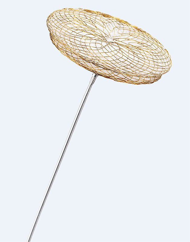
The CeraFlexTM ASD occluder (see Figure 1) is a double-disk device for transcatheter secundum ASD closure. Polyethylene terephthalate (PET) membranes are sewn into each disc and the waist as foundation for growth of tissue over the occluder after placement and sealing of the hole (14). It has bioceramic titanium nitride (TiN) coating and no left atrial hub to reduce nickel ion elution and the risk of clot formation, and improve endothelialization and biocompatibility (13,14). The occluder has a 360° flexible rotation feature between the device and delivery cable, enabling accurate positioning without tension and a better adaptation to the interatrial septum (12). This device is available up to 42 mm waist diameters with 2 mm increments, and delivery sheath sizes from 8 to 14 Fr (14).
Figure 1.

LifeTech CeraFlexTM ASD occluder. ASD, atrial septal defect.
Implantation procedure and FU
Interventional procedures were conducted according to instructions for use and the standard approach of the respective study site. Transcatheter treatment was performed using standard intraprocedural imaging techniques such as TEE with fluoroscopic guidance if necessary. Device size was determined mainly on the basis of the stretched defect diameter using the “stop-flow technique” or frequently using the “waist technique” (15). Successful implantation was defined as stable and correct placement of the occluder within the defect.
All patients were admitted to the hospital 1 day preceding the intervention for all necessary examinations and were discharged the day after ASD-closure to ensure a good procedural result. Efficacy and safety were assessed by physical examination, vital signs, electrocardiogram (ECG), transthoracic echocardiography (TTE), performed during admission and intervention, before discharge and at 6-month FU. Serious adverse events (SAEs) that occurred during the procedure, until discharge or FU were documented and treated as appropriate (see Table 1).
Table 1. Schedule of assessments.
| Assessments | Admission | Procedure in cardiac catheter | Before discharge | Six-month FU (±1 month) |
|---|---|---|---|---|
| Informed consent | √ | |||
| Inclusion/exclusion criteria | √ | |||
| Medical history | √ | |||
| Demographic data† | √ | √ | ||
| Examination | √ | √ | √ | |
| Vital signs‡ | √ | √ | √ | |
| ECG | √ | √ | √ | |
| Transthoracic echocardiogram | à | à | ä | |
| Transesophageal echocardiogram | à | ä | ||
| Data from cardiac catheter | √ | |||
| Anticoagulation | √ | √ | √ | |
| SAE§§ | √ | √ | √ |
†, sex, age (years), height (cm), weight (kg); ‡, vital signs: blood pressure, heart rate; §, patients <15 years: transthoracic echocardiogram, patients >15 years: in accordance with clinical routine transesophageal echocardiogram or transthoracic echocardiogram; §§, documentation of SAEs. ECG, electrocardiogram; SAE, serious adverse event; FU, follow-up.
Primary endpoint was defined as successful placement of the occluder and successful closure of the defect without severe complications [embolization of the occluder, surgical re-intervention, moderate or large residual shunt, new onset of atrioventricular (AV) block II° or III°]. Secondary endpoint was defined as clinical efficacy 6-month after the intervention, implicating a stable occlusion and a successful closure of the defect without severe complications (erosions, embolization of the occluder, surgical re-intervention, moderate or large residual shunt, new onset of arrhythmia or AV block II° or III°) in the FU period. The size of the residual shunt was classified into trivial (<1 mm), small (1–2 mm), moderate (>2–4 mm) and large (>4 mm).
Quality assurance was ensured by an external monitor at the three participating study sites. Two to eleven monitoring visits, depending on the number of patients enrolled, were performed per study site. All study specific activities and the regular inspections of the collected data by the certified monitor were performed in accordance with DIN EN ISO 14155:2011 (German version).
Statistics
Data of intention to treat analysis are presented as median and interquartile range (IQR 25; 75). Categorical variables are expressed as absolute numbers or percentage.
Statistical analyses were performed using SPSS (version 23.0, IBM Corporation, Armonk, NY, USA).
Results
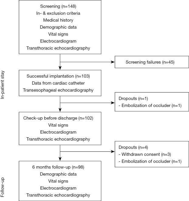
Between April 2016 and December 2019, a total of 148 patients were identified as potential study patients in the three participating study sites. Forty-five patients had to be excluded due to multiple defects or too large defect size. Course of Study, study characteristics and procedure related data of the patients are displayed in detail in Figure 2, Tables 2,3.
Figure 2.
Course of study.
Table 2. Anthropometric data of patients with a successfully closed ASD using CeraFlexTM occluder.
| Anthropometric data | Successfully closed ASDs (n=103) | |
|---|---|---|
| Median [IQR 25-75] | Min/max | |
| Sex, female, n (%) | 75 (72%) | – |
| Age at procedure (years) | 9 [6-47] | 3/80 |
| Weight (kg) | 29 [21-64] | 13/120 |
| Height (cm) | 135 [117-167] | 97/192 |
| Defect size (mm) | 12 [10-16] | 4/30 |
ASD, atrial septal defect; IQR, interquartile range.
Table 3. Procedural data of patients with a successfully closed ASD using CeraFlexTM occluder.
| Procedural data | Successfully closed ASDs (n=103) | |
|---|---|---|
| Median [IQR 25-75] | Min/max | |
| Defect size (mm) | 12 [10-16] | 4/30 |
| Device size (mm) | 12 [10-16] | 6/32 |
| Procedure duration (min) | 42 [28-65] | 15/221 |
| Fluoroscopy time (min) | 0.9 [0-5] | 0/34 |
ASD, atrial septal defect; IQR, interquartile range.
Due to the flexible delivery system placement of the occluders was simple and the occluder did not change its position in the heart during and after detachment from the delivery cable. The primary endpoint (successful ASD closure without severe complications) was reached by 102/103 patients (99%; see Figure 2). One device embolized early and was removed with a snare catheter on the first post interventional day. Surgical ASD closure was performed in this patient, and he did not reach the primary endpoint.
Another patient was found to have late device embolization into the aortic arch 4 weeks after device closure. The occluder was removed with a snare via the femoral artery from the aorta and the ASD was then closed with a larger CeraFlexTM device. This patient did not reach the secondary endpoint. Four patients developed clinically non-relevant supraventricular tachycardias (atrial flutter/fibrillation) during the FU period and were treated with antiarrhythmic medication. Atrial flutter, paroxysmal atrial fibrillation, supraventricular extra systoles and palpitations, as well as ECG abnormalities in 24-hour ECG were diagnosed for the first time in a total of four patients (4%) at 6-month FU. Accordingly, these four patients did not reach the secondary endpoint.
In the 6-month FU period three patients withdrew their consent and were excluded from the study. Hence, 98 patients completed the 6-month FU and 94 patients reached the secondary endpoint. One patient showed moderate residual shunt at 6-month FU. Since this shunt was newly detected and not related to the occluder a previously not diagnosed second ASD was postulated and the patient was not excluded. In 92/98 patients (94%) the defect was completely closed, without residual shunt at TTE (6-month FU). In 5/98 patients (5%) a trivial residual shunt was visible adjacent to the occluder.
Preceding secundum ASD closure incomplete right bundle branch block (iRBBB) was diagnosed in 31/103 patients (30%). At 6-month FU it was diagnosed in 17/98 patients (17%) no new ECG changes were noted.
Known minor side effects (adverse events) such as groin hematoma, hemorrhage, vertigo, nausea, vomiting, headache and migraine occurred only temporarily without permanent sequelae after intervention. SAEs like embolizations (n=2), vessel complications (n=1), syncope (n=1), vasovagal reaction (n=1), and arrhythmia (n=1) were gathered after transcatheter treatment and during FU period. In all SAEs the original state of health could be recovered.
No erosion, perforation or death occurred in any of the study patients during study period.
Device embolization
First patient
A 19-year-old woman presented an 11 mm ASD II in echocardiography (TTE and TEE). During balloon sizing a balloon occlusion diameter (BOD with “stop flow” technique) of 12 mm was assessed. A 12 mm CeraFlexTM ASD device was positioned. It was not stable and, hence, a 16 mm CeraFlexTM ASD occluder was implanted. On the next day this occluder had migrated to the left ventricle, from where it was removed by gentle retrieval into the descending aorta. It was withdrawn through a 12 F sheath from the right femoral artery with a Maslanka biotome. Six months later, during a second catheter interventional procedure, the defect now had a diameter of 16 mm in TEE and a BOD of 19 mm. A 21 mm Occlutech ASD device was positioned, but during stability testing, preceeding device release, the device fell easily into the left atrium. It was retrieved and surgical closure was scheduled. During surgery an oval shaped ASD II 16×25 mm was seen, which was closed with a GoreTex patch.
Due to device embolization before discharge the primary endpoint was not reached by this patient.
Second patient
A 63-year-old male patient, who was treated previously for severe stenosis of the right coronary artery with a drug eluting stent, presented an 18 mm ASD II with thin interatrial septum combined with an atrial septum aneurysm. Balloon sizing revealed a BOD (“stop flow” technique) of 20–22 mm. To exclude severe diastolic dysfunction, left ventricular end-diastolic pressure (LVEDP) was assessed during test occlusion of the ASD. LVEDP remained unchanged at 17 mmHg. A 20 mm CeraFlexTM ASD occluder slipped through at the first attempt, but was positioned correctly at the second try.
During an examination 4 weeks after ASDO, not considering our study, the patient had no cardiac symptoms, however, the occluder had migrated to the proximal aortic arch. It was removed with a 6 F Multisnare 20 mm through a 14 F sheath introduced into the right femoral artery. At the same session the ASD was closed with a 28 mm CeraFlexTM ASD occluder, which remained stable until the last routine examination.
Due to device embolization during 6-month FU the secondary endpoint was not reached by this patient.
Discussion
This is a German prospective multi-center trial evaluating the efficacy and safety of the LifeTech CeraFlexTM ASD occluder for transcatheter closure in patients with secundum ASD. Unique strengths of this trial compared to published studies (12,13,16) are a prospective multi-center study design and a sample size of more than 100 patients that underwent ASD closure with the LifeTech CeraFlexTM ASD occluder. The results of this intention to treat analysis demonstrate efficient and safe ASDO and extend these positive results to the first visit 6 months after ASDO. The primary endpoint (successful placement of the occluder and successful closure of the defect without severe complications) was reached by 102/103 patients and the secondary endpoint (clinical efficacy 6 months after the intervention) was reached by 94 patients.
Advantages/features
The advent of the Amplatzer Septal occluder (Abbott) in 1997 enabled catheter interventional closure of a secundum ASD in most patients. A recently published systematic review and meta-analysis, including 2,972 patients, reported safe use with adverse event rates for 1 in 20 patients (17). Studies documented significantly lower morbidity and shorter hospital stay after interventional ASD-closure with the Amplatzer ASD occluder compared to surgical ASD-closure (8,18). The Amplatzer ASD occluder is self-centering, easy to handle and fully retrievable until release. Even after device release the right atrial hub can be snared and the occluder can be removed safely through an adequately sized longsheath. However, the stiff connection of the Amplatzer ASD occluder to the delivery cable may cause difficult device placement, especially in patients with larger ASDs and deficient aortic septal rims. Just recently Abbott introduced a more flexible delivery cable (Treviso) (19), which improves the flexibility at device delivery. However, the effectivity of this modification still needs to be documented in larger patient series. The CeraFlexTM ASD occluder is comparable to the Amplatzer ASD occluder, as it is a nitinol double-disk device with PET membranes sewn into each disk. It is comparable to the Amplatzer ASD occluder with good outcomes and low complication rates (20). However, the 360° flexible rotation features between the device and delivery cable facilitate safe device implantation. Accurate positioning, even in difficult defects, is reported by Apostolopoulou et al. (12) and was also achieved in our study. Deployment without tension of the delivery catheter was confirmed by our study (13).
Correct device placement (primary endpoint of the study) on the first attempt was reached in 98 of 103 patients, the results being comparable with the use of the Occlutech Figulla Flex II Occluder (21). Both occluders have enhanced flexibility and a softer left atrial disc, due to the lack of a left atrial hub, resulting in a reduced risk of prolapse of the left atrial disc into the right atrium (22,23). Additionally, the missing left atrial hub may lower the risk for left atrial thrombus formation on the left disc of the device in the long-term.
A second implantation trial during the same procedure was necessary in five patients. One had an obvious residual shunt, one device could not be positioned properly. Repositioning of the same occluder was performed in one patient and one occluder was removed because of contact with the mitral valve. No additional maneuvers, such as deployment of the left disk in a pulmonary vein and additional balloon during deployment, were needed. Another potential advantage of the CeraFlexTM ASD occluder is the bioceramic TiN coating of the double disk, done to reduce nickel ion elution (14). Although sensitization to nickel is not a contra indication to ASDO with a nitinol device, increased levels of nickel were assessed in the blood of patients after ASDO with the Amplatzer device (24). Few case reports exist on the necessity to remove a nitinol device for suspected overreaction to nickel (25,26). Since at present there are no reports on nickel levels after ASD-closure with the CeraFlexTM ASD occluder, the advantage in this respect is speculative.
Device embolization
In 2/103 patients (2%) the device embolized. One device was seen in the left ventricle the day after the initial closure and an additional device was noticed in the aortic arch during an examination, not related to our study. Both patients were described in detail above. Device embolization occurs at a rate of 0.6–2% and usually, as in our cases, device retrieval is successful (22,27-29). Obviously in both patients the actual defect size was wrongly underestimated, although in both patients balloon sizing with the “stop flow” technique was employed. After successful retrieval it was tried in both patients to close the defect with a larger occluder. A 5 mm larger Occlutech device was not stable in the first patient and the defect was closed surgically, where a large defect (16×25 mm) was seen and closed with a patch. The other defect was successfully closed with a CeraFlexTM ASD occluder 5 mm larger than the first one.
Removing large CeraFlexTM occluders with a snare may be difficult since it may slip of the right atrial hub. Georgiev et al. reported on a viable method of interventional occluder removal using the Maslanka biopsy forceps assisting the snare (28). Left ventricle, abdominal aorta and femoral vessels are familiar embolization locations if the device embolizes to the systemic circulation (30,31). An overall embolization rate of 2% is rather high although in concordance with other study reports, accounting an embolization rate of 0.2–2.2% (21,22,28,29,32-35). Undersized device and large defects with either deficient or floppy septal tissues are also associated with these complications (15,36,37). It is assumed that embolizations are more related to anatomy than to specific devices (38). Nevertheless, operator-related technical issues like excessive tension on delivery cable during device deployment and malposition during the “push-pull” maneuver have to be taken into account (37).
Residual shunts
The presence of residual shunts after device implantation maybe caused by a second defect which previously was not diagnosed (22). Kaya et al. reported that residual shunting before discharge was present in 8 of 205 patients (4%) receiving Cera septal occluder, in 3 patients (1%) at 6-month FU and in 1 patient (1%) at 12-month FU (38). Decreasing shunts was confirmed by Haas et al. reporting that in 1,291 patients trivial residual shunting was present in 17% before discharge and in 4% at 6-month FU after ASD closure with the Occlutech occluder device. At 5-year FU a residual shunt was diagnosed solely in 2% (22). In 1 patient (1%) a moderate residual shunt was diagnosed not related to the occluder at the 6-month FU examination, while no residual shunt was apparent before discharge. This may be explained by a second ASD which had not been detected previously.
Arrhythmia and RBBB
Atrial arrhythmia is a frequent complication following transcatheter ASD closure (39). Occurrence of atrial tachyarrhythmia after ASD closure is associated with a prevalence of 2–4.8% (15,34,40). This data was confirmed by our study, since new-onset cardiac arrhythmia was detected in 4% (4 patients) with an average patient age of 64 years. Affected patients were diagnosed with new onset atrial flutter, paroxysmal atrial fibrillation, supraventricular extra systoles and palpitations, as well as ECG abnormalities in 24-hour ECG. Arrhythmia resolved after medical treatment in all patients. Particularly the occurrence of atrial fibrillation and flutter as well as ventricular extra systoles in this cohort is common. A relationship with the device is possible in all affected patients. In one patient hemodynamically relevant bradycardia occurred. This vasovagal reaction was handled with emergency treatment. A relationship with the procedure was considered possible. However, the symptoms completely resolved and the patient remained clinically stable.
In our study, iRBBB was diagnosed in 31 patients before secundum ASD closure. When comparing the QRS-time before the transcatheter treatment and at 6-month FU, the QRS-time was normalized in 14 patients. Moreover, no AV block occurred, confirming previously reported prevalence of advanced heart block as being below 1% (35,41,42). Careful monitoring of the development of conduction disturbance is recommended (43).
Study limitation
All three participating centers have 20 years and more experience with catheter interventional closure of secundum ASDs with self-centering double disc devices. Hence, no detailed implantation protocol was applied for this very standardized procedure. The implantations were conducted according to the CeraFlexTM ASD occluder instructions for use.
Conclusions
Interventional secundum ASD closure with the CeraFlexTM ASD occluder proved to be feasible, effective and safe. Prospective, randomized long-term studies with larger patient cohorts are needed to fully evaluate the advantages of this occluder comparing its results with other devices and capture possible long-term benefits.
Supplementary
The article’s supplementary files as
Acknowledgments
The maintenance of the patient database was sponsored by LifeTech (Lifetech Scientific Co., Shenzhen, China) and initially Medtronic (Medtronic PLC, Dublin, Ireland). Leon Brudy provided language help.
Funding: This study was supported financially by grants from Medtronic and LifeTech.
Ethical Statement: The authors are accountable for all aspects of the work in ensuring that questions related to the accuracy or integrity of any part of the work are appropriately investigated and resolved. The study was conducted in accordance with the Declaration of Helsinki (as revised in 2013) and the Good Clinical Practice guidelines, DIN EN ISO 14155:2011 (German version) and the current national regulations including the Medical Devices Act. The study protocol was approved by the local ethical board of the Technical University Munich (project number 451/15S) and registered at the German Clinical Trial Register (https://www.drks.de/drks_web/; registration ID: DRKS00010233). All participants gave written informed consent for study participation after being provided with information about the study protocol.
Footnotes
Provenance and Peer Review: This article was commissioned by the Guest Editors (Yskert von Kodolitsch, Harald Kaemmerer and Koichiro Niwa) for the series “Current Management Aspects in Adult Congenital Heart Disease (ACHD): Part V” published in Cardiovascular Diagnosis and Therapy. The article has undergone external peer review.
Reporting Checklist: The authors have completed the TREND reporting checklist. Available at https://cdt.amegroups.com/article/view/10.21037/cdt-21-798/rc
Peer Review File: Available at https://cdt.amegroups.com/article/view/10.21037/cdt-21-798/prf
Conflicts of Interest: All authors have completed the ICMJE uniform disclosure form (available at https://cdt.amegroups.com/article/view/10.21037/cdt-21-798/coif). The series “Current Management Aspects in Adult Congenital Heart Disease (ACHD): Part V” was commissioned by the editorial office without any funding or sponsorship. A Engelhardt and DT report grants from Medtronic and LifeTech. ID reports that he was paid by the German Heart Center a reimbursement. A Eicken reports that he has acted as proctor for LifeTech ASD-Occlusion. The authors have no other conflicts of interest to declare.
References
- 1.Fuster V, Brandenburg RO, McGoon DC, et al. Clinical approach and management of congenital heart disease in the adolescent and adult. Cardiovasc Clin 1980;10:161-97. [PubMed] [Google Scholar]
- 2.Warnes CA, Williams RG, Bashore TM, et al. ACC/AHA 2008 guidelines for the management of adults with congenital heart disease: a report of the American College of Cardiology/American Heart Association Task Force on Practice Guidelines (Writing Committee to Develop Guidelines on the Management of Adults With Congenital Heart Disease). Developed in Collaboration With the American Society of Echocardiography, Heart Rhythm Society, International Society for Adult Congenital Heart Disease, Society for Cardiovascular Angiography and Interventions, and Society of Thoracic Surgeons. J Am Coll Cardiol 2008;52:e143-263. 10.1016/j.jacc.2008.10.001 [DOI] [PubMed] [Google Scholar]
- 3.Alexi-Meskishvili VV, Konstantinov IE. Surgery for atrial septal defect: from the first experiments to clinical practice. Ann Thorac Surg 2003;76:322-7. 10.1016/S0003-4975(03)00508-3 [DOI] [PubMed] [Google Scholar]
- 4.Hörer J, Eicken A, Müller S, et al. Risk factors for prolonged intensive care treatment following atrial septal defect closure in adults. Int J Cardiol 2008;125:57-61. 10.1016/j.ijcard.2007.02.022 [DOI] [PubMed] [Google Scholar]
- 5.King TD, Thompson SL, Steiner C, et al. Secundum atrial septal defect. Nonoperative closure during cardiac catheterization. JAMA 1976;235:2506-9. 10.1001/jama.1976.03260490024013 [DOI] [PubMed] [Google Scholar]
- 6.Nassif M, Abdelghani M, Bouma BJ, et al. Historical developments of atrial septal defect closure devices: what we learn from the past. Expert Rev Med Devices 2016;13:555-68. 10.1080/17434440.2016.1182860 [DOI] [PubMed] [Google Scholar]
- 7.Cowley CG, Lloyd TR, Bove EL, et al. Comparison of results of closure of secundum atrial septal defect by surgery versus Amplatzer septal occluder. Am J Cardiol 2001;88:589-91. 10.1016/S0002-9149(01)01750-7 [DOI] [PubMed] [Google Scholar]
- 8.Du ZD, Hijazi ZM, Kleinman CS, et al. Comparison between transcatheter and surgical closure of secundum atrial septal defect in children and adults: results of a multicenter nonrandomized trial. J Am Coll Cardiol 2002;39:1836-44. 10.1016/S0735-1097(02)01862-4 [DOI] [PubMed] [Google Scholar]
- 9.Butera G, Biondi-Zoccai G, Sangiorgi G, et al. Percutaneous versus surgical closure of secundum atrial septal defects: a systematic review and meta-analysis of currently available clinical evidence. EuroIntervention 2011;7:377-85. 10.4244/EIJV7I3A63 [DOI] [PubMed] [Google Scholar]
- 10.Chen TH, Hsiao YC, Cheng CC, et al. In-Hospital and 4-Year Clinical Outcomes Following Transcatheter Versus Surgical Closure for Secundum Atrial Septal Defect in Adults: A National Cohort Propensity Score Analysis. Medicine (Baltimore) 2015;94:e1524. 10.1097/MD.0000000000001524 [DOI] [PMC free article] [PubMed] [Google Scholar]
- 11.Jung SY, Choi JY. Transcatheter closure of atrial septal defect: principles and available devices. J Thorac Dis 2018;10:S2909-22. 10.21037/jtd.2018.02.19 [DOI] [PMC free article] [PubMed] [Google Scholar]
- 12.Apostolopoulou SC, Tsoutsinos A, Laskari C, et al. Large single centre experience with the Cera™ and CeraFlex™ occluders for closure of interatrial communications: usefulness of the flexible rotation feature. Cardiovasc Interv Ther 2018;33:70-6. 10.1007/s12928-016-0440-y [DOI] [PubMed] [Google Scholar]
- 13.Astarcioglu MA, Kalcik M, Sen T, et al. Ceraflex versus Amplatzer occluder for secundum atrial septal defect closure. Multicenter clinical experience. Herz 2015;40 Suppl 2:146-50. 10.1007/s00059-014-4192-0 [DOI] [PubMed] [Google Scholar]
- 14.LifeTech Scientific (Shenzhen) Co., Ltd. Instructions for Use. CeraFlexTM ASD Closure System. 2016.
- 15.Amin Z, Hijazi ZM, Bass JL, et al. Erosion of Amplatzer septal occluder device after closure of secundum atrial septal defects: review of registry of complications and recommendations to minimize future risk. Catheter Cardiovasc Interv 2004;63:496-502. 10.1002/ccd.20211 [DOI] [PubMed] [Google Scholar]
- 16.Leong MC, Karyostyko B, Ramli MNH, et al. The Ceraflex and Figulla atrial septal occluders: early and intermediate-term safety and efficacy study. Cardiol Young 2022. [Epub ahead of print]. doi: . 10.1017/S1047951121004728 [DOI] [PubMed] [Google Scholar]
- 17.Heaton JN, Okoh AK, Suh S, et al. Safety and Efficacy of the Amplatzer Septal Occluder: A Systematic Review and Meta-Analysis. Cardiovasc Revasc Med 2022;37:52-60. 10.1016/j.carrev.2021.06.002 [DOI] [PMC free article] [PubMed] [Google Scholar]
- 18.Masura J, Gavora P, Formanek A, et al. Transcatheter closure of secundum atrial septal defects using the new self-centering amplatzer septal occluder: initial human experience. Cathet Cardiovasc Diagn 1997;42:388-93. [DOI] [PubMed] [Google Scholar]
- 19.Haddad RN, Khraiche D, Bonnet D, et al. Preliminary Experience With the New Amplatzer™ Trevisio™ Delivery System in Transcatheter Atrial Septal Defect Closures in Children. Front Pediatr 2021;9:641742. 10.3389/fped.2021.641742 [DOI] [PMC free article] [PubMed] [Google Scholar]
- 20.Bhattacharjya S, Pillai LS, Doraiswamy V, et al. Prospective concurrent head-to head comparison of three different types of nitinol occluder device for transcatheter closure of secundum atrial septal defects. EuroIntervention 2019;15:e321-8. 10.4244/EIJ-D-18-01016 [DOI] [PubMed] [Google Scholar]
- 21.Kenny D, Eicken A, Dähnert I, et al. A randomized, controlled, multi-center trial of the efficacy and safety of the Occlutech Figulla Flex-II Occluder compared to the Amplatzer Septal Occluder for transcatheter closure of secundum atrial septal defects. Catheter Cardiovasc Interv 2019;93:316-21. 10.1002/ccd.27899 [DOI] [PubMed] [Google Scholar]
- 22.Haas NA, Soetemann DB, Ates I, et al. Closure of Secundum Atrial Septal Defects by Using the Occlutech Occluder Devices in More Than 1300 Patients: The IRFACODE Project: A Retrospective Case Series. Catheter Cardiovasc Interv 2016;88:571-81. 10.1002/ccd.26497 [DOI] [PubMed] [Google Scholar]
- 23.Haas NA, Happel CM, Soetemann DB, et al. Optimal septum alignment of the Figulla Flex occluder to the atrial septum in patients with secundum atrial septal defects. EuroIntervention 2016;11:1153-60. 10.4244/EIJY14M12_09 [DOI] [PubMed] [Google Scholar]
- 24.Ries MW, Kampmann C, Rupprecht HJ, et al. Nickel release after implantation of the Amplatzer occluder. Am Heart J 2003;145:737-41. 10.1067/mhj.2003.7 [DOI] [PubMed] [Google Scholar]
- 25.Singh HR, Turner DR, Forbes TJ. Nickel allergy and the amplatzer septal occluder. J Invasive Cardiol 2004;16:681-2. [PubMed] [Google Scholar]
- 26.Vincent RN, Raviele AA, Diehl HJ. Single-center experience with the HELEX septal occluder for closure of atrial septal defects in children. J Interv Cardiol 2003;16:79-82. 10.1046/j.1540-8183.2003.08005.x [DOI] [PubMed] [Google Scholar]
- 27.Diab KA, Cao QL, Bacha EA, et al. Device closure of atrial septal defects with the Amplatzer septal occluder: safety and outcome in infants. J Thorac Cardiovasc Surg 2007;134:960-6. 10.1016/j.jtcvs.2007.06.018 [DOI] [PubMed] [Google Scholar]
- 28.Georgiev S, Tanase D, Genz T, et al. Retrieval of large Occlutech Figula Flex septal defect occluders using a commercially available bioptome: proof of concept. Cardiol Young 2018;28:955-60. 10.1017/S1047951118000586 [DOI] [PubMed] [Google Scholar]
- 29.Takaya Y, Akagi T, Kijima Y, et al. Long-term outcome after transcatheter closure of atrial septal defect in older patients: impact of age at procedure. JACC Cardiovasc Interv 2015;8:600-6. 10.1016/j.jcin.2015.02.002 [DOI] [PubMed] [Google Scholar]
- 30.Durmuş G, Kalyoncuoğlu M, Belen E, et al. Retrival of Embolized Atrial Septal Defekt Closure Device in the Left Iliac Artery. Am J Cardiol 2018;121:e157. 10.1016/j.amjcard.2018.03.341 [DOI] [Google Scholar]
- 31.Crawford DA, Naidu SG, Shah AA, et al. Endovascular Retrieval of an Embolized Atrial Septal Occluder Device From the Abdominal Aorta. Vasc Endovascular Surg 2018;52:669-73. 10.1177/1538574418790393 [DOI] [PubMed] [Google Scholar]
- 32.El-Said H, Hegde S, Foerster S, et al. Device therapy for atrial septal defects in a multicenter cohort: acute outcomes and adverse events. Catheter Cardiovasc Interv 2015;85:227-33. 10.1002/ccd.25684 [DOI] [PubMed] [Google Scholar]
- 33.O'Byrne ML, Gillespie MJ, Kennedy KF, et al. The influence of deficient retro-aortic rim on technical success and early adverse events following device closure of secundum atrial septal defects: An Analysis of the IMPACT Registry®. Catheter Cardiovasc Interv 2017;89:102-11. 10.1002/ccd.26585 [DOI] [PMC free article] [PubMed] [Google Scholar]
- 34.Levi DS, Moore JW. Embolization and retrieval of the Amplatzer septal occluder. Catheter Cardiovasc Interv 2004;61:543-7. 10.1002/ccd.20011 [DOI] [PubMed] [Google Scholar]
- 35.DiBardino DJ, McElhinney DB, Kaza AK, et al. Analysis of the US Food and Drug Administration Manufacturer and User Facility Device Experience database for adverse events involving Amplatzer septal occluder devices and comparison with the Society of Thoracic Surgery congenital cardiac surgery database. J Thorac Cardiovasc Surg 2009;137:1334-41. 10.1016/j.jtcvs.2009.02.032 [DOI] [PMC free article] [PubMed] [Google Scholar]
- 36.Chessa M, Carminati M, Butera G, et al. Early and late complications associated with transcatheter occlusion of secundum atrial septal defect. J Am Coll Cardiol 2002;39:1061-5. 10.1016/S0735-1097(02)01711-4 [DOI] [PubMed] [Google Scholar]
- 37.Moore J, Hegde S, El-Said H, et al. Transcatheter device closure of atrial septal defects: a safety review. JACC Cardiovasc Interv 2013;6:433-42. 10.1016/j.jcin.2013.02.005 [DOI] [PubMed] [Google Scholar]
- 38.Kaya MG, Akpek M, Celebi A, et al. A multicentre, comparative study of Cera septal occluder versus AMPLATZER Septal Occluder in transcatheter closure of secundum atrial septal defects. EuroIntervention 2014;10:626-31. 10.4244/EIJY14M07_04 [DOI] [PubMed] [Google Scholar]
- 39.Suda K, Raboisson MJ, Piette E, et al. Reversible atrioventricular block associated with closure of atrial septal defects using the Amplatzer device. J Am Coll Cardiol 2004;43:1677-82. 10.1016/j.jacc.2003.12.042 [DOI] [PubMed] [Google Scholar]
- 40.Johnson JN, Marquardt ML, Ackerman MJ, et al. Electrocardiographic changes and arrhythmias following percutaneous atrial septal defect and patent foramen ovale device closure. Catheter Cardiovasc Interv 2011;78:254-61. 10.1002/ccd.23028 [DOI] [PubMed] [Google Scholar]
- 41.Abaci A, Unlu S, Alsancak Y, et al. Short and long term complications of device closure of atrial septal defect and patent foramen ovale: meta-analysis of 28,142 patients from 203 studies. Catheter Cardiovasc Interv 2013;82:1123-38. 10.1002/ccd.24875 [DOI] [PubMed] [Google Scholar]
- 42.Reddy BT, Patel JB, Powell DL, et al. Interatrial shunt closure devices in patients with nickel allergy. Catheter Cardiovasc Interv 2009;74:647-51. 10.1002/ccd.22155 [DOI] [PubMed] [Google Scholar]
- 43.Yang MC, Wu JR. Recent review of transcatheter closure of atrial septal defect. Kaohsiung J Med Sci 2018;34:363-9. 10.1016/j.kjms.2018.05.001 [DOI] [PubMed] [Google Scholar]
Associated Data
This section collects any data citations, data availability statements, or supplementary materials included in this article.
Supplementary Materials
The article’s supplementary files as


