Abstract
Background
Most patients with advanced non-small-cell lung cancer (NSCLC) fail to derive significant benefit from programmed cell death protein-1 (PD-1) axis blockade, and new biomarkers of response are needed. In this study, we aimed to discover and validate spatially resolved protein markers associated with sensitivity to PD-1 axis inhibition in NSCLC.
Methods
We initially assessed a discovery cohort of 56 patients with NSCLC treated with PD-1 axis inhibitors at Yale Cancer Center. Using the GeoMx Digital Spatial Profiling (DSP) system, 71 proteins were measured in spatial context on each spot in a tissue microarray. We used the AQUA method of quantitative immunofluorescence (QIF) to orthogonally validate candidate biomarkers. For external independent validation, we assessed whole tissue sections derived from 128 patients with NSCLC treated with single-agent PD-1 axis inhibitors at the 12 de Octubre Hospital (Madrid) using DSP. We further analyzed two immunotherapy untreated cohorts to address prognostic significance (n=252 from Yale Cancer Center; n=124 from University Clinic of Navarra) using QIF and DSP, respectively.
Results
Using continuous log-scaled data, we identified CD44 expression in the tumor compartment (pan-cytokeratin (CK)+) as a novel predictor of prolonged progression-free survival (PFS) (multivariate HR=0.68, p=0.043) in the discovery set. We validated by QIF that tumor CD44 levels assessed as continuous QIF scores were associated with longer PFS (multivariate HR=0.31, p=0.022) and overall survival (multivariate HR=0.29, p=0.038). Using DSP in an independent immunotherapy treated cohort, we validated that CD44 levels in the tumor compartment, but not in the immune compartment (panCK–/CD45+), were associated with clinical benefit (OR=1.22, p=0.018) and extended PFS under PD-1 axis inhibition using the highest tertile cutpoint (multivariate HR=0.62, p=0.03). The effect of tumor cell CD44 in predicting PFS remained significant after correcting for programmed death-ligand 1 (PD-L1) Tumor Proportion Score (TPS) in both cohorts. High tumor cell CD44 was not prognostic in the absence of immunotherapy. Using DSP data, intratumoral regions with elevated tumor cell CD44 expression showed prominent (fold change>1.5, adjusted p<0.05) upregulation of PD-L1, TIM-3, ICOS, and CD40 in two independent cohorts.
Conclusions
This work highlights CD44 as a novel indicative biomarker of sensitivity to PD-1 axis blockade that might help to improve immunotherapy strategies for NSCLC.
Keywords: immunotherapy; biomarkers, tumor; tumor microenvironment; lung neoplasms; programmed cell death 1 receptor
WHAT IS ALREADY KNOWN ON THIS TOPIC
CD44 has been identified as a programmed death-ligand 1 (PD-L1) regulator at both the protein and the messenger RNA levels, and it has been shown to contribute to PD-L1-mediated T cell suppression in preclinical models. However, the association between tumor cell CD44 overexpression and improved clinical outcomes to programmed cell death protein-1 (PD-1) axis blockade has not been previously reported.
WHAT THIS STUDY ADDS
This study identifies and validates that CD44, only when expressed by tumor cells and not immune cells, is a marker of sensitivity to PD-1 blockade that is non-redundant with PD-L1 expression.
HOW THIS STUDY MIGHT AFFECT RESEARCH, PRACTICE OR POLICY
CD44 could complement existing biomarkers for optimal patient stratification, and potentially open new therapeutic strategies to improve precision immunotherapy in lung cancer. Further mechanistic studies are needed to understand the interplay between CD44 positive cancer stem cell phenotype and mechanisms of immune evasion.
Introduction
Programmed cell death protein-1 (PD-1) axis blockade has transformed the treatment landscape of advanced non-small cell lung cancer (NSCLC). Antibodies blocking the PD-1/programmed death-ligand 1 (PD-L1) interaction have shown improved survival as single agents, in combination with chemotherapy, or combined with cytotoxic T-lymphocytes-associated protein 4 (CTLA-4) blockade.1 2 However, most patients fail to develop durable responses particularly when these drugs are given as monotherapies in unselected patients.1 2 Moreover, there is a limited understanding of the mechanisms of sensitivity and resistance to PD-1 axis inhibitors, and robust biomarkers of response are also lacking.2 3
The characterization of tumor cells and their surrounding microenvironment by direct analysis of tumor specimens has shown utility to predict outcomes from PD-1 axis blockade. Notably, in a meta-analysis involving 10 different tumor types and more than 8000 tumor specimens, multiplexed immunohistochemistry (IHC)/immunofluorescence showed significantly higher performance than PD-L1 expression (assessed by IHC), tumor mutational burden, or bulk interferon (IFN)-γ-based gene expression signatures for discriminating between responders and non-responders to immune checkpoint blockade.4 These findings underscore the importance of assessing immunotherapy biomarkers in a quantitative manner and in their spatial context, and illustrate the potential of these technologies to identify new mechanisms of response to immunotherapy.
CD44 is a transmembrane glycoprotein expressed as a wide variety of isoforms in most cells of the human body,5 and has been implicated in multiple pathways that are essential for tumor maintenance and progression. Most notably, CD44 participates in key aspects of stemness,6 7 particularly epithelial to mesenchymal transition (EMT),8–11 migration,9 11 12 and apoptosis resistance.5 Beyond its roles in regulating tumor-intrinsic activities, CD44 can also modulate antitumor immunity. For instance, CD44 has been identified as a PD-L1 regulator at both the protein and the messenger RNA (mRNA) levels, and it has been shown to contribute to PD-L1-mediated T cell suppression in preclinical models.13 14 However, the distribution and patterns of CD44 protein expression and its potential role in promoting sensitivity to PD-1 axis blockade in human NSCLC has not been reported.
In this study, we aimed to discover and validate spatially resolved protein markers associated with sensitivity to PD-1 axis inhibition in advanced NSCLC. Using two orthogonal spatial proteomic technologies in immunotherapy treated and immunotherapy untreated NSCLC cohorts from different institutions, we identify and validate tumor cell CD44 as a novel indicative biomarker of sensitivity to PD-1 axis blockade. In addition, we show that intratumoral regions with elevated expression of CD44 in the tumor compartment display a unique immune microenvironment, characterized by prominent upregulation of multiple immunomodulatory molecules.
Methods
Patient cohorts
We analyzed retrospectively collected, formalin-fixed, paraffin-embedded tumor specimens from four independent multi-institutional NSCLC cohorts.
We used two immunotherapy treated NSCLC cohorts as discovery and validation sets. The discovery set (YTMA471) contained 56 patients that received immune checkpoint blockade for advanced disease between 2012 and 2019 at Yale Cancer Center (New Haven, Connecticut, USA), whose pre-treatment tumors were represented in a tissue microarray (TMA) format. The validation cohort (H12O_ITX1) was composed with 128 patients that were treated with single-agent PD-1 axis inhibitors between 2013 and 2019 at the 12 de Octubre University Hospital (Madrid, Spain), whose pre-treatment tumors were assessed as whole tissue sections. The baseline characteristics of these cohorts are summarized in table 1.
Table 1.
Baseline patient characteristics of the immunotherapy treated NSCLC cohorts
| Characteristic | YTMA471 cohort N (%) |
H12O_ITX1 cohort N (%) |
| Total | 56 | 128 |
| Gender | ||
| Male | 28 (50) | 100 (78.1) |
| Female | 28 (50) | 28 (21.9) |
| Age | ||
| <70 years old | 30 (53.6) | 68 (53.1) |
| ≥70 years old | 26 (46.4) | 60 (46.9) |
| Smoking history | ||
| Active smoker | 10 (18) | 34 (26.6) |
| Former smoker | 44 (79) | 88 (68.7) |
| Never smoker | 2 (3.6) | 6 (4.7) |
| Histology | ||
| Adenocarcinoma | 41 (73) | 54 (42.2) |
| Large-cell carcinoma | – | 16 (12.5) |
| Squamous cell carcinoma | 9 (16) | 58 (45.3) |
| Adenosquamous carcinoma | 2 (3.6) | – |
| NSCLC-NOS | 4 (7.1) | – |
| Stage | ||
| III | 5 (8.9) | 5 (3.9) |
| IV | 51 (91.1) | 123 (96.1) |
| Actionable drivers | ||
| EGFR/ALK/ROS1/RET alterations | 1 (1.7) | 7 (5.4) |
| No EGFR/ALK/ROS1/RET alterations | 48 (85.7) | 63 (49.2) |
| Unknown genetic alterations | 7 (12.5) | 58 (45.3) |
| Central nervous system metastasis | ||
| Yes | 21 (38) | 24 (18.7) |
| No | 35 (62) | 104 (81.3) |
| Liver metastasis | ||
| Yes | 11 (20) | 25 (19.5) |
| No | 45 (80) | 103 (80.5) |
| LIPI score | ||
| Good | 9 (16.1) | 27 (21.1) |
| Intermediate | 22 (39.3) | 54 (42.2) |
| Poor | 3 (5.4) | 42 (32.8) |
| Unknown | 22 (39.3) | 5 (3.9) |
| PD-L1 TPS by IHC | ||
| ≥50% | 15 (26.8) | 32 (25) |
| 1%–49% | 20 (35.7) | 44 (34.4) |
| <1% | 11 (19.6) | 46 (36) |
| Not evaluable | 10 (17.9) | 6 (4.6) |
| Immunotherapy line | ||
| 1 | 39 (70) | 20 (15.6) |
| 2 | 14 (25) | 79 (61.7) |
| ≥3 | 3 (5.4) | 29 (12.5) |
| Type of immunotherapy | ||
| Chemotherapy +PD-1 axis blockade | 22 (39.3) | 0 |
| Dual PD-1 +CTLA4 blockade | 1 (1.8) | 0 |
| Other PD-1 axis-based combinations | 5 (8.9) | 0 |
| Single-agent PD-1 axis blockade | 28 (50) | 128 (100) |
| Best response to immunotherapy | ||
| Complete or partial response | 17 (33.5) | 22 (17.1) |
| Stable disease | 22 (39) | 32 (25) |
| Progressive disease | 15 (27) | 74 (57.8) |
| Not evaluable | 1 | 0 |
| Clinical benefit group | ||
| Clinical benefit | 36 (64.3) | 37 (28.9) |
| No clinical benefit | 19 (33.9) | 90 (70.3) |
| Not evaluable | 1 (1.8) | 1 (0.8) |
CTLA-4, cytotoxic T-lymphocytes-associated protein 4; IHC, immunohistochemistry; LIPI, Lung Immune Prognostic Index; NSCLC-NOS, non-small cell lung cancer not otherwise specified; PD-1, programmed cell death protein-1; PD-L1, programmed death-ligand 1; TPS, Tumor Proportion Score.
To rule out prognostic significance of the validated predictors, we analyzed two control cohorts of immunotherapy untreated patients with NSCLC, whose tumors were represented in TMA formats: YTMA423 (Yale control cohort), which contained 252 evaluable tumors resected between 2011 and 2016 at Yale Cancer Center; and University Clinic of Navarra (CUN) cohort (CIMA-CUN control cohort), containing 124 evaluable tumors resected between 2000 and 2013 at CUN (Pamplona, Spain). The baseline characteristics of the two control cohorts are summarized in online supplemental table S1. The details for TMA construction are summarized in the online supplemental materials and methods.
jitc-2022-004757supp001.pdf (1,012.4KB, pdf)
Digital spatial profiling
Tissue slides from YTMA471, H12O_ITX1, and CIMA-CUN cohorts were subjected to digital spatial profiling (DSP) experiments. Two slides derived from independent YTMA471 blocks, each block containing two non-adjacent tumor cores per NSCLC case, were profiled at Yale (Rimm Lab). One hundred and twenty-eight whole tissue sections from H12O_ITX1, and 6 TMA slides from CIMA-CUN cohort, each containing three tumor cores per NSCLC case, were profiled at the 12 de Octubre Research Facilities (Zugazagoitia Lab).
Briefly, the slides were first deparaffinized and subjected to antigen retrieval procedures, then we co-incubated them with fluorescent labeled antibodies (morphology markers), together with photocleavable oligonucleotide-labeled primary antibodies (profiling antibodies). In YTMA471 cohort, we used three morphology markers to detect tumor cells (pan-cytokeratin (CK)), all immune cells (CD45), and macrophages (CD68), together with a 71-plex profiling antibody panel. In cohorts H12O_ITX1 and CIMA-CUN, we used two morphology markers (panCK and CD45), together with a 36-plex profiling antibody panel (online supplemental table S2). Once the staining step was completed, we loaded the slides on the GeoMx DSP instruments (NanoString), where they were scanned to produce a digital fluorescent image of the tissue. Next, we generated circular regions of interest (ROIs) of a maximum of 660 µm in each slide. In TMA slides, ROIs were restricted to the tissue cores (generally one ROI per core was generated) (figure 1). For whole tissue slides, we selected multiple ROIs from intratumoral immune-enriched areas (panCK+/CD45+) distributed across the entire tissue (figure 2). Then, to obtain compartment-specific protein measurements, we generated molecularly defined tissue compartments in each ROI by fluorescent marker colocalization. In YTMA471, three compartments were generated: tumor compartment (panCK+), macrophage compartment (CD68+), and lymphocyte compartment (panCK−/CD45+/CD68−). An additional fourth compartment containing the summation of the protein counts from lymphocyte and macrophage compartments (panCK−/CD45+/CD68+) was defined as the immune compartment in this cohort (figure 1). In H12O_ITX1 and CIMA-CUN cohorts, the tumor compartment (panCK+) and the immune compartment (panCK−/CD45+) were generated (figure 2). Subsequently, oligos from these compartments were released on ultraviolet light exposure, dispensed in a 96-well plate, hybridized to 4-color, 6-spot optical barcodes, and digitally counted in the nCounter system (NanoString). Using the GeoMx software (NanoString), digital counts were first normalized to internal spike-in controls (ERCCs), and then normalized to the counts of internal housekeeper protein probes. A more detailed description of the DSP protocol can be found in online supplemental materials and methods.
Figure 1.
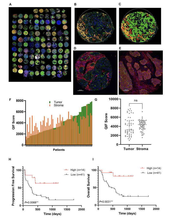
Identification of CD44 expression in the tumor compartment as a predictor of survival in YTMA471 discovery cohort. (A) Representative image of YTMA471 acquired using the GeoMx DSP system. (B–C) Representative TMA spot showing the fluorescence image (B) and the compartmentalized image created by fluorescence colocalization (C) using the GeoMx DSP system; panCK (green), CD45 (yellow), CD68 (red), SYTO13 (blue). (D–E) Representative TMA spot of low CD44 expression in panCK +tumor cells (D) and high CD44 expression in panCK +tumor cells (E) using QIF; panCK (green), CD44 (red), DAPI (blue). (F) Dynamic range of CD44 expression in the tumor compartment (panCK+) and in the stromal compartment (panCK–) using QIF. (G) Comparative analysis of CD44 levels measured by QIF in the tumor compartment and the stromal compartment. (H–I) Kaplan-Meier PFS curve (H) and OS curve (I) according to CD44 expression in the tumor compartment using QIF (optimal quartile cutpoint). CK, cytokeratin; DSP, digital spatial profiling; ns, not significant; OS, overall survival; PFS, progression-free survival; QIF, quantitative immunofluorescence; TMA, tissue microarray.
Figure 2.
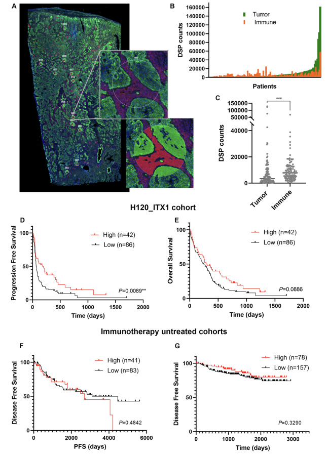
Validation of CD44 expression in the tumor compartment as an indicative biomarker of sensitivity to single-agent PD-1 axis blockade in NSCLC. (A) Representative image of a whole tissue section with 12 ROIs selected from immune-enriched (panCK+/CD45+) intratumoral areas using DSP. Fluorescence image is shown on the top, and the compartmentalized image at the bottom; panCK (green), CD45 (red), SYTO13 (blue). (B), Dynamic range of CD44 expression in the tumor compartment (panCK+) and in the immune compartment (panCK–/CD45+) using DSP; (C) Comparative analysis of CD44 levels measured by DSP in the tumor compartment and in the immune compartment. (D–E) Kaplan-Meier PFS curve (D) and OS curve (E) according to CD44 expression in the tumor compartment using DSP (tertile cutpoint). (F–G) Kaplan-Meier disease-free survival curves according to CD44 expression using DSP (F) or QIF (G) in immunotherapy untreated cohorts (CIMA-CUN cohort and YTMA423 cohort as F and G, respectively) (tertile cutpoint). CK, cytokeratin; DSP, digital spatial profiling; ns, not significant; NSCLC, non-small-cell lung cancer; OS, overall survival; PD-1, programmed cell death protein-1; PFS, progression-free survival; QIF, quantitative immunofluorescence; ROI, regions of interest.
Quantitative immunofluorescence
We developed a quantitative immunofluorescence (QIF) protocol (CD44/CK/DAPI) to detect and quantify CD44 using slides from YTMA471 and YTMA423 cohorts. Two slides derived from independent TMA blocks, each block containing two non-adjacent tumor cores per NSCLC case, were used in both cohorts. The QIF staining protocol is detailed in online supplemental materials and methods.
We used the AQUA system (Navigate BP) to acquire and quantify the fluorescence signal of CD44 as previously described.15 CD44 was measured from two compartments: the CK +tumor compartment, created by binarizing the CK signal, and the stromal compartment, created by excluding the tumor mask from a dilated DAPI mask representing the total tissue. CD44 QIF scores were calculated by dividing the target pixel intensity by the area of the compartment, and then normalized to the exposure time and bit depth at which the images were captured.15
CD44 antibody validation
To assess antibody specificity, we followed the guidelines for pillars of validation proposed by Uhlen et al.16 We tested two independent anti-CD44 antibodies (clone 156–3 C11 (Cell Signaling) and clone EPR1013Y, (Abcam)) targeting non-overlapping epitopes (online supplemental table S3) in an NSCLC test array (YTMA295), which contained 35 lung tumor cores with variable CD44 expression. For each antibody, first we assessed membrane localization, then optimized the titration, and then compared the CD44 QIF scores obtained with the optimal antibody concentration of each clone. A high correlation in the quantitative measurements of CD44 between independent antibodies would prove specificity for recognizing the target.16 17
Immunotherapy efficacy assessment
We used Response Evaluation Criteria in Solid Tumors V.1.1 to retrospectively evaluate treatment response to immune checkpoint blockade. We defined clinical benefit as having experienced complete or partial response, or stable disease lasting ≥6 months, whereas non-clinical benefit was defined as primary progressive disease or stable disease lasting <6 months. Patients with stable disease who did not progress and were censored before 6 months of follow-up were non-evaluable. Overall survival (OS) and progression-free survival (PFS) were calculated from the treatment start date to the date of death or loss of follow-up, or the date of disease progression, death, or loss of follow-up, respectively. For those patients who did not die or progress during the study period, the outcome was considered left-censored.
Data processing and statistical analysis
To analyze the association between marker expression and patients’ clinical-pathological characteristics or survival, normalized counts or QIF scores were averaged across all ROIs of each tissue sample to derive a single value per patient. We used non-parametric tests to compare CD44 counts between patient clinical subgroups or tissue compartments. For survival prediction analysis (PFS and OS), we used the Cox proportional hazards model, first utilizing averaged counts or QIF scores as continuous log2-transformed data. We used the X-tile software18 to explore the optimal cutpoint for patients’ survival stratification. Survival curves were computed with the Kaplan-Meier method and compared using the log-rank test. For prediction of clinical benefit (binary outcome) in the validation set, we utilized log2-transformed counts from each ROI independently (without averaging across ROIs) and fitted a regular logistic regression model with clustering by patient using Stata V.17.0 software. To test the association between CD44 counts in the tumor compartment with other immune-related proteins measured in the same compartment, we selected those ROIs containing matched tumor and immune compartment protein measurements and considered each ROI as an independent sample, since we aimed to assess the immune microenvironment features in each of the independently profiled intratumoral regions. Then, we used two complementary analyses. First, we used protein counts as continuous variables and tested the association between tumor cell CD44 with each of the other immune related markers using Pearson correlation analysis. Second, we dichotomized tumor cell CD44 expression using a clinically relevant cutpoint (upper tertile) to analyze what proteins were differentially expressed in tumor cell CD44 high versus tumor cell CD44 low ROIs. For this differential expression analysis, we quantified the fold change (FC) of all protein levels in ROIs with elevated CD44 expression relative to ROIs with low CD44 expression, and tested the significance with non-parametric tests accounting for multiple comparisons (Benjamini-Hochberg false discovery rate (FDR) method) using MetaboAnalyst V.5.0 software. All hypothesis testing was performed at a two-sided significance level of α=0.05.
Results
We first assessed the YTMA471 discovery cohort using the DSP system. One-hundred and ten ROIs were generated in 56 NSCLC cases. Seventy-one proteins were measured selectively from four compartments (figure 1A–C), resulting in a total of 284 protein variables per ROI. Using continuous log-scaled data, PD-L1 expression in the tumor compartment (HR=0.67, p=0.017) and in the immune compartment (HR=0.52, p=0.008) was predictive of longer PFS, thus validating the cohort. Among the novel candidate predictors, CD44 expression measured in the tumor compartment, but not in stromal compartments, was associated with longer PFS both in the univariate (HR=0.76, 95% CI 0.61 to 0.96; p=0.024) and in the multivariate analysis after controlling for stage, baseline liver metastasis, and Lung Immune Prognostic Index (LIPI) score19 (HR=0.68, 95% CI 0.46 to 0.99; p=0.043) (online supplemental table S4), and was selected for further study and validation.
In the quantitative analysis using DSP, CD44 levels followed a continuous distribution. CD44 counts were significantly higher in the immune compartment than in the tumor compartment (Wilcoxon paired test, p<0.001) (online supplemental figure S1A). CD44 levels in the tumor compartment were significantly higher in patients with squamous-cell carcinomas (SCC) (Kruskal-Wallis test, p=0.0014) and patients with no baseline liver metastasis (Mann-Whitney test, p=0.0026). Tumor CD44 expression was not significantly different among other clinical factors analyzed (online supplemental figure S1B–H).
As the initial step for validation, we aimed to reproduce the DSP findings using an orthogonal QIF-based method in the same cohort. First, we sought to prove specificity for detecting CD44. For this purpose, we tested two different anti-CD44 antibodies that recognized non-overlapping epitopes in an NSCLC test array (YTMA295). We observed a good dynamic range (online supplemental figure S2A, B) and a specific membranous CD44 staining pattern with both antibodies (online supplemental figure S2C, D). Then, we compared the CD44 QIF scores obtained with the two clones, showing a high correlation coefficient (R2=0.88) (online supplemental figure S2E). This provided evidence that both antibodies specifically recognized CD44 without significant cross reactivity, and we selected clone 156–3 C11 as a validated antibody for the remainder of the QIF studies. CD44 quantitative measurements were concordant between QIF and DSP methods (R2=0.44) (online supplemental figure S2F).
Then, we evaluated CD44 expression patterns and outcome performance using QIF in YTMA471 cohort. CD44 displayed predominantly membranous staining pattern and was detected both in tumor cells and stromal cells (figure 1D, E). Virtually all tumors were visually positive for CD44 in the stroma, while 70% of NSCLCs had CD44 staining in tumor cells. CD44 QIF scores also followed a continuous distribution and were numerically higher in the stromal compartment than in the tumor compartment (Wilcoxon paired test, p=0.13) (figure 1F, G). Consistent with DSP findings, CD44 levels in the tumor compartment assessed as continuous QIF scores were significantly associated with longer PFS (multivariate HR=0.31, 95% CI 0.11 to 0.87; p=0.022) and OS (multivariate HR=0.29, 95% CI 0.09 to 0.97; p=0.038). Using the X-tile software, the highest quartile was determined as the optimal single cutpoint that significantly predicted both PFS (log-rank test, p=0.0068) and OS (log-rank test, p=0.0031) in this cohort (figure 1H, I). CD44 QIF levels in the stromal compartment did not predict outcomes, either as continuous data or using median, tertile, or quartile cutpoints (online supplemental figure S3A–F).
Next, after having validated tumor cell CD44 as a candidate predictor using two orthogonal methods in the discovery set, we sought for external validation in the H12O_ITX1 cohort using DSP. We generated a total of 662 ROIs from 128 patient-derived whole tissue sections (average ROI per tissue sample=6 (1–12)), each ROI was then segmented in the tumor compartment and the immune compartment (figure 2A). In this validation set, PD-L1 as well as other known markers such as CD3 or CD8 were associated with outcomes from PD-1 axis blockade (online supplemental figure S4A–F), thus providing validity to the cohort and the ROI selection approach.
Consistent with the results in the YTMA471 discovery cohort, CD44 expression in the tumor compartment followed a continuous distribution (figure 2B) and was significantly higher in the immune compartment than in the tumor compartment (Wilcoxon paired test, p<0.001) (figure 2C). As seen in the training set, tumor cell CD44 expression was significantly higher in patients with SCC (Kruskal-Wallis test, p<0.001) (online supplemental figure S5A–F). In terms of outcome, CD44 expression in the tumor compartment validated as a predictor of clinical outcomes in the H12O_ITX1 cohort. Assessed as a continuous variable, CD44 levels in the tumor compartment were predictive of clinical benefit (OR=1.22, 95% CI 1.03 to 1.45; p=0.018). Using the top tertile cutpoint, patients with high CD44 expression in the tumor compartment showed significantly longer PFS under single-agent PD-1 axis blockade (HR=0.58, 95% CI 0.38 to 0.87; p=0.01) (figure 2D). In the multivariate analysis, CD44 expression in the tumor compartment remained as an independent predictor of PFS after adjusting for performance status, baseline liver metastasis, and LIPI score (HR=0.62, 95% CI 0.40 to 0.96; p=0.035). The effect of tumor cell CD44 in predicting PFS remained significant after correcting for PD-L1 Tumor Proportion Score (TPS), either using the PD-L1 ≥1% TPS cutpoint (HR=0.25, p=0.003 in the discovery set; HR=0.59, p=0.015 in the validation set), or the PD-L1 ≥50% TPS cutpoint (HR=0.26, p=0.004 in the discovery set; HR=0.62, p=0.032 in the validation set) (online supplemental table S5). In this regard, when stratifying patients with NSCLC by clinically relevant cutpoints for tumor cell CD44 and PD-L1 TPS, there were approximately 45% of discordant cases when using the PD-L1 ≥1% TPS cutpoint, and about 30% of discordant cases when using the PD-L1 ≥50% TPS cutpoint consistently in both cohorts (online supplemental figure S6A–D). In the validation set, the differences in OS according to tumor cell CD44 levels were not statistically significant (log-rank test, p=0.08) (figure 2E). CD44 expression in the immune compartment showed no association with clinical outcomes (online supplemental figure S7A, B).
Then, to assess the prognostic value of tumor cell CD44 expression, we analyzed two control cohorts of patients with NSCLC that never received PD-1 axis inhibitors during the whole course of their disease. The quantitative analysis of CD44 expression in these cohorts confirmed significantly higher CD44 levels in the immune compartment, and higher CD44 expression in the tumor compartment in patients with SCC (online supplemental figure S8A–D). In contrast to the immunotherapy treated cohorts, CD44 expression in the tumor compartment, assessed either by DSP (CIMA-CUN), or by QIF (YTMA423), was not predictive of survival outcomes in the absence of PD-1 axis blockade (figure 2F, G).
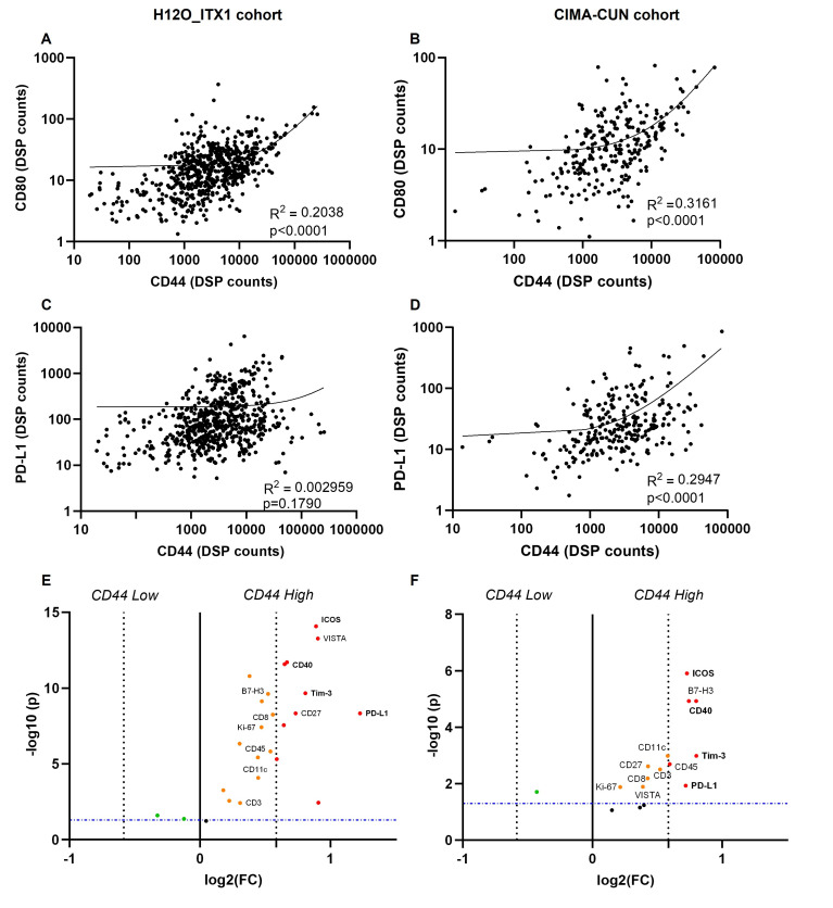
Finally, we explored whether a distinct tumor microenvironment could potentially explain a higher responsiveness to PD-1 axis blockade of NSCLC cases with high tumor cell CD44 expression. For this analysis, we used ROI-level counts in the tumor compartment from two cohorts, H12O_ITX1 and CIMA-CUN, as they were assessed with the same DSP assay and covered a greater number of ROIs in each tissue sample. First, we tested the correlation between tumor cell CD44 expression and each individual marker as continuous variables. Among all the markers with statistically significant results (online supplemental table S6), CD80 showed the highest, although modest, positive correlation (R2 >0.2) consistently in both cohorts (figure 3A, B). There was a statistically significant (p<0.0001) but modest correlation (R2 <0.3) between tumor cell CD44 counts and PD-L1 counts in the CIMA-CUN cohort, but this was not reproduced in the H12O_ITX1 cohort (figure 3C, D). In the H12O_ITX1 data set, the association between tumor cell CD44 and PD-L1 was significant (p=0.005) when removing extreme CD44 and PD-L1 values falling outside the upper limits of expression in the CIMA-CUN data set, but still with a loose correlation coefficient (R2=0.01) (online supplemental figure S9). Then, we interrogated what proteins were differentially expressed in the ROIs with high CD44 expression in the tumor compartment (top tertile) as compared with those with low CD44 expression (rest). Consistently in the two cohorts, CD44 high ROIs showed prominent and statistically significant upregulation (FC >1.5, FDR-adjusted p<0.05) of PD-L1, TIM-3, ICOS, and CD40. Other immune cell markers (CD45, CD8, CD11c), co-inhibitory molecules (VISTA, B7-H3), and co-stimulatory molecules (CD27), were also enriched in ROIs with elevated CD44 expression in both cohorts, but were less prominently upregulated (figure 3E, F and online supplemental table S7).
Figure 3.
CD44 levels in the tumor compartment and immune microenvironment features in human NSCLC. (A–B) Correlation between CD44 and CD80 levels in the tumor compartment in H12O_ITX1 cohort (A) and CIMA-CUN cohort (B). (C–D) Correlation between CD44 and PD-L1 levels in the tumor compartment in H12O_ITX1 cohort (C) and CIMA-CUN cohort (D). (E–F) Differentially expressed protein markers in ROIs with high CD44 expression in the tumor compartment (top tertile) relative to ROIs with low CD44 expression in the tumor compartment (rest) in H12O_ITX1 cohort (E) and CIMA-CUN cohort (F). The significance (FDR-adjusted p values) is represented relative to the FC in protein levels in CD44 high relative to CD44 low ROIs. Only statistically significant markers are highlighted. Markers with a FC >1.5 and FDR-adjusted p values<0.05 in the two cohorts are marked in red bold. DSP, digital spatial profiling; FC, fold change; FDR, false discovery rate; PD-L1, programmed death-ligand 1; NSCLC, non-small-cell lung cancer; ROI, regions of interest.
Discussion
In the present study, we have discovered and validated tumor cell CD44 as a new biomarker associated with sensitivity to PD-1 axis blockade across multiple NSCLC cohorts. Using two quantitative and spatially informed technologies in independent NSCLC cohorts, we show that CD44 overexpression in the tumor compartment, but not in the immune compartment, predicts clinical outcomes from PD-1 axis blockade. By analyzing two additional immunotherapy untreated cohorts, we found that the association between tumor cell CD44 and outcome was specific for anti-PD-1/PD-L1 treatment. Finally, we show that NSCLCs with CD44 overexpression in the tumor compartment are associated with a unique immune microenvironment.
CD44 plays multifaceted roles in cancer biology, including modulation of antitumor immunity. It is a surface marker commonly overexpressed by tumor cells with cancer initiating properties,6 7 20 and has therefore been repeatedly associated with cancer stem cells particularly in breast cancer.20–24 To our knowledge, the association between tumor cell CD44 overexpression and improved clinical outcomes to PD-1 axis blockade has not been previously reported. In the present study, we have performed rigorous validation of this novel finding, including orthogonal biomarker assessment with a second QIF-based technology (with prior validation of an anti-CD44 antibody following recommended guidelines16), and external validation utilizing whole tissue sections by independent investigators. It is possible that the use of a technology that provides quantitative data considering the cell compartment of expression might have increased the sensitivity to detect predictive information in our study. For instance, there are several studies that have evaluated the association between bulk tumor transcriptomes and response to PD-1 axis blockade, where CD44 is not among the list of overexpressed genes linked to response and PFS.25 However, as CD44 protein is quantitatively more abundant in the immune compartment than in the epithelial compartment, the predictive capacity of tumor cell CD44 might have been diluted when using bulk mRNA analysis25 or more subjective and semiquantitative methods such as IHC. Using spatially resolved quantitative protein measurements, we also excluded prognostic relevance of tumor cell CD44 expression. Although some studies have reported that CD44 overexpression is associated with poor prognosis (particularly overexpression of variant isoform 6 (CD44v6)) in some tumor types,26 27 these studies have not used quantitative assays enabling cell compartment specific protein measurements.
The higher sensitivity to PD-1 axis inhibition observed in NSCLCs with CD44 overexpression by cancer cells in this study could be at least partially explained by the role of CD44 in regulating antitumor immunity. CD44 has been identified as a PD-L1 modulator in preclinical models.13 14 Notably, CD44 expressing cancer cells derived from human squamous cell carcinoma of the head and neck showed higher responsiveness to IFN-γ than CD44 negative cells, which resulted in greater inducible PD-L1 expression following IFN-γ exposure in vitro.13 Consistent with these preclinical findings, we did observe a statistically significant association between tumor cell CD44 and PD-L1, but the strength of this association, particularly the linear relationship between both markers when assessed as continuous values, is low-to-modest at best. From a clinical perspective, this means that there is non-redundancy or at least some degree of complementarity between both biomarkers for patient stratification. Beyond its potential role in PD-L1 modulation, and further supporting a role for CD44 in adaptive immune resistance, it has been shown that tumor initiating cancer cells expressing CD44 can selectively upregulate CD80 under immune pressure, thereby suppressing T cell immunity in a CTLA4-dependent manner.28 This is also concordant with the significant correlation observed between CD44 and CD80 counts in the tumor compartment in our study. On the other hand, CD44 is a necessary protein for EMT,5–9 and EMT has been associated with a broad inflammatory microenvironment characterized by coexistent upregulation of co-stimulatory and co-inhibitory checkpoint molecules in human lung cancer.24 29–31 We also observed that intratumoral regions with elevated tumor cell CD44 had prominent upregulation of co-inhibitory (TIM-3, B7-H3, PD-L1) and co-stimulatory (ICOS, CD40, CD27) molecules in two independent NSCLC cohorts. Of note, although EMT has been generally associated with poorer outcomes and treatment resistance in several cancers,6 7 some tumor types with more mesenchymal features such as sarcomatoid malignant pleural mesotheliomas32 or certain subtypes of small cell lung cancers (SCLC),33 also display an inflammatory microenvironment and show higher sensitivity to checkpoint blockade therapies.33 34 Whether tumor cell CD44 plays a role in determining sensitivity to immunotherapy in these tumor types remains unknown, but might deserve to be explored in future studies. Collectively, these data suggest that NSCLCs with CD44 overexpression in cancer cells contain a distinct tumor microenvironment that is potentially more prone to immune checkpoint blockade responsiveness.
From a clinical standpoint, it needs to be considered that with the current use of chemoimmunotherapy in the front-line setting of advanced NSCLC, where even PD-L1 has no or limited utility to select best candidates to receive PD-1 axis inhibitors, it is unlikely that tumor cell CD44 will have an immediate major clinical impact as a biomarker for patient stratification. However, as quantitative tumor cell CD44 expression appears non-redundant with PD-L1 expression, there is potential to use them as complementary biomarkers. This possibility seems particularly appealing in tumor types where PD-L1 offers limited predictive information.34–37 On the other hand, CD44 is a surface glycoprotein potentially amenable for therapeutic antibody-based blockade. Some attempts to target CD44 using monoclonal antibodies have shown little clinical activity,27 38 but based on our findings, CD44 could be reconsidered as a target for tumor-cell-specific antibody-based strategies particularly in combination with PD-L1 blockade (ie, bi-specific antibodies with cis co-engagement39 or anti-CD44 probody formats40), and preferentially in those cases with specifically tumor cell CD44 overexpression.
This study needs to be interpreted in the context of a number of limitations. First, we analyzed tumor samples collected from retrospective immunotherapy treated cohorts, not randomized clinical trials. Retrospective studies have limitations including the possibility of selection bias, so despite having found tumor cell CD44 overexpression as a marker associated with sensitivity to PD-1 axis blockade in two independent cohorts, these results still need to be interpreted with caution and can only be considered hypothesis generating. In this same line, in the absence of randomization, we could not perform an interaction test between CD44 and PD-1 axis inhibitor treatment in a statistical model to prove that CD44 is a predictive biomarker. We were able to show absence of prognostic value and thus have used the term ‘indicative’ rather than predictive. Second, the tumor samples used for this study were archived samples from different tumor sites, not collected immediately preceding immunotherapy in most of the cases. The optimal time to assess immunotherapy biomarkers is still undefined, and some studies suggest that the key biological information is reasonably preserved in archived tissue.41 Another limitation is that we do not fully validate an identical cutpoint in the training and validation sets for optimal patient stratification. Future studies addressing the predictive value of CD44 will require the validation of an optimal and reproducible cutpoint in larger well-powered cohorts. It is perhaps also a limitation that we could not correct the CD44 survival prediction analysis by TMB since most of the patients in our cohorts did not have their tumors assessed for TMB status. Finally, this study does not prove the mechanism by which NSCLCs with CD44 overexpression in the tumor compartment are more sensitive to PD-1 axis blockade. Although the unique immune microenvironment of these tumors might partially explain a higher responsiveness, further functional studies are needed to understand the interplay between CD44 positive cancer stem cell phenotype and mechanisms of immune evasion.
In conclusion, this works highlights a previously underappreciated role for tumor cell CD44 and sensitivity to PD-1 axis blockade in NSCLC. With further validation, CD44 could complement existing biomarkers for optimal patient stratification, and potentially open new therapeutic strategies to improve precision immunotherapy in lung cancer.
Acknowledgments
The authors thank Lori A Charette and the staff of Yale Pathology tissue services for expert histology services, and Teresa Muñoz for her help in reviewing tumor specimens for adequacy for Digital Spatial Profiling analysis.
Footnotes
Twitter: @mkmoutafi
MKM and MM contributed equally.
Contributors: MKM, MM, DLR, and JZ conceived the study. MKM, MM, JB, VA, LC-L performed experiments and/or data analysis. Clinical information and specimens were provided by MKM, JB, VA, DO, KV, AC, LMM, SPA, LP-A, DLR, and JZ. Manuscript preparation was performed by MKM, MM, DLR, and JZ. Manuscript revisions were performed by all authors. All authors approved the final manuscript. JZ is responsible for the overall content of the manuscript as the guarantor.
Funding: This work was supported by grants from the Lung Cancer Foundation of America (LCFA/IASLC/BMS research award), Spanish Association Against Cancer (AECC, LABAE2044), and Carlos III Health Institute (ISCIII, PI20/01494) to JZ, and Yale SPORE in Lung Cancer (P50 CA196530) to DLR and RSH. MKM is receiving a scholarship from the Hellenic Society of Medical Oncologists (HESMO). LMM is supported by FIMA, AECC Scientific Foundation (GCB14-2170), Fundación Ramón Areces, Spanish Ministry of Economy and Innovation, Fondo de Investigación Sanitaria-Fondo Europeo de Desarrollo Regional (PI19/00098), and Fundación Roberto Arnal Planelles. LP-A is supported by CIBERONC (CB16/12/00442). DLR is supported by the Yale SPORE in Lung Cancer (P50 CA196530) and has a sponsored research agreement from Konica/Minolta. JZ is supported by a Juan Rodes contract from the Carlos III Health Institute (JR19/00028).
Competing interests: SMM is currently a Boehringer Ingelheim employee. LMM reports speakers honoraria from AstraZeneca and research grants from AstraZeneca and BMS. KAS reports consulting or advisory roles for Shattuck Labs, Pierre Fabre, EMD Serono, Clinica Alemana de Santiago, Genmab, Takeda, Merck Sharpe & Dohme, Bristol Myers Squibb, AstraZeneca, Agenus, Repertoire Therapeutics, OnCusp and Ariagen. Reports grants or research funding from Navigate Biopharma, Tesaro/GSK, Moderna, Takeda, Surface Oncology, Pierre Fabre Research Institute, Merck Sharpe & Dohme, Bristol Myers Squibb, AstraZeneca, Ribon Therapeutics, Akoya Biosciences, Boehringer Ingelheim and Eli Lilly. RSH has served as Non-Executive Director for Immunocore and is a member of the Board of Directors (non-executive/independent) for Junshi Biosciences; is a consultant for AbbVie, Armo Biosciences, AstraZeneca, Bristol Myers Squibb, Bayer HealthCare Pharmaceuticals, Bolt Biotherapeutics, Candel Therapeutics, Checkpoint Therapeutics, Cybrexa Therapeutics, DynamiCure Biotechnology, Eli Lilly and Company, eFFECTOR Therapeutics, EMD Serono, Foundation Medicine, Genentech/Roche, Genmab, Gilead, HiberCell, I‐Mab Biopharma, Immune‐Onc Therapeutics, Immunocore, Johnson & Johnson, Loxo Oncology, Merck and Company, Mirati Therapeutics, NextCure, Novartis, Ocean Biomedical, Oncocyte, Oncternal Therapeutics, Pfizer, Refactor Health, Ribbon Therapeutics, Sanofi, STCube Pharmaceuticals, Takeda, WindMIL Therapeutics, Xencor; has received research support from AstraZeneca, Eli Lilly and Company, Genentech/Roche, and Merck and Company; is a committee chair in American Association for Cancer Research, International Association for the Study of Lung Cancer, Society for Immunotherapy of Cancer, Southwest Oncology Group LP-A reports receiving honoraria from Amgen, AstraZeneca, Bayer, Blueprint Medicines, Bristol Myers Squibb, Celgene, Ipsen, Eli Lilly, Merck Serono, Mirati Therapeutics, Merck Sharp & Dohme, Novartis, Pfizer, PharmaMar, Roche/Genentech, Sanofi, Servier, and Takeda; leadership fees from Genomica and ALTUM Sequencing; research funding from AstraZeneca, Bristol Myers Squibb, Kura Oncology, PharmaMar, and Merck Sharp & Dohme; speaker fees from Bristol Myers Squibb, Eli Lilly, Merck Serono, Merck Sharp & Dohme Oncology, Pfizer, and Roche/Genentech; and travel, accommodation, and expenses from AstraZeneca, Bristol Myers Squibb, Merck Sharp & Dohme, Pfizer, Roche, and Takeda. DLR reports grants from Navigate Biopharma and Konica/Minolta/Invicro during the conduct of the study. Activities outside this work include honoraria and/or grants and/or instrument support from Akoya, Amgen, AstraZeneca, BMS, Cell Signaling Technology, Cepheid, Danaher, Konica/Minolta, Lilly, Merck, NanoString, NextCure, Odonate, Paige.AI, Roche, Sanofi, and Ventana. JZ has served as a consultant for AstraZeneca, BMS, Roche, Pfizer, Novartis, and Guardant Health. Reports speakers honoraria from BMS, Pfizer, Roche, AstraZeneca, NanoString and Guardant Health. Reports travel honoraria from BMS, Pfizer, Roche, AstraZeneca, and NanoString. Receives research support/funds from BMS, AstraZeneca, and Roche. The rest of the authors declare no conflicts of interest.
Provenance and peer review: Not commissioned; externally peer reviewed.
Supplemental material: This content has been supplied by the author(s). It has not been vetted by BMJ Publishing Group Limited (BMJ) and may not have been peer-reviewed. Any opinions or recommendations discussed are solely those of the author(s) and are not endorsed by BMJ. BMJ disclaims all liability and responsibility arising from any reliance placed on the content. Where the content includes any translated material, BMJ does not warrant the accuracy and reliability of the translations (including but not limited to local regulations, clinical guidelines, terminology, drug names and drug dosages), and is not responsible for any error and/or omissions arising from translation and adaptation or otherwise.
Data availability statement
All data relevant to the study are included in the article or uploaded as supplemental information. Data are available upon reasonable request.
Ethics statements
Patient consent for publication
Not applicable.
Ethics approval
All study procedures were done under the approval of the Yale Human Investigation Committee protocol #9505008219, 12 de Octubre Ethics Committee protocols #18/062 and #20/153, and University of Navarra Research Ethics Committee protocol #19/150. Investigation committees approved patients’ informed consent or in some cases waiver of consent all in accordance with ethical guidelines.
References
- 1.Doroshow DB, Sanmamed MF, Hastings K, et al. Immunotherapy in non-small cell lung cancer: Facts and hopes. Clin Cancer Res 2019;25:4592–602. 10.1158/1078-0432.CCR-18-1538 [DOI] [PMC free article] [PubMed] [Google Scholar]
- 2.Grant MJ, Herbst RS, Goldberg SB. Selecting the optimal immunotherapy regimen in driver-negative metastatic NSCLC. Nat Rev Clin Oncol 2021;18:625–44. 10.1038/s41571-021-00520-1 [DOI] [PubMed] [Google Scholar]
- 3.Morad G, Helmink BA, Sharma P, et al. Hallmarks of response, resistance, and toxicity to immune checkpoint blockade. Cell 2021;184:5309–37. 10.1016/j.cell.2021.09.020 [DOI] [PMC free article] [PubMed] [Google Scholar]
- 4.Lu S, Stein JE, Rimm DL, et al. Comparison of biomarker modalities for predicting response to PD-1/PD-L1 checkpoint blockade: a systematic review and meta-analysis. JAMA Oncol 2019;5:1195–204. [DOI] [PMC free article] [PubMed] [Google Scholar]
- 5.Zöller M. CD44: can a cancer-initiating cell profit from an abundantly expressed molecule? Nat Rev Cancer 2011;11:254–67. 10.1038/nrc3023 [DOI] [PubMed] [Google Scholar]
- 6.Lambert AW, Weinberg RA. Linking EMT programmes to normal and neoplastic epithelial stem cells. Nat Rev Cancer 2021;21:325–38. 10.1038/s41568-021-00332-6 [DOI] [PubMed] [Google Scholar]
- 7.Dongre A, Weinberg RA. New insights into the mechanisms of epithelial–mesenchymal transition and implications for cancer. Nat Rev Mol Cell Biol 2019;20:69–84. 10.1038/s41580-018-0080-4 [DOI] [PubMed] [Google Scholar]
- 8.Brown RL, Reinke LM, Damerow MS, et al. CD44 splice isoform switching in human and mouse epithelium is essential for epithelial-mesenchymal transition and breast cancer progression. J Clin Invest 2011;121:1064–74. 10.1172/JCI44540 [DOI] [PMC free article] [PubMed] [Google Scholar]
- 9.Larsen JE, Nathan V, Osborne JK, et al. ZEB1 drives epithelial-to-mesenchymal transition in lung cancer. J Clin Invest 2016;126:3219–35. 10.1172/JCI76725 [DOI] [PMC free article] [PubMed] [Google Scholar]
- 10.Natsuizaka M, Whelan KA, Kagawa S, et al. Interplay between Notch1 and Notch3 promotes EMT and tumor initiation in squamous cell carcinoma. Nat Commun 2017;8:1758. 10.1038/s41467-017-01500-9 [DOI] [PMC free article] [PubMed] [Google Scholar]
- 11.Wang Y-Y, Vadhan A, Chen P-H, et al. Cd44 promotes lung cancer cell metastasis through erk–zeb1 signaling. Cancers 2021;13:4057–15. 10.3390/cancers13164057 [DOI] [PMC free article] [PubMed] [Google Scholar]
- 12.Guo J-Y, Hsu H-S, Tyan S-W, et al. Serglycin in tumor microenvironment promotes non-small cell lung cancer aggressiveness in a CD44-dependent manner. Oncogene 2017;36:2457–71. 10.1038/onc.2016.404 [DOI] [PMC free article] [PubMed] [Google Scholar]
- 13.Lee Y, Shin JH, Longmire M, et al. Cd44+ cells in head and neck squamous cell carcinoma suppress T-cell-mediated immunity by selective constitutive and inducible expression of PD-L1. Clin Cancer Res 2016;22:3571–81. 10.1158/1078-0432.CCR-15-2665 [DOI] [PMC free article] [PubMed] [Google Scholar]
- 14.Kong T, Ahn R, Yang K, et al. CD44 promotes PD-L1 expression and its tumor-intrinsic function in breast and lung cancers. Cancer Res 2020;80:444–57. 10.1158/0008-5472.CAN-19-1108 [DOI] [PubMed] [Google Scholar]
- 15.Camp RL, Chung GG, Rimm DL. Automated subcellular localization and quantification of protein expression in tissue microarrays. Nat Med 2002;8:1323–8. 10.1038/nm791 [DOI] [PubMed] [Google Scholar]
- 16.Uhlen M, Bandrowski A, Carr S, et al. A proposal for validation of antibodies. Nat Methods 2016;13:823–7. 10.1038/nmeth.3995 [DOI] [PMC free article] [PubMed] [Google Scholar]
- 17.MacNeil T, Vathiotis IA, Martinez-Morilla S, et al. Antibody validation for protein expression on tissue slides: a protocol for immunohistochemistry. Biotechniques 2020;69:460–8. 10.2144/btn-2020-0095 [DOI] [PMC free article] [PubMed] [Google Scholar]
- 18.Camp RL, Dolled-Filhart M, Rimm DL. X-tile: a new bio-informatics tool for biomarker assessment and outcome-based cut-point optimization. Clin Cancer Res 2004;10:7252–9. 10.1158/1078-0432.CCR-04-0713 [DOI] [PubMed] [Google Scholar]
- 19.Mezquita L, Auclin E, Ferrara R, et al. Association of the lung immune prognostic index with immune checkpoint inhibitor outcomes in patients with advanced non-small cell lung cancer. JAMA Oncol 2018;4:351–7. 10.1001/jamaoncol.2017.4771 [DOI] [PMC free article] [PubMed] [Google Scholar]
- 20.Creighton CJ, Li X, Landis M, et al. Residual breast cancers after conventional therapy display mesenchymal as well as tumor-initiating features. Proc Natl Acad Sci U S A 2009;106:13820–5. 10.1073/pnas.0905718106 [DOI] [PMC free article] [PubMed] [Google Scholar]
- 21.Al-Hajj M, Wicha MS, Benito-Hernandez A, et al. Prospective identification of tumorigenic breast cancer cells. Proc Natl Acad Sci U S A 2003;100:3983–8. 10.1073/pnas.0530291100 [DOI] [PMC free article] [PubMed] [Google Scholar]
- 22.Prince ME, Kaczorowski RSA, Wolf GT. Identification of a subpopulation of cells with cancer stem cell properties. Proc Natl Acad Sci U S A 2007;104:973–8. [DOI] [PMC free article] [PubMed] [Google Scholar]
- 23.Nishino M, Ozaki M, Hegab AE, et al. Variant CD44 expression is enriching for a cell population with cancer stem cell-like characteristics in human lung adenocarcinoma. J Cancer 2017;8:1774–85. 10.7150/jca.19732 [DOI] [PMC free article] [PubMed] [Google Scholar]
- 24.Kursunel MA, Taskiran EZ, Tavukcuoglu E, et al. Small cell lung cancer stem cells display mesenchymal properties and exploit immune checkpoint pathways in activated cytotoxic T lymphocytes. Cancer Immunol Immunother 2022;71:445–59. 10.1007/s00262-021-02998-1 [DOI] [PMC free article] [PubMed] [Google Scholar]
- 25.Prat A, Navarro A, Paré L, et al. Immune-related gene expression profiling after PD-1 blockade in non–small cell lung carcinoma, head and neck squamous cell carcinoma, and melanoma. Cancer Res 2017;77:3540–50. 10.1158/0008-5472.CAN-16-3556 [DOI] [PubMed] [Google Scholar]
- 26.Zhao S, He J-L, Qiu Z-X, et al. Prognostic value of CD44 variant exon 6 expression in non-small cell lung cancer: a meta-analysis. Asian Pacific J Cancer Prev 2014;15:6761–6. 10.7314/APJCP.2014.15.16.6761 [DOI] [PubMed] [Google Scholar]
- 27.Chen C, Zhao S, Karnad A, et al. The biology and role of CD44 in cancer progression: therapeutic implications. J Hematol Oncol 2018;11:1–23. 10.1186/s13045-018-0605-5 [DOI] [PMC free article] [PubMed] [Google Scholar]
- 28.Miao Y, Yang H, Levorse J, et al. Adaptive immune resistance emerges from tumor-initiating stem cells. Cell 2019;177:1172–86. 10.1016/j.cell.2019.03.025 [DOI] [PMC free article] [PubMed] [Google Scholar]
- 29.Chen L, Gibbons DL, Goswami S, et al. Metastasis is regulated via microRNA-200/ZEB1 axis control of tumour cell PD-L1 expression and intratumoral immunosuppression. Nat Commun 2014;5:5241. 10.1038/ncomms6241 [DOI] [PMC free article] [PubMed] [Google Scholar]
- 30.Lou Y, Diao L, Cuentas ERP, et al. Epithelial-mesenchymal transition is associated with a distinct tumor microenvironment including elevation of inflammatory signals and multiple immune checkpoints in lung adenocarcinoma. Clinical Cancer Research 2016;22:3630–42. 10.1158/1078-0432.CCR-15-1434 [DOI] [PMC free article] [PubMed] [Google Scholar]
- 31.Kim S, Koh J, Kim M-Y, et al. PD-L1 expression is associated with epithelial-to-mesenchymal transition in adenocarcinoma of the lung. Hum Pathol 2016;58:7–14. 10.1016/j.humpath.2016.07.007 [DOI] [PubMed] [Google Scholar]
- 32.Hmeljak J, Sanchez-Vega F, Hoadley KA, et al. Integrative molecular characterization of malignant pleural mesothelioma. Cancer Discov 2018;8:1548–65. 10.1158/2159-8290.CD-18-0804 [DOI] [PMC free article] [PubMed] [Google Scholar]
- 33.Gay CM, Stewart CA, Park EM, et al. Patterns of transcription factor programs and immune pathway activation define four major subtypes of SCLC with distinct therapeutic vulnerabilities. Cancer Cell 2021;39:346–60. 10.1016/j.ccell.2020.12.014 [DOI] [PMC free article] [PubMed] [Google Scholar]
- 34.Baas P, Scherpereel A, Nowak AK, et al. First-line nivolumab plus ipilimumab in unresectable malignant pleural mesothelioma (CheckMate 743): a multicentre, randomised, open-label, phase 3 trial. Lancet 2021;397:375–86. 10.1016/S0140-6736(20)32714-8 [DOI] [PubMed] [Google Scholar]
- 35.Goldman JW, Dvorkin M, Chen Y, et al. Durvalumab, with or without tremelimumab, plus platinum–etoposide versus platinum–etoposide alone in first-line treatment of extensive-stage small-cell lung cancer (CASPIAN): updated results from a randomised, controlled, open-label, phase 3 trial. Lancet Oncol 2021;22:51–65. 10.1016/S1470-2045(20)30539-8 [DOI] [PubMed] [Google Scholar]
- 36.Liu SV, Reck M, Mansfield AS, et al. Updated overall survival and PD-L1 subgroup analysis of patients with extensive-stage small-cell lung cancer treated with Atezolizumab, carboplatin, and etoposide (IMpower133). J Clin Oncol 2021;39:619–30. 10.1200/JCO.20.01055 [DOI] [PMC free article] [PubMed] [Google Scholar]
- 37.Fennell DA, Ewings S, Ottensmeier C, et al. Nivolumab versus placebo in patients with relapsed malignant mesothelioma (CONFIRM): a multicentre, double-blind, randomised, phase 3 trial. Lancet Oncol 2021;22:1530–40. 10.1016/S1470-2045(21)00471-X [DOI] [PMC free article] [PubMed] [Google Scholar]
- 38.Menke-van der Houven van Oordt CW, Gomez-Roca C, van Herpen C, et al. First-in-human phase I clinical trial of RG7356, an anti-CD44 humanized antibody, in patients with advanced, CD44-expressing solid tumors. Oncotarget 2016;7:80046–58. 10.18632/oncotarget.11098 [DOI] [PMC free article] [PubMed] [Google Scholar]
- 39.Blanco B, Domínguez-Alonso C, Alvarez-Vallina L. Bispecific immunomodulatory antibodies for cancer immunotherapy. Cli Cancer Res 2021;27:5457–64. 10.1158/1078-0432.CCR-20-3770 [DOI] [PMC free article] [PubMed] [Google Scholar]
- 40.Autio KA, Boni V, Humphrey RW, et al. Probody therapeutics: an emerging class of therapies designed to enhance on-target effects with reduced off-tumor toxicity for use in immuno-oncology. Clin Cancer Res 2020;26:984–9. 10.1158/1078-0432.CCR-19-1457 [DOI] [PMC free article] [PubMed] [Google Scholar]
- 41.Herbst RS, Baas P, Perez-Gracia JL, et al. Use of archival versus newly collected tumor samples for assessing PD-L1 expression and overall survival: an updated analysis of KEYNOTE-010 trial. Ann Oncol 2019;30:281–9. 10.1093/annonc/mdy545 [DOI] [PMC free article] [PubMed] [Google Scholar]
Associated Data
This section collects any data citations, data availability statements, or supplementary materials included in this article.
Supplementary Materials
jitc-2022-004757supp001.pdf (1,012.4KB, pdf)
Data Availability Statement
All data relevant to the study are included in the article or uploaded as supplemental information. Data are available upon reasonable request.


