



Abstract
A 3-y-old male pigtailed macaque (Macaca nemestrina) presented for swelling of the left distal forearm and decreased use of the arm. The monkey had been raised at an indoor-outdoor facility in Arizona and transferred to an indoor facility in Washington 2 mo prior to presentation. A preliminary diagnosis of fungal osteomyelitis of the radius was made based on radiographs and Coccidioides titers. In addition to systemic antifungal treatment, surgery was performed to debride the bony lesion and implant polymethylmethacrylate beads impregnanted with the anti-fungal fluconazole. Histologic examination of the debrided material confirmed the diagnosis of fungal osteomyelitis. The surgical procedure resulted in clinical improvement, as evidenced by weight gain and decreased Coccidioides titers. The beads were removed in a second surgery, and the bony lesion completely resolved. With continued systemic fluconazole treatment, the monkey remained healthy with no further evidence of osteomyelitis. Coccidioides is an emerging pathogen that causes significant morbidity and mortality in both humans and animals. Bone infections can be resistant to systemic treatment, and the implantation of fluconazole-impregnated beads may offer a successful treatment strategy for fungal osteomyelitis.
Abbreviations: PMMA, polymethylmethacrylate; VF, Valley Fever
Introduction
Coccidioidomycosis, commonly known as Valley Fever (VF), is a fungal disease caused by Coccidioides immitis or Coccidioides posadasii.41 VF has been reported in a wide variety of domestic species including dogs, cats, and horses,3,9,12,20,21,39,40 and in wildlife species such as bats, wallabies and kangaroos, tapirs, and dolphins.37,40 Naturally-occurring VF infections have also been found in multiple species of nonhuman primates29 (NHP), including rhesus,7,31 cynomolgus,35 and Japanese macaques17 in Texas, and rhesus and bonnet macaques in California.4 NHP have also been used as experimental models to study various aspects of VF,5 including evaluation of potential VF vaccines.25 Recrudescence of VF was recently reported after treatment of a pigtailed macaque.18 When a macaque’s history includes housing in a VF-endemic area, coccidioidomycosis must be considered in the differential diagnosis of lesions consistent with this diagnosis.
Osteomyelitis is a difficult condition to treat because of the poor penetration of systemic antibiotics into bone.26 One option for the treatment of chronic osteomyelitis is the implantation of antibiotic-impregnated polymethyl methacrylate (PMMA) spheres.6,26 While initial recommendations included the removal of the PMMA implants at 6 to 8 wk in humans,26 some patients have shown clinical improvement when the PMMA spheres remain in place.6 Moreover, the use of antibiotic-impregnated PMMA has been used successfully to treat osteomyelitis in a rhesus macaque (Macaca mulatta).27 Nevertheless, because of concerns regarding bone healing, the use of biodegradable materials to replace the PMMA has also been studied in both human and veterinary medicine.16,26
Antibiotic-impregnated calcium sulfate beads are frequently used in the management of bacterial infections associated with prosthetic implants in humans.1,2 The beads are more successful if the implant is removed than if it remains in place.1 Fungal infection is rare around these implants and is usually treated with systemic antifungal agents2,10,24,28 either with or without local antifungals.24 Long-term antifungal therapy both before and after surgical debridement is necessary for successful treatment.43 Voriconazole-impregnated calcium sulfate beads32 and amphotericin B-impregnated PMMA44 have been used to successfully treat fungal infections associated with total knee arthroplasty. We are not aware of any reports of antifungal-impregnated implants for the treatment of osteomyelitis in veterinary medicine.
Case Report
A 3-y-old male pigtailed macaque (Macaca nemestrina) presented for swelling of the left distal forearm and decreased use of the arm. The monkey had been raised at an indoor-outdoor facility in Arizona as part of a specific pathogen free breeding colony on a University of Washington IACUC-approved protocol. The macaque was confirmed negative for tuberculosis, simian immunodeficiency virus, simian retrovirus, simian lymphotropic virus, and Macacine alphaherpesvirus-1 and then transferred to an indoor facility in Washington 2 mo before clinical presentation. At both AAALAC-accredited facilities, the monkey was maintained under ABSL-2 conditions, fed a commercial diet (LabDiet 5045 or 5049, St. Louis, MO) supplemented with fresh produce twice a day, and given water ad libitum. Environmental enrichment consisted of social contact (full or protected contact depending on compatibility with partner), manipulanda, and foraging opportunities. Five months before presentation, the macaque experienced an episode of respiratory illness characterized by rales on auscultation. At that time, radiographs revealed a mild bronchial pattern in the dorsocaudal lung lobes; serologic titers for Coccidioides were negative at that time. Treatment included albuterol (0.66 mg/kg PO BID) and nutritional support, and clinical signs resolved within 2 wk.
At the time of presentation for the forearm swelling, physical exam findings included 10% weight loss (from 3.0 to 2.7 kg) and intermittent expiratory stridor and stertor. A complete blood count revealed a mild monocytosis (1.8 thou/mL) and serum chemistry displayed a moderate hyperglobulinemia (6.7 g/dL) and hypoalbuminemia (2.1 g/dL). Radiographs of the left arm revealed a lytic lesion in the distal radius (Figure 1A). Analgesics (buprenorphine-SR at 0.2 mg/kg SC and meloxicam at 0.2 mg/kg PO loading dose followed by 0.1 mg/kg SID) and antibiotics (amoxicillin/clavulanate at 12 mg/kg PO BID) were administered initially. Serum samples were submitted to a commercial laboratory (ProtaTek Reference Laboratory, Mesa, AZ). Titers for Coccidioides were positive at 1:64 (IgG) and 1:16 (IgM). Based on these results, a preliminary diagnosis of fungal osteomyelitis was made, and therapy with fluconazole (8 mg/kg PO BID) was initiated.
Figure 1.

Fungal osteomyelitis in the distal radius of a M. nemestrina. A) Initial presentation. A single osteolytic lesion is seen within the left radius, with cortical thinning and periosteal reaction on the lateral surface. B) After 4 wk of systemic fluconazole treatment, there was increased periosteal proliferation with loss of cortical definition on the lateral surface, irregular density within the lesion, and decreased definition at the margins of the lesion.
Although the use of the arm improved with treatment, the swelling did not resolve. Weight loss stopped, but body weight did not increase as expected for a young, growing macaque. Whole body radiographs were then obtained to determine if any other bony lesions were present. All other bones appeared to be unaffected; however, the lesion in the left radius had progressed (Figure 1B). Therefore, the decision was made to surgically debride the lesion and implant PMMA beads containing fluconazole. This technique had been used in humans for treatment of Candida infection after hip replacement.8 Appropriate materials were identified, ordered, and received, and surgery was performed approximately 6 wk later.
Materials and Methods
On the day of surgery, anesthesia was induced with ketamine (15 mg/kg IM) and maintained with inhalational isoflurane. A 3 cm skin incision was made over the dorsal aspect of the left radius at the level of the distal metaphysis centered approximately on the firm swelling on the radius. The subcutaneous tissue was dissected between the second and third extensor tendon compartments, and through the periosteum to the bone. During subcutaneous dissection, a cystic soft tissue was seen protruding from a cortical defect in the dorsal surface of the radius (Figure 2A, B). A fine needle aspirate of the lesion was obtained and submitted to the UW Research Testing Services for routine aerobic bacterial culture. Prior to submission, a fresh smear was made of the aspirate and stained with Wrights-Giemsa for cytology. The soft tissue structure was excised, fixed in formalin, and submitted for routine H and E histopathology. The bone was burred away at the site of the cortical defect to expose the medullary cavity (Figure 2C). The cancellous bone was curetted, and the medullary cavity was debrided of all visible abnormal tissue (Figure 2D).
Figure 2.

Bead implantation surgery. A) Incision over dorsal surface of distal radius showing both bony and soft tissue swelling. Solid arrow = radius bone, double arrow = soft tissue in bony defect. B) Pyogranulomatous tissue with reactive fibroplasia (arrow) partially removed from the medullary cavity of the distal radius, C) Expansion of the cortical bone defect using a surgical drill, D) Medullary cavity (arrow) after debridement, E) Fluconazole-impregnated PMMA beads (arrow) within the medullary cavity.
Fluconazole (3 g powder) was mixed aseptically with one package (40 g) of bone cement powder (Palacos LV, Heraeus Medical, Yardley, PA). Aliquots of this mixture were catalyzed by the addition of methylmethacrylate/toluidine per the manufacturer’s instructions and formed into PMMA beads (approximately 0.2 g/bead) by hand. Two beads were inserted into the excavated medullary cavity (Figure 2E) and all the subcutaneous tissues were closed in a single layer with absorbable suture. The skin incision was closed with simple continuous absorbable suture and infiltrated with bupivacaine. Radiographs were obtained immediately before and after surgery (Figure 3A, B). Analgesics (one injection of buprenorphine-SR at 0.2 mg/kg SC and meloxicam at a loading dose of 0.2 mg/kg PO, followed by 0.1 mg/kg SID) and antibiotics (cephalexin at 25 mg/kg PO BID) were administered after surgery. The arm was bandaged for a total of 5 d after surgery, with a bandage change at day 3.
Figure 3.

Radiographs associated with bead insertion. A) Preoperative, approximately 10 wk after initial presentation. After additional systemic fluconazole treatment while preparing for surgery, the periosteal reaction appears to be resolving. However, the osteolytic lesion in the radius is unchanged in size. B) Immediately after surgery. Placement of 2 fluconazole-impregnated beads within the radius was confirmed. An area of radiolucency is present proximal to the beads. C) 4 wk after implantation. Radiodensity has increased around the beads. D) 8 wk after implantation. Radiodensity has further increased around the beads and has the appearance of trabecular bone.
Fifteen weeks after the initial surgery, a second surgery was performed to remove the nonresorbable fluconazole-cement beads. Surgical preparation and approach were essentially the same as in the initial surgery. Additional blunt dissection was required due to the presence of scar tissue. The beads were partially exposed on the dorsal surface of the radius (Figure 4A), as indicated in preoperative radiographs (Figure 5A). A surgical drill was used to remove bead material, taking care not to damage healthy bone. Once each bead was loosened, the remaining bead fragments were removed using a periosteal elevator (Figure 4B). The resulting cavity was flushed copiously with sterile saline, and the subcutaneous tissue and skin were closed routinely and were infiltrated with bupivacaine. Analgesics (one injection of buprenorphine-SR at 0.2 mg/kg SC and meloxicam at 0.2 mg/kg PO loading dose followed by 0.1 mg/kg SID) and antibiotics (cephalexin at 25 mg/kg PO BID) were administered after surgery. Recovery from surgery was uneventful.
Figure 4.

Bead removal surgery. A) Beads visible in situ within radius, B) Partial bead that had been removed shown next to cavity after bead removal. Note ridge in center of cavity where bone had remodeled around beads.
Figure 5.

Radiographs associated with bead removal. A) Preoperative on the day of bead removal, 15 wk after the initial surgery. Beads appear unchanged and are surrounded by trabecular bone. B) 4 wk after bead removal. The void where the beads were persists, but the surrounding bone has normal trabecular architecture. C) 4 mo after bead removal. The defect where the beads had been has closed over. Some irregularity of the cortical/trabecular margins persists. D) 8 mo after bead removal. The bone shows normal trabecular architecture and smooth cortical contours.
Results
The monkey did well after the first surgery with no evidence of discomfort and was using the arm normally within 2 wk afterwards. Bacterial culture of the soft tissue cyst was negative. Histopathology revealed multifocal granulomas and pyogranulomas with reactive fibroplasia. Fragments of spherules were seen on histopathology, and intact, variably sized, double-walled, nonbudding spherules were seen on cytology, consistent with Coccidioides organisms. Fungal culture was not attempted, because germination of arthroconidia could pose a laboratory hazard.36 While BSL-2 practices are appropriate for handling animal tissues that may contain Coccidioides, BSL-3 practices are recommended for manipulating cultures.36 The cytologic appearance, in combination with the serologic titers and history of residence in an endemic area, combined for a presumptive diagnosis of coccidioidomycosis. Follow up examination one month after surgery revealed healing of the surgical incision and a decrease in external swelling of the arm. Serum globulin had returned to normal (3 g/dL), and albumin was higher (2.5 g/dL), although still below the reference range for this species. Coccidioides titers were measured periodically and fell over time, although some regression occurred, and a cure, as indicated by negative titers, was not achieved within one year (Table 1). Radiographs (Figure 3C, D) showed a consistent increase in bone density around the beads over the 8 wk period after surgery.
Table 1.
Changes in Coccidioides titers over time.
| Day | IgG Titer Result | IgG Titer Value | IgM Titer Result | IgM Titer Value |
|---|---|---|---|---|
| Prior to presentation | − | < 1:1 | − | < 1:1 |
| Presentation | + | 1:64 | + | 1:16 |
|
Implant + 30 |
+ | 1:16 | + | 1:2 |
| Implant + 63 | + | 1:4 | + | 1:1 |
| Implant removal (Implant + 106) | + | 1:2 | − | < 1:1 |
| Removal + 33 | + | 1:2 | − | < 1:1 |
| Removal + 128 | + | 1:2 | − | < 1:1 |
| Removal + 177 | + | 1:4 | − | < 1:1 |
| Removal + 239 | + | 1:1 | − | < 1:1 |
| Removal + 338 | − | < 1:1 | − | < 1:1 |
| Removal + 350 | − | < 1:1 | − | < 1:1 |
| Removal + 510 | + | 1:2 | − | < 1:1 |
| The first sample shown was collected 5 mo prior to presentation. Later sample time points are indicated by the number of days after specific events. | ||||
After the surgery to remove the beads, the monkey showed no evidence of discomfort, was using the arm normally within one week, and remained in good health, as evidenced by steady growth after surgery (Figure 6). The animal was euthanized more than a year later (510 d after bead removal) as part of an unrelated research project. At necropsy, the bone appeared radiographically and histologically normal, although a positive VF titer at that time indicated a reservoir of Coccidioides organisms within the animal.
Figure 6.
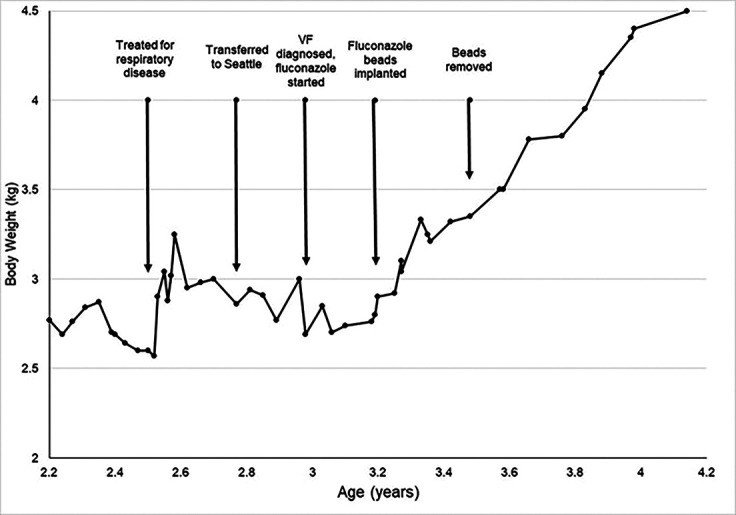
Body weight changes over time in relation to clinical events. VF = Valley Fever. A juvenile M. nemestrina is expected to steadily gain weight between 2 and 4 y of age. Weight gain stopped during clinical respiratory illness but rebounded after symptomatic treatment. Weight gain stopped again around the time of transfer to Seattle, and the diagnosis of VF was made after weight loss. Systemic treatment with fluconazole stabilized weight but weight gain still did not occur. After surgery to debride the fungal bony lesion and implant fluconazole-impregnated beads, normal weight gain resumed. Weight gain continued uninterrupted after surgical removal of the beads.
Discussion
We used surgical implantation of fluconazole-impregnated PMMA beads to treat fungal osteomyelitis in a pigtailed macaque, which resulted in resolution of the bone lesion. In humans, most deaths due to VF occur in California and Arizona.23 thus indicating a risk of naturally occurring VF infections in a captive population of pigtailed macaques in Arizona. Because the primary route of exposure is inhalation, the organ most commonly affected by VF in all species is the lung, although dissemination to multiple organs may occur. Bone is frequently reported as a secondary site of infection; in dogs, the appendicular skeleton is more often affected, while in humans and NHPs, the axial skeleton appears to be more commonly affected.29
Treatment for VF most commonly consists of antifungal medication and supportive care. Fluconazole is the most frequently prescribed antifungal in both humans and animals, based on its effectiveness, cost, and the feasibility of once-a-day dosing.40,42 However, fluconazole is less effective in treating disseminated VF as compared with pulmonary VF, and surgical intervention is often necessary to treat skeletal VF.22,38,41 Surgical procedures used in humans with VF include incision and drainage, surgical debridement, synovectomy, and bone resection.22,38
Although coccidioidomycosis is most prevalent in southwestern state of the U.S., its prevalence is increasing in Washington state.13,33 The animal in this report was housed indoors in the western part of the state, where the causative organism is not known to be naturally present, making it unlikely that the initial infection was acquired in Washington and more likely that the initial infection was acquired in Arizona prior to transport. No clinical signs of VF were apparent on physical examination during the first month after arrival in Washington. This is consistent with a previous report that VF can remain latent for several months prior to development of clinical signs in macaques.31 In humans, extrapulmonary dissemination of VF is more common in immunocompromised patients30 and in monkeys, shipping stress can result in immune dysregulation,15 suggesting that the shipment from Arizona to Seattle may have contributed to development of the bony VF lesion. Travel history must always be considered when developing differential diagnoses.
The standard treatment for VF is systemic antifungal medication. In our case, the initial treatment arrested weight loss but did not result in resumption of normal growth, and did not improve the osteomyelitic lesion. After failure of medical therapy, we determined that surgery would be necessary to address the bone infection. Options for surgery included debridement, debridement with implantation of an antifungal agent, or amputation. We elected to perform debridement with implantation of an antifungal agent to balance the probability of success with the least impact to the animal. Our approach was successful, as measured by improvement in bone integrity both radiographically and as seen directly at surgery, as well as by the decrease in VF titers and resumption of normal weight gain.
We cannot determine how much of the clinical improvement (decreased VF titers, increased weight gain, improved the radiographic appearance of the radius) can be attributed to the implantation of fluconazole-impregnated beads as compared with manual debridement of infected bone. When a similar procedure was used in humans, the local concentration of fluconazole was measured in fluid collected from external drains.8 However implanted drains are not feasible in a NHP due to the risk of interference by the patient. Therefore, we were not able to confirm that the beads provided an effective local concentration of fluconazole. However, we can state that we saw no adverse effects with this procedure.
PMMA bone cement was used as the vehicle to provide fluconazole locally. We chose this method based on published data8 and our experience using it in other NHP implants. We removed the beads to allow regrowth of cancellous bone to maximize bone integrity at the site of the lesion, as the distal radius is the most common site for fractures in humans.11 Removal of the implants required a second surgery, which is a source of stress that could affect the infection. Identification of a biocompatible material that could be used as a vehicle to maintain fluconazole locally, but would be resorbed slowly over time, would be a significant refinement of the described technique. Calcium sulfate has been used for antibiotic delivery,14,19,34,45 and we tried this approach in a different macaque with a similar history. Unfortunately, that animal suffered a fracture after surgery, and we cannot determine whether that was related to excessive debridement in a smaller bone (the ulna) or to a factor related to the implant material. Given the effect of technique on bone integrity, we do not recommend the bead implantation technique for multifocal bone infections.
In conclusion, this report describes treatment of isolated osteomyelitis due to coccidioidomycosis in a pigtailed macaque by the use of implanted fluconazole-impregnated beads. While the lack of a negative titer suggests that the infection was not completely eradicated, the osteomyelitis lesion identified by radiology was successfully resolved. This technique is likely to be successful in other species as well and may provide an alternative to euthanasia or amputation.
Acknowledgments
Support for the animal in this report was provided by NIH grants P51 OD 010425 and U42 OD011123. The authors are grateful for the assistance of Dennis Raines in preparing the figures for publication.
References
- 1.Abosala A, Ali M. 2020. The Use of Calcium Sulphate beads in Periprosthetic Joint Infection, a systematic review. J Bone Jt Infect 5:43–49. 10.7150/jbji.41743. [DOI] [PMC free article] [PubMed] [Google Scholar]
- 2.Anagnostakos K, Kelm J, Schmitt E, Jung J. 2012. Fungal Periprosthetic Hip and Knee Joint Infections: Clinical Experience With a 2-Stage Treatment Protocol. J Arthroplasty 27:293–298. 10.1016/j.arth.2011.04.044. [DOI] [PubMed] [Google Scholar]
- 3.Arbona N, Butkiewicz CD, Keyes M, Shubitz LF. 2019. Clinical features of cats diagnosed with coccidioidomycosis in Arizona, 2004-2018. J Feline Med Surg 22:129–137. 10.1177/1098612x19829910. [DOI] [PMC free article] [PubMed] [Google Scholar]
- 4.Beaman L, Holmberg C, Henrickson R, Osburn B. 1980. The incidence of coccidioidomycosis among nonhuman primates housed outdoors at the California Primate Research Center. J Med Primatol 9:254–261. 10.1159/000460147. [DOI] [PubMed] [Google Scholar]
- 5.Blundell GP, Castleberry MW, Lowe EP, Converse JL. 1961. The pathology of Coccidioides immitis in the Macaca mulatta. Am J Pathol 39:613–630. [PMC free article] [PubMed] [Google Scholar]
- 6.Bor N, Dujovny E, Rinat B, Rozen N, Rubin G. 2022. Treatment of chronic osteomyelitis with antibiotic-impregnated polymethyl methacrylate (PMMA) - the Cierny approach: is the second stage necessary? BMC Musculoskelet Disord 23:38. 10.1186/s12891-021-04979-y. [DOI] [PMC free article] [PubMed] [Google Scholar]
- 7.Breznock AW, Henrickson RV, Silverman S, Schwartz LW. 1975. Coccidioidomycosis in a rhesus monkey. J Am Vet Med Assoc 167:657–661. [PubMed] [Google Scholar]
- 8.Bruce AS, Kerry RM, Norman P, Stockley I. 2001. Fluconazole-impregnated beads in the management of fungal infection of prosthetic joints. J Bone Joint Surg Br 83-B:183–184. 10.1302/0301-620X.83B2.0830183. [DOI] [PubMed] [Google Scholar]
- 9.Butkiewicz CD, Shubitz LE, Dial SM. 2005. Risk factors associated with Coccidioides infection in dogs. J Am Vet Med Assoc 226:1851–1854. 10.2460/javma.2005.226.1851. [DOI] [PubMed] [Google Scholar]
- 10.Cobo F, Rodríguez-Granger J, Sampedro A, Aliaga-Martínez L, Navarro-Marí JM. 2017. Candida Prosthetic Joint Infection. A Review of Treatment Methods. J Bone Jt Infect 2:114–121. 10.7150/jbji.17699. [DOI] [PMC free article] [PubMed] [Google Scholar]
- 11.Court-Brown CM, Caesar B. 2006. Epidemiology of adult fractures: A review. Injury 37:691–697. 10.1016/j.injury.2006.04.130. [DOI] [PubMed] [Google Scholar]
- 12.Davidson AP, Shubitz LF, Alcott CJ, Sykes JE. 2019. Selected Clinical Features of Coccidioidomycosis in Dogs. Med Mycol 57 Supplement_1:S67–S75. 10.1093/mmy/myy113. [DOI] [PubMed] [Google Scholar]
- 13.Engelthaler DM, Roe CC, Hepp CM, Teixeira M, Driebe EM, Schupp JM, Gade L, Waddell V, Komatsu K, Arathoon E, Logemann H, Thompson GR, 3rd, Chiller T, Barker B, Keim P, Litvintseva AP. 2016. Local Population Structure and Patterns of Western Hemisphere Dispersal for Coccidioides spp., the Fungal Cause of Valley Fever. MBio 7:e00550-16. 10.1128/mBio.00550-16. [DOI] [PMC free article] [PubMed] [Google Scholar]
- 14.Gauland C. 2011. Managing lower-extremity osteomyelitis locally with surgical debridement and synthetic calcium sulfate antibiotic tablets. Adv Skin Wound Care 24:515–523. 10.1097/01.ASW.0000407647.12832.6c. [DOI] [PubMed] [Google Scholar]
- 15.Glaser R, Kiecolt-Glaser JK. 2005. Stress-induced immune dysfunction: implications for health. Nat Rev Immunol 5:243–251. 10.1038/nri1571. [DOI] [PubMed] [Google Scholar]
- 16.González-Martín M, Silva V, Poeta P, Corbera JA, Tejedor- Junco MT. 2022. Microbiological aspects of osteomyelitis in veterinary medicine: drawing parallels to the infection in human medicine. Vet Q 42:1–11. 10.1080/01652176.2021.2022244. [DOI] [PMC free article] [PubMed] [Google Scholar]
- 17.Graybill JR, Griffith L, Sun SH. 1990. Fluconazole therapy for coccidioidomycosis in Japanese macaques. Rev Infect Dis 12 Suppl 3:S286–S290. 10.1093/clinids/12.Supplement_3.S286. [DOI] [PubMed] [Google Scholar]
- 18.Guerriero KA, Murnane RD, Lewis TB, Brown B, Baldessari A, Jeffery DA, Malinowski CM, Fuller DH, O’Connor MA. 2021. Recrudescence of Natural Coccidioidomycosis During Combination Antiretroviral Therapy in a Pigtail Macaque Experimentally Infected with Simian Immunodeficiency Virus. AIDS Res Hum Retroviruses 37:505–509. 10.1089/aid.2020.0228. [DOI] [PMC free article] [PubMed] [Google Scholar]
- 19.Ham K, Griffon D, Seddighi M, Johnson AL. 2008. Clinical application of tobramycin-impregnated calcium sulfate beads in six dogs (2002-2004). J Am Anim Hosp Assoc 44:320–326. 10.5326/0440320. [DOI] [PubMed] [Google Scholar]
- 20.Higgins JC, Leith GS, Pappagianis D, Pusterla N. 2006. Treatment of Coccidioides immitis pneumonia in two horses with fluconazole. Vet Rec 159:349–351. 10.1136/vr.159.11.349. [DOI] [PubMed] [Google Scholar]
- 21.Higgins JC, Leith GS, Voss ED, Pappagianis D. 2005. Seroprevalence of antibodies against Coccidioides immitis in healthy horses. J Am Vet Med Assoc 226:1888–1892. 10.2460/javma.2005.226.1888. [DOI] [PubMed] [Google Scholar]
- 22.Ho AK, Shrader MW, Falk MN, Segal LS. 2014. Diagnosis and initial management of musculoskeletal coccidioidomycosis in children. J Pediatr Orthop 34:571–577. 10.1097/BPO.0000000000000147. [DOI] [PubMed] [Google Scholar]
- 23.Huang JY, Bristow B, Shafir S, Sorvillo F. 2012. Coccidioidomycosis-associated Deaths, United States, 1990-2008. Emerg Infect Dis 18:1723–1728. 10.3201/eid1811.120752. [DOI] [PMC free article] [PubMed] [Google Scholar]
- 24.Ji B, Zhang X, Xu B, Guo W, Mu W, Cao L. 2017. Single-Stage Revision for Chronic Fungal Periprosthetic Joint Infection: An Average of 5 Years of Follow-Up. J Arthroplasty 32:2523–2530. 10.1016/j.arth.2017.03.030. [DOI] [PubMed] [Google Scholar]
- 25.Johnson SM, Lerche NW, Pappagianis D, Yee JL, Galgiani JN, Hector RF. 2007. Safety, antigenicity, and efficacy of a recombinant coccidioidomycosis vaccine in cynomolgus macaques (Macaca fascicularis). Ann N Y Acad Sci 1111:290–300. 10.1196/annals.1406.042. [DOI] [PubMed] [Google Scholar]
- 26.Kasza K, Gurnani P, Hardie KR, Cámara M, Alexander C. 2021. Challenges and solutions in polymer drug delivery for bacterial biofilm treatment: A tissue-by-tissue account. Adv Drug Deliv Rev 178:113973. 10.1016/j.addr.2021.113973. [DOI] [PubMed] [Google Scholar]
- 27.Kelly KR, Kapatkin AR, Zwingenberger AL, Christe KL. 2012. Efficacy of antibiotic-impregnated polymethylmethacrylate beads in a rhesus macaque (Macaca mulatta) with osteomyelitis. Comp Med 62:311–315. [PMC free article] [PubMed] [Google Scholar]
- 28.Kim J-K, Lee D-Y, Kang D-W, Ro D-H, Lee MC, Han H-S. 2018. Efficacy of antifungal-impregnated cement spacer against chronic fungal periprosthetic joint infections after total knee arthroplasty. Knee 25:631–637. 10.1016/j.knee.2018.04.004. [DOI] [PubMed] [Google Scholar]
- 29.Koistinen K, Mullaney L, Bell T, Zaki S, Nalca A, Frick O, Livingston V, Robinson CG, Estep JS, Batey KL, Dick EJ, Jr, Owston MA. 2018. Coccidioidomycosis in Nonhuman Primates: Pathologic and Clinical Findings. Vet Pathol 55:905–915. 10.1177/0300985818787306. [DOI] [PMC free article] [PubMed] [Google Scholar]
- 30.Krogstad P, Johnson R, Garcia-Lloret MI, Heidari A, Butte MJ. 2019. Host-Pathogen Interactions in Coccidioidomycosis: Prognostic Clues and Opportunities for Novel Therapies. Clin Ther 41:1939–1954. 10.1016/j.clinthera.2019.08.011. [DOI] [PMC free article] [PubMed] [Google Scholar]
- 31.Kundu MC, Ringenberg MA, d’Epagnier DL, Haag HL, Maguire S. 2017. Coccidioidomycosis in an Indoor-housed Rhesus Macaque (Macaca mulatta). Comp Med 67:452–455. [PMC free article] [PubMed] [Google Scholar]
- 32.Kurmis AP. 2021. Eradicating Fungal Periprosthetic TKA “Super-infection”: Review of the Contemporary Literature and Consideration of Antibiotic-Impregnated Dissolving Calcium Sulfate Beads as a Novel PJI Treatment Adjunct. Arthroplast Today 8:163–170. 10.1016/j.artd.2021.02.009. [DOI] [PMC free article] [PubMed] [Google Scholar]
- 33.Litvintseva AP, Marsden-Haug N, Hurst S, Hill H, Gade L, Driebe EM, Ralston C, Roe C, Barker BM, Goldoft M, Keim P, Wohrle R, Thompson GR, 3rd, Engelthaler DM, Brandt ME, Chiller T. 2015. Valley fever: finding new places for an old disease: Coccidioides immitis found in Washington State soil associated with recent human infection. Clin Infect Dis 60:e1–e3. 10.1093/cid/ciu681. [DOI] [PMC free article] [PubMed] [Google Scholar]
- 34.McKee MD, Li-Bland EA, Wild LM, Schemitsch EH. 2010. A prospective, randomized clinical trial comparing an antibiotic-impregnated bioabsorbable bone substitute with standard antibiotic-impregnated cement beads in the treatment of chronic osteomyelitis and infected nonunion. J Orthop Trauma 24:483–490. 10.1097/BOT.0b013e3181df91d9. [DOI] [PubMed] [Google Scholar]
- 35.Mense MG, Batey KL, Estep S, Armstrong K, Fleurie G, Suttie AW. 2013. Disseminated coccidioidomycosis in a cynomolgus monkey (Macaca fascicularis). J Primatol 2:111. [Google Scholar]
- 36.PHS-CDC-NIH. 2020. Biosafety in Microbiological and Biomedical Laboratories, 6th ed. [Google Scholar]
- 37.Reidarson TH, Griner LA, Pappagianis D, McBain J. 1998. Coccidioidomycosis in a bottlenose dolphin. J Wildl Dis 34: 629–631. 10.7589/0090-3558-34.3.629. [DOI] [PubMed] [Google Scholar]
- 38.Ricciotti RW, Shekhel TA, Blair JE, Colby TV, Sobonya RE, Larsen BT. 2014. Surgical pathology of skeletal coccidioidomycosis: a clinical and histopathologic analysis of 25 cases. Am J Surg Pathol 38:1672–1680. 10.1097/PAS.0000000000000284. [DOI] [PubMed] [Google Scholar]
- 39.Shubitz LE, Butkiewicz CD, Dial SM, Lindan CP. 2005. Incidence of coccidioides infection among dogs residing in a region in which the organism is endemic. J Am Vet Med Assoc 226:1846–1850. 10.2460/javma.2005.226.1846. [DOI] [PubMed] [Google Scholar]
- 40.Shubitz LF. 2007. Comparative aspects of coccidioidomycosis in animals and humans. Ann N Y Acad Sci 1111:395–403. 10.1196/annals.1406.007. [DOI] [PubMed] [Google Scholar]
- 41.Taljanovic MS, Adam RD. 2011. Musculoskeletal coccidioidomycosis. Semin Musculoskelet Radiol 15:511–526. 10.1055/s-0031-1293497. [DOI] [PubMed] [Google Scholar]
- 42.Thompson GR, 3rd, Lewis JS, 2nd, Nix DE, Patterson TF. 2019. Current Concepts and Future Directions in the Pharmacology and Treatment of Coccidioidomycosis. Med Mycol 57 Supplement_1:S76–S84. 10.1093/mmy/myy029. [DOI] [PubMed] [Google Scholar]
- 43.Ueng SWN, Lee C-Y, Hu C, Hsieh P-H, Chang Y. 2013. What is the success of treatment of hip and knee candidal periprosthetic joint infection? Clin Orthop Relat Res 471:3002–3009. 10.1007/s11999-013-3007-6. [DOI] [PMC free article] [PubMed] [Google Scholar]
- 44.Wang QJ, Shen H, Zhang XL, Jiang Y, Wang Q, Chen YS, Shao JJ. 2015. Staged reimplantation for the treatment of fungal peri-prosthetic joint infection following primary total knee arthroplasty. Orthop Traumatol Surg Res 101:151–156. 10.1016/j.otsr.2014.11.014. [DOI] [PubMed] [Google Scholar]
- 45.Wenke JC, Owens BD, Svoboda SJ, Brooks DE. 2006. Effectiveness of commercially-available antibiotic-impregnated implants. J Bone Joint Surg Br 88-B:1102–1104. 10.1302/0301-620X.88B8.17368. [DOI] [PubMed] [Google Scholar]