Abstract
Simple Summary
This review is an attempt to gather, analyze, and systemize data about the antimicrobial, anticancer, antioxidant, anthelmintic, pesticidal, and nematocidal properties of coumarin-derived imine–metal complexes. With these properties is a discussion of the progress medicinal chemistry has made on these complexes, and the findings presented here show a promising future for this field. We hope that our review will provide medicinal chemists and biologists with a thorough and accurate review of coumarin-derived imine–metal complexes and encourage them to identify investigational new drug (IND) candidates. With those IND candidates there is the hope of biologically active drugs and a healthier, happier future for humanity.
Abstract
Coumarins are fused six-membered oxygen-containing benzoheterocycles that join two synthetically useful rings: α-pyrone and benzene. A survey of the literature shows that coumarins and their metal complexes have received great interest from synthetic chemists, medicinal scientists, and pharmacists due to their wide spectrum of biological applications. For instance, coumarin and its derivatives have been used as precursors to prepare a large variety of medicinal agents. Likewise, coumarin-derived imine–metal complexes have been found to display a variety of therapeutic applications, such as antibacterial, antifungal, anticancer, antioxidant, anthelmintic, pesticidal, and nematocidal activities. This review highlights the current synthetic methodologies and known bioactivities of coumarin-derived imine–metal complexes that make this molecule a more attractive scaffold for the discovery of newer drugs.
Keywords: coumarin-derived imine–metal complexes, antimicrobial, anticancer, antioxidant, anthelmintic, pesticidal
1. Introduction
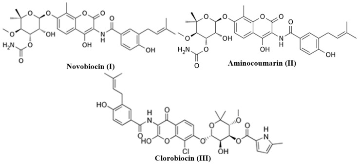
The oxygen-containing benzo-fused heterocycles, such as coumarins (2H-chromen-2-ones), are one of the most important and considered classes of compounds in medicinal chemistry because of their widespread pharmacological activities [1]. The simplest member of the coumarin family of compounds, “coumarin”, was isolated by Vogel in the year 1920 and eventually prepared by Sir William Henry Perkin through the Perkin reaction in 1868 [2,3]. Over the last few decades, researchers have explored the coumarin derivatives for various important biological activities such as anti-inflammatory, antioxidant, antithrombotic, antiallergic, antiviral, and anticancer [4,5,6,7,8,9,10,11]. Medicinal chemists continue to discover novel coumarins of natural (plant extracts) and unnatural (synthetic) analogs to further improve the currently identified biological activities while uncovering new medicinal uses. Coumarins have been considered as ideal small molecule candidates for the drug discovery and development process because they possess drug-like properties such as high solubility, low molecular weight, high bioavailability, and low toxicity along with their diverse biological activities [12,13]. Some notable coumarin analogs such as novobiocin (I), aminocoumarin (II), and clorobiocin (III) have been clinically used as antibiotic drugs [14,15,16] (Figure 1). At present, coumarin pharmacophore has been considered a privileged scaffold because of its medicinal aspects.
Figure 1.
Clinically used coumarin-based antibiotic drugs.
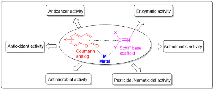
In recent years, Schiff bases have been used as multipurpose scaffolds to obtain biologically important molecules. Particularly, Schiff bases complexed with metal will have further enhanced biological activity. Therefore, the combination of Schiff base metal complexes with pharmacologically important small-molecule organic ligands such as coumarins is one of the important medicinal chemistry strategies to obtain ideal drug candidates. [17] (Figure 2).
Figure 2.
Important biological activities of the coumarin-derived imine–metal complexes.
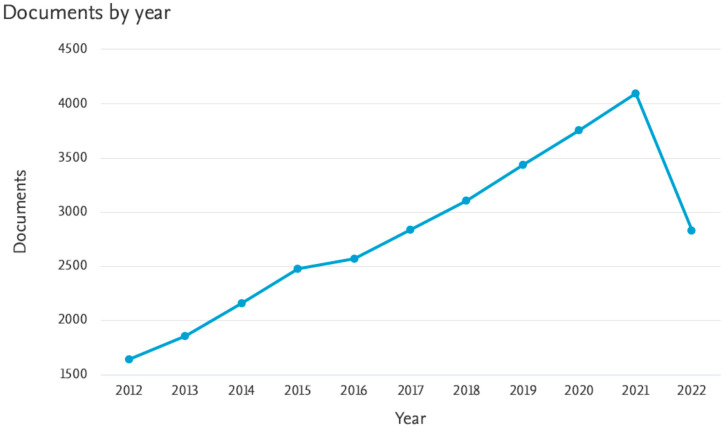
Coumarin pharmacophores are a part of several active drugs, such as the vitamin K antagonist, Warfarin. Because it can be assumed that coumarin–metal complexes will have important medical applications, we have provided a chart (Figure 3) that includes the articles published on these complexes indexed in Scopus for the past ten years (2012–2022). We recently reviewed medicinal applications of coumarins bearing azetidinone and thiazolidinone moieties [18]. In continuation of our effort on coumarin chemistry, we have mainly focused on its Schiff base–metal complexes as possible pharmacological agents in this review.
Figure 3.
Articles published on coumarin–metal complexes from 2012–2022.
2. Chemistry
Coumarins are therapeutically active members of the benzopyran-2-one family. Coumarins are extensively dispersed in nature and can be found in both naturally occurring and synthetic medicinally active compounds. In recent years there has been considerable growth in the chemistry of coumarins as a keystone for the design and development of a considerable number of compounds [19,20]. Nowadays, coumarin and its derivatives, especially Schiff bases derived from coumarins, belong to the most active classes of compounds and possess a wide spectrum of biological activity [21,22]. On the other hand, metal complexes derived from Schiff base ligands of coumarin show tremendous potential in numerous fields such as fluorescent probes, optical brighteners, antioxidants, antimicrobials, anthelmintics, hypotensive, and inhibitors of platelet aggregation and cytotoxic activity [23,24,25,26,27,28,29,30]. Hence, the synthesis, structural identification, and biological activity evaluation of new derivatives of coumarin-derived imine–metal complexes continually pique research interest across the world.
3. Medicinal Applications of Coumarin-Derived Imine–Metal Complexes
3.1. Antimicrobial Activity of Coumarin-Derived Imine–Metal Complexes
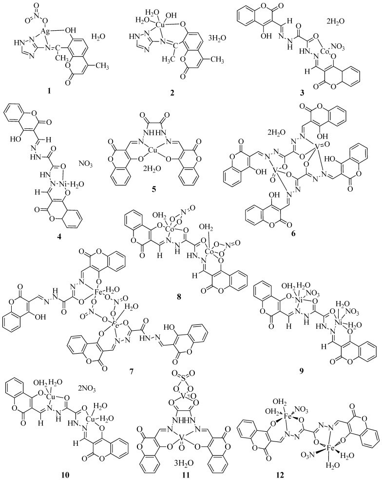
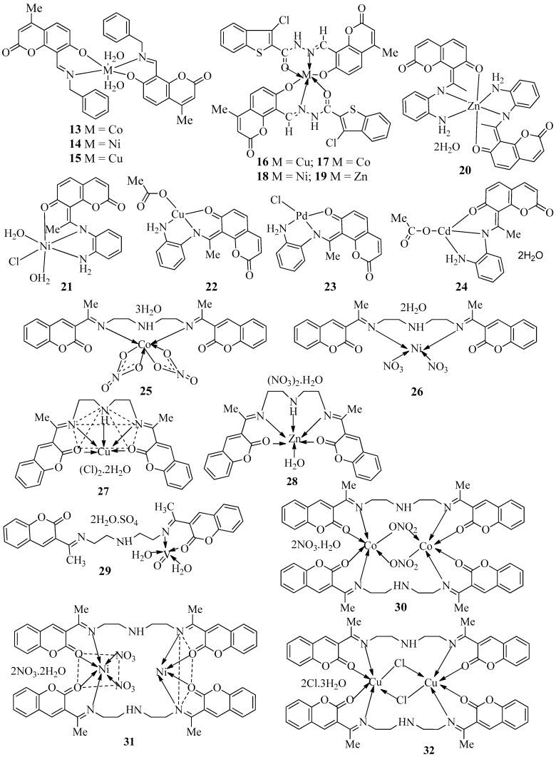
Farghaly et al. synthesized a Schiff base (Sbat) using 8-acetyl-7-hydroxy-4-methyl coumarin and 3-amion-1,2,4-triazole [31]. The reaction of the Schiff base with Ag(I) and Cu(II) metal ions produced the [Ag(Sbat)(NO3)]H2O (1) and [Cu(Sbat)(OH)(H2O)2].3H2O (2) complexes (Figure 4). The antimicrobial properties of Gram-positive bacteria Staphylococcus aureus (S. aureus), Staphylococcus faecalis (S. faecalis), and Bacillus subtilis (B. subtilis) were evaluated using the standard agar diffusion test and Gram-negative bacteria used included Escherichia coli (E. coli), Neisseria gonorrhoeae (N. gonorrhoeae), and Pseudomonas aeruginosa (P. aeruginosa) (Table 1). Antifungal activity against Aspergillus flavus (A. flavus) and Candida albicans (C. albicans) were also studied with the prepared complexes (Table 1). From the activity studies obtained, it was understood that the Schiff base had no activity against the two fungus species. Compounds 1 and 2 show no inhibitory action against the fungus. Sbat was found to be active against E. coli, N. gonorrhoeae, and S. faecalis. Also, 1 exhibited better antibacterial activity than the Schiff base. Complex 2 did not show any antibacterial activity against S. faecalis and N. gonorrhoeae. Against all microorganisms, 1 showed the maximum activity. The fact that the silver(I) complex was identified in a nanostructure might explain its biological activity because small silver nanoparticles with large surface area interact with bacteria better than larger ones. Also, it is worth noting that the reduced lipophilicity of 2 explains why it has weaker antibacterial activity than its parent ligands.
Figure 4.
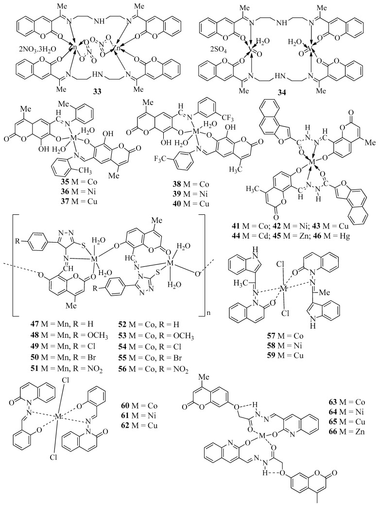
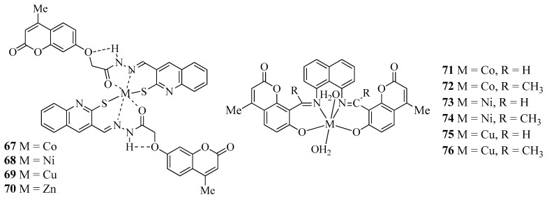
Structures of coumarin-derived imine–metal complexes (1–76).
Table 1.
Antimicrobial activity of coumarin-derived imine–metal complexes.
| Compound | Bacteria Screened (n) | Fungi Screened (n) | Highest Activity Against | Concentration | Activity Inhibition Zone (mm) | Ref. |
|---|---|---|---|---|---|---|
| 1 | 5 | 3 | B. subtilis, S. aureus, S. faecalis and P. aeruginosa | NA | 13 | [31] |
| 2 | 5 | 3 | S. aureus | NA | 11 | [31] |
| 3 | 2 | 1 | F. oxysporum | NA | 26 ± 0.2 | [32] |
| 4 | 2 | 1 | P. phaseolicola | NA | 28 ± 0.3 | [32] |
| 5 | 2 | 1 | S. aureus | NA | 36 ± 0.3 | [32] |
| 6 | 2 | 1 | S. aureus | NA | 33 ± 0.1 | [32] |
| 7 | 2 | 1 | S. aureus | NA | 37 ± 0.4 | [32] |
| 8 | 2 | 1 | S. aureus | NA | 27 ± 0.4 | [32] |
| 9 | 2 | 1 | F. oxysporum | NA | 29 ± 0.3 | [32] |
| 10 | 2 | 1 | S. aureus | NA | 31 ± 0.1 | [32] |
| 11 | 2 | 1 | F. oxysporum | NA | 34 ± 0.3 | [32] |
| 12 | 2 | 1 | S. aureus | NA | 32 ± 0.2 | [32] |
| 13 | 4 | 3 | E. coli | 200 μg/mL | NA | [33] |
| 14 | 4 | 3 | S. aureus | 200 μg/mL | NA | [33] |
| 15 | 4 | 3 | S. aureus | 200 μg/mL | NA | [33] |
| 16 | 4 | 3 | B. subtilis, E. coli and A. niger | 12.5 μg/mL | MIC | [34] |
| 17 | 4 | 3 | B. subtilis and C. oxysporum | 12.5 μg/mL | MIC | [34] |
| 18 | 4 | 3 | B. subtilis | 12.5 μg/mL | MIC | [34] |
| 19 | 4 | 3 | B. subtilis, E. coli, A. niger and C. oxysporum | 12.5 μg/mL | MIC | [34] |
| 20 | 8 | 0 | B. subtilis | NA | MP | [35] |
| 21 | 8 | 0 | B. subtilis | NA | MP | [35] |
| 22 | 8 | 0 | B. subtilis | NA | MP | [35] |
| 23 | 8 | 0 | B. subtilis | NA | MP | [35] |
| 24 | 8 | 0 | B. subtilis | NA | MP | [35] |
| 25 | 0 | 8 | B. cinerea | NA | RI | [35] |
| 21 | 0 | 8 | A. alternate, A. flavus, F. moniliforme, H. tetramera and V. alboatrum | NA | RI | [35] |
| 22 | 0 | 8 | A. flavus | NA | RI | [35] |
| 23 | 0 | 8 | A. flavus, F. moniliforme, R. stolonifera and V. alboatrum | NA | RI | [35] |
| 24 | 0 | 8 | A. flavus, F. moniliforme and H. tetramera | NA | RI | [35] |
| 25 | 2 | 1 | P. phaseolicola | NA | 26 ± 0.2 | [36] |
| 26 | 2 | 1 | P. phaseolicola | NA | 22 ± 0.2 | [36] |
| 27 | 2 | 1 | P. phaseolicola | NA | 27 ± 0.2 | [36] |
| 28 | 2 | 1 | P. phaseolicola | NA | 23 ± 0.3 | [36] |
| 29 | 2 | 1 | P. phaseolicola | NA | 20 ± 0.1 | [36] |
| 30 | 2 | 1 | P. phaseolicola | NA | 32 ± 0.3 | [36] |
| 31 | 2 | 1 | P. phaseolicola | NA | 30 ± 0.1 | [36] |
| 32 | 2 | 1 | S. aureus | NA | 37 ± 0.1 | [36] |
| 33 | 2 | 1 | P. phaseolicola | NA | 32 ± 0.2 | [36] |
| 34 | 2 | 1 | P. phaseolicola | NA | 39 ± 0.1 | [36] |
| 35 | 6 | 3 | Klebsiella and Salmonella | 200 μg/mL | 11 | [37] |
| 36 | 6 | 3 | Klebsiella and A. niger | 200 μg/mL | 13 | [37] |
| 37 | 6 | 3 | S. aureus | 200 μg/mL | 14 | [37] |
| 38 | 6 | 3 | S. aureus | 200 μg/mL | 13 | [37] |
| 39 | 6 | 3 | A. niger | 200 μg/mL | 14 | [37] |
| 40 | 6 | 3 | A. niger | 200 μg/mL | 15 | [37] |
| 41 | 4 | 4 | P. aeruginosa, A. niger and Cladosporium | 12.5 μg/mL | MIC | [38] |
| 42 | 4 | 4 | E. coli, S. aureus and A. flavus | 12.5 μg/mL | MIC | [38] |
| 43 | 4 | 4 | B. subtilis, A. niger and C. albicans | 12.5 μg/mL | MIC | [38] |
| 44 | 4 | 4 | E. coli, P. aeruginosa, A. niger and Cladosporium | 12.5 μg/mL | MIC | [38] |
| 45 | 4 | 4 | S. aureus and A. flavus | 12.5 μg/mL | MIC | [38] |
| 46 | 4 | 4 | S. aureus, P. aeruginosa, A. niger, A. flavus and C. albicans | 12.5 μg/mL | MIC | [38] |
| 47 | 5 | 2 | S. typhi | 100 μg/mL | 68.13 | [39] |
| 48 | 5 | 2 | S. typhi | 100 μg/mL | 72.00 | [39] |
| 49 | 5 | 2 | S. typhi | 100 μg/mL | 79.36 | [39] |
| 50 | 5 | 2 | S. typhi | 100 μg/mL | 76.44 | [39] |
| 51 | 5 | 2 | S. typhi | 100 μg/mL | 82.05 | [39] |
| 52 | 5 | 2 | S. typhi | 100 μg/mL | 65.00 | [39] |
| 53 | 5 | 2 | C. albicans | 100 μg/mL | 71.32 | [39] |
| 54 | 5 | 2 | S. typhi | 100 μg/mL | 75.66 | [39] |
| 55 | 5 | 2 | S. typhi | 100 μg/mL | 72.22 | [39] |
| 56 | 5 | 2 | S. typhi | 100 μg/mL | 80.00 | [39] |
| 57 | 3 | 0 | P. aeruginosa | 200 μg/mL | 21 | [40] |
| 58 | 3 | 0 | P. aeruginosa | 200 μg/mL | 21 | [40] |
| 59 | 3 | 0 | P. aeruginosa | 200 μg/mL | 22 | [40] |
| 60 | 3 | 0 | P. aeruginosa | 200 μg/mL | 23 | [40] |
| 61 | 3 | 0 | E. coli | 200 μg/mL | 24 | [40] |
| 62 | 3 | 0 | P. aeruginosa | 200 μg/mL | 23 | [40] |
| 63 | 2 | 2 | E. coli | 100 μg/mL | 26 | [41] |
| 64 | 2 | 2 | E. coli | 100 μg/mL | 27 | [41] |
| 65 | 2 | 2 | S. aureus | 100 μg/mL | 21 | [41] |
| 66 | 2 | 2 | S. aureus | 100 μg/mL | 25 | [41] |
| 67 | 2 | 2 | E. coli | 100 μg/mL | 22 | [41] |
| 68 | 2 | 2 | S. aureus | 100 μg/mL | 28 | [41] |
| 69 | 2 | 2 | S. aureus | 100 μg/mL | 23 | [41] |
| 70 | 2 | 2 | E. coli, S. aureus | 100 μg/mL | 28 | [41] |
| 71 | 4 | 3 | E. coli, A. flavus | 500 μg/mL | 24 | [42] |
| 72 | 4 | 3 | Cladosporium | 500 μg/mL | 28 | [42] |
| 73 | 4 | 3 | A. niger | 500 μg/mL | 27 | [42] |
| 74 | 4 | 3 | A. flavus | 500 μg/mL | 26 | [42] |
| 75 | 4 | 3 | A. flavus | 500 μg/mL | 28 | [42] |
| 76 | 4 | 3 | A. flavus | 500 μg/mL | 29 | [42] |
| 77 | 4 | 3 |
P. aeroginosa
A. flavus A. niger |
10 10 10 |
80% 81% 82% |
[43] |
| 78 | 4 | 3 | A. niger | 10 | 80% | [43] |
| 79 | 4 | 3 | Cladosporium | 10 | 90% | [43] |
| 80 | 4 | 3 | Cladosporium | 10 | 82% | [43] |
| 81 | 4 | 3 | Cladosporium | >100 | >93% | [43] |
| 82 | 4 | 3 | Cladosporium | >100 | >96% | [43] |
| Gentamycin | 4 | 0 | All four | 100 | 100% | [43] |
| Fluconozole | 3 | 0 | All three | 100 | 100% | [43] |
| 83 | 2 | 2 | P. aeroginosa | NA | 17 | [44] |
| 84 | 2 | 2 | P. aeroginosa | NA | 18 | [44] |
| 85 | 2 | 2 | E. coli | NA | 20 | [44] |
| 86 | 2 | 2 | P. aeroginosa | NA | 13 | [44] |
| 87 | 2 | 2 | P. aeroginosa | NA | 16 | [44] |
| 88 | 2 | 2 | P. aeroginosa | NA | 17 | [44] |
| 89 | 2 | 2 | P. aeroginosa | NA | 22 | [44] |
| 90 | 2 | 2 | P. aeroginosa | NA | 16 | [44] |
| 91 | 2 | 2 |
E. coli P. aeruginosa and C. albicans |
NA | 15 | [44] |
| 92 | 2 | 2 |
A.niger
C.albicans |
NA | 18 | [44] |
| 93 | 2 | 2 | P. aeroginosa | NA | 24 | [44] |
| 94 | 2 | 2 | E. coli | NA | 17 | [44] |
| 95 | 2 | 2 | E. coli | NA | 18 | [44] |
| 96 | 2 | 2 |
E. coli
C. albicans |
NA | 17 | [44] |
| Fluconozole | 0 | 2 | A. niger | NA | 24 | [44] |
| Ciprofloxacin | 2 | 0 | P. aeroginosa | NA | 30 | [44] |
| 97 | 2 | 2 | S. aureus | 3.12 | 21 | [45] |
| 98 | 2 | 2 | S. aureus | 19 | [45] | |
| 99 | 2 | 2 | S. aureus | 1.56 | 22 | [45] |
| 100 | 2 | 2 |
S. aureus
E. coli |
18 | [45] | |
| 101 | 2 | 2 |
S. aureus
E. coli |
3.12 | 21 | [45] |
| 102 | 2 | 2 | S. aureus | 20 | [45] | |
| 103 | 2 | 2 | S. aureus | 1.56 | 25 | [45] |
| 104 | 2 | 2 | S. aureus | 20 | [45] | |
| Norfloxacin | 2 | 0 |
S. aureus
E. coli |
1.56 | 26 | [45] |
| Griseofulvin | 0 | 2 |
A. niger and C. albicans |
1.56 | 26 | [45] |
| 105 | 4 | 3 | Cladosporium | 10 | >59% | [46] |
| 106 | 4 | 3 | Cladosporium | 10 | >55% | [46] |
| 107 | 4 | 3 | Cladosporium | 10 | >65% | [46] |
| 108 | 4 | 3 | Cladosporium | 10 | >61% | [46] |
| 109 | 4 | 3 | E. coli and A. flavus | 25 | >20% >16% |
[46] |
| 110 | 4 | 3 | E. coli | 25 | [46] | |
| Gentamycin | 4 | 0 | E. coli | 25 | 86% | [46] |
| Flucanozole | 0 | 3 | Cladosporium | 25 | 92% | [46] |
| 111 | 4 | 3 | A. flavus | 25 | 80% | [47] |
| 112 | 4 | 3 | A. flavus | 25 | 77% | [47] |
| 113 | 4 | 3 |
A. niger
A. flavus |
25 25 |
83% 71% |
[47] |
| 114 | 4 | 3 |
A. niger and S. aureus |
10 | [47] | |
| 115 | 4 | 3 | A. niger | 25 | 79% | [47] |
| 116 | 4 | 3 | A. flavus | 25 | 72% | [47] |
| 117 | 4 | 3 |
A. niger and P. aeruginosa |
25 25 |
82% 66% |
[47] |
| 118 | 4 | 3 | A. niger | 25 | 70% | [47] |
| 119 | 4 | 3 | A. flavus | 25 | 78% | [47] |
| 120 | 4 | 3 | A. flavus | 10 | [47] | |
| 121 | 4 | 3 |
A. flavus
A. niger |
25 25 |
80% 74% |
[47] |
| 122 | 4 | 3 | A. flavus | 25 | 85% | [47] |
| Gentamycin | 4 | 3 | All four | 25 | 100% | [47] |
| Flucanozole | 4 | 3 | All three | 25 | 100% | [47] |
| 123 | 4 | 0 | B. subtili, E. coli and P. aeruginosa | NA | 13 mm | [48] |
| 124 | 4 | 0 |
B. subtili S. aureus and P. aeruginosa |
NA | 18 mm | [48] |
| 125 | 4 | 0 |
B. subtili and P. aeruginosa |
NA | 17 mm | [48] |
| 126 | 4 | 0 | E. coli | NA | 17 mm | [48] |
| 127 | 4 | 0 |
S. aureus and E. coli |
NA | 18 mm | [48] |
| 128 | 4 | 0 | B. subtili | NA | 18 mm | [48] |
| 129 | 4 | 3 |
F. oxysporum and S. aureus |
100 ppm 500 ppm |
79 ± 1.4% 15 ± 0.03% |
[49] |
| 130 | 4 | 3 |
F. oxysporum
S. aureus |
100 ppm 500 ppm |
71 ± 0.4% 13.9 ± 0.09% |
[49] |
| 131 | 4 | 3 |
F. oxysporum and S. aureus |
100 ppm 500 ppm |
84 ± 0.6% 14.4 ± 0.1% |
[49] |
| 132 | 4 | 3 |
C. albicans and S. aureus |
100 ppm 500 ppm |
59 ± 0.7% 12.6 ± 0.09% |
[49] |
| 133 | 4 | 3 |
C. albicans and P. aeruginosa |
100 ppm 500 ppm |
60 ± 0.4% 12.0 ± 0.03% |
[49] |
| 134 | 4 | 3 |
F. oxysporum and E. coli |
100 ppm 500 ppm |
69 ± 0.2% 10.5 ± 0.09% |
[49] |
| Ciprofloxacin | 4 | 0 | S. aureus | 500 ppm | 18.6 ± 0.03% | [49] |
| Fluconazole | 0 | 3 | F. oxysporum, C. albicans and A. Niger | 500 ppm | 100 | [49] |
| 135 | 1 | 0 | F. psychrophilum | 32 | 16.1 ± 0.9% | [50] |
| 136 | 4 | 1 | A. Niger | NA | 4.6 mm | [51] |
| 137 | 4 | 1 | A. Niger | NA | 1.15 mm | [51] |
| 138 | 4 | 1 | S. aureus | NA | 0.81 mm | [51] |
| 139 | 4 | 1 | S. aureus | NA | 1.72 mm | [51] |
| 140 | 4 | 1 | S. aureus | NA | 1.72 mm | [51] |
| 141 | 4 | 2 | R. oryzae | 200 | 10 mm | [11] |
| 142 | 4 | 2 | A. niger | 200 | 11 mm | [11] |
| 143 | 4 | 2 |
P. auregenosa
A. niger R. oryzae |
200 200 200 |
12 mm 12 mm 12 mm |
[52] |
| 144 | 4 | 2 | A. niger | 200 | 11mm | [52] |
| 145 | 4 | 2 | A. niger | 200 | 14 mm | [52] |
| 146 | 4 | 2 | A. niger | 200 | 15 mm | [52] |
| Gentamicin | 2 | 0 | P. auregenosa | 200 | 15 mm | [52] |
| Fluconazole | 0 | 2 | A. niger | 200 | 16 mm | [52] |
| 147 | 4 | 0 | K. pneumonia | 100 | 22 mm | [53] |
| 148 | 0 | 2 | O. latemarginatus | 100 | 82.2 ± 1.1 | [54] |
| 149 | 0 | 2 | O. latemarginatus | 100 | 74.4 ± 1.1 | [54] |
| 150 | 0 | 2 | O. latemarginatus | 100 | 83.7 ± 0.6 | [54] |
| 151 | 0 | 2 | O. latemarginatus | 100 | 76.7 ± 1.1 | [54] |
| 152 | 0 | 2 | O. latemarginatus | 100 | 67.8 ± 1.1 | [54] |
| 153 | 1 | 1 |
E. coli and A. niger |
20 µg/mL | MIC | [55] |
| 154 | 1 | 1 |
E. coli and A. niger |
20 µg/mL | MIC | [55] |
| 155 | 1 | 1 | E. coli and A. niger | 20 µg/mL | MIC | [55] |
| 156 | 1 | 1 | E. coli and A. niger | 20 µg/mL | MIC | [55] |
| 157 | 0 | 1 | Candida | 5.2 µM | MIC | [56] |
| 158 | 0 | 1 | Candida | 10.4 µM | MIC | [56] |
| 159 | 0 | 1 | Candida | 16.7 µM | MIC | [56] |
| 160 | 0 | 1 | Candida | 8.2 µM | MIC | [56] |
| 161 | 0 | 1 | Candida | NA | MIC | [56] |
| 162 | 0 | 1 | Candida | 14.5 µM | MIC | [56] |
| 163 | 0 | 1 | Candida | 3.6 µM | MIC | [56] |
| 164 | 0 | 1 | Candida | 4.4 µM | MIC | [56] |
| 165 | 0 | 1 | Candida | 0.7 µM | MIC | [56] |
| 166 | 0 | 1 | Candida | 9.8 µM | MIC | [56] |
| 167 | 0 | 1 | Candida | 12.6 µM | MIC | [56] |
| Amphotericin B | 1 | Candida | 0.7 µM | MIC | [56] | |
| Ketoconazole | 1 | Candida | 4.7 µM | MIC | [56] | |
| 168 | 3 | 0 | K. pneumoniae and S. aureus | 100 µg/mL | 19 ± 0 | [57] |
| 169 | 3 | 0 | K. pneumoniae and S. aureus | 100 µg/mL | 18 ± 0 | [57] |
| 170 | 3 | 0 | K. pneumoniae, and S. aureus | 100 µg/mL | 17 ± 0 | [57] |
| 171 | 3 | 0 | K. pneumoniae, E. coli, and S. aureus | 100 µg/mL | 16 ± 0 | [57] |
MIC: Minimum inhibitory concentration; MP: Mortality percentage; RI: Radial inhibition; NA: Not applicable.
By condensing 3-formyl-4-hydroxycoumarin and oxalyldihydrazide in a 2:1 molar ratio, Linert et al. produced a hydrazone Schiff base ligand. In molar ratios of 1:1 or 2:1 (M:L), the Schiff base ligand interacted as a mono-, bi-, tri-, or even tetradentate ligand with metal cations to form mono- or binuclear complexes as keto or enol isomers, where M = Co(II), Ni(II), Cu(II), VO(IV), and Fe(III) (3–12) (Figure 4) [32]. Both the Schiff base ligand and its metal complexes were tested against Gram-positive and Gram-negative bacteria along with one type of fungi. Against these microorganisms, the synthesized complexes showed modest antibacterial and antifungal activity (Table 1). Chelation tends to make the ligands much more effective and potent antibacterial agents, hence, complexes outperform the ligands in terms of activity. The results show that Cu (II), Fe (III), and VO2+ complexes inhibited the growth of the selected bacteria and fungi the most. On the other hand, the Co(II) and Ni(II) complexes demonstrated modest activity. According to available data, binuclear complexes outperform acyclic complexes in terms of antibacterial activity, demonstrating that all these complexes are physiologically more efficient and implying that the chemical geometry of compounds is significant in explaining the complexes’ biological activity.
Badami et al. prepared Schiff base (HL) from 8-formyl-7-hydroxy-4-methylcoumarin and benzylamine, which they subsequently used to synthesize complexes of Co(II) (13), Ni(II) (14), and Cu(II) (15) (Figure 4) [33]. The Schiff base metal complexes were investigated for their antibacterial (E. coli, P. aeruginosa, Klebsiella pneumoniae (K. pneumoniae), and S. aureus) and antifungal (Penicillium chrysogenum (P. chrysogenum), and Aspergillus niger (A. niger)) activities, and it was discovered that the metal complexes had a more deadly impact on bacteria and fungus development than their parent ligand (Table 1). Against all strains, compound 15 demonstrated potential antibacterial and antifungal action. Solubility, conductivity, dipole moment, and cell permeability factors are thought to be the probable causes for the enhancement in the activity.
In another report, Schiff base ligand 3-chloro-N-((7-hydroxy-4-methyl-2-oxo-2H-chromen-8-yl)methylene)benzo[b]thiophene-2-carbohydrazide and its Cu(II) (16), Co(II) (17), Ni(II) (18), and Zn(II) (19) (Figure 4) complexes with octahedral geometries were synthesized by Mruthyunjayaswamy et al. [34]. The compounds’ antibacterial activity was evaluated in vitro against two Gram-negative bacteria (E. coli (MTCC 46) and Salmonella typhi (S. typhi) (MTCC 98)) and two Gram-positive bacteria (B. subtilis (MTCC 736) and S. aureus (MTCC 3160)) (Table 1). C. albicans (MTCC 227), Cladosporium oxysporum (C. oxysporum) (MTCC 1777), and A. niger (MTCC 1881) (Table 1) were tested for antifungal activity in vitro. All of the synthesized compounds showed antimicrobial capabilities in antimicrobial screening, and as expected, the metal complexes exhibited a greater inhibitory impact than their parent ligands and metal chlorides. Apart from the chelation theory, the increase in activity can be explained by the fact that most ligands have an azomethine link. Additionally, in metal complexes the positive charge of the metal ion is shared partially with the hetero donor atoms found in the ligand, and p-electron delocalization may occur throughout the chelating system. As a result, increasing the lipophilic character of metal chelates facilitates their passage through the lipoid layer of bacterial membranes and the blockage of metal binding sites in microorganism enzymes. Other factors that promote activity include conductivity, solubility, and the length of the link between the ligand and the metal ions.
From the coumarin–imine ligand, 8-[(1E)-1-(2-aminophenyliminio)ethyl]-2-oxo-2H-chromen-7-olate, Al-Amri et al. synthesized a series of metal complexes of Zn(II) (20), Ni(II) (21), Cu(II) (22), Pd(II) (23), and Cd(II) (24) (Figure 4) [35]. Compound 22 had the maximum efficacy and the lowest inhibitory concentration on the enzymatic activities of the investigated microbial species (Table 1). The agar plate antifungal activity of coumarin imine ligand and its metal complexes was evaluated against eight plant pathogenic fungi species (Alternaria alternata (A. alternate), A. flavus, Botrytis cinerea (B. cinerea), Fusarium verticillioides (F. moniliforme) and Verticillium albo atrum (V. alboatrum)) (Table 1). The copper complex outperformed the three most commonly used antifungal drugs. It had 100% radial inhibition (RI) against the most susceptible fungus, A. flavus. The RI of the tetrahedral cadmium complex against A. alternata, A. flavus, F. moniliforme, and V. alboatrum was 90%. However, the octahedral 20 possessed a 95% RI against B. cinerea. The square planar 23 has antifungal activity of 57–80%, with the highest activity against A. flavus and F. moniliforme. The octahedral 21 had the least antifungal activity, comparable to the drug Nystatin. The antibacterial activity of the investigated compounds was evaluated against four Gram-positive bacteria (S. citrus, Streptococcus pneumoniae (S. pneumoniae), B. subtilis, and Micrococcus luteus (M. luteus)) and four Gram-negative bacteria (Enterobacter aerogenes (E. aerogenes), E. coli, P. aeruginosa, and S. typhi). Even though the complexes under examination stopped bacteria from growing, they had a stronger antibacterial impact against Gram-positive bacteria than Gram-negative strains. Also, antibacterial activity was lowest for 21. The antibacterial and antifungal activities increased with metal chelation. The square planar copper complex has excellent antibacterial activity, which can be attributed to its structure.
Linert et al. prepared two new mono and binuclear complexes with a Schiff base ligand obtained from the condensation of 3-acetylcoumarine and diethylenetriamine [36]. With cobalt(II), nickel(II), copper(II), zinc(II), and oxovanadium(II), the Schiff base ligand formed mono- or bi-nuclear cyclic or macrocyclic complexes, depending on the metal-to-ligand mole ratio and preparation method (25–34) (Figure 4). The Schiff base, HL, ligand, and its metal complexes were investigated for antibacterial activity against Gram-positive and Gram-negative bacteria, as well as a pathogenic fungus, Fusarium oxysporum (F. oxysporum) (Table 1). The findings were compared with the gram-negative and gram-positive antibiotics, chloramphenicol and cephalothin. The antifungal standard was cycloheximide. In vitro antibacterial and antifungal activity showed that complexes outperform ligands. The π-electron delocalization and partial sharing of the positive charge with a donor group lower the metal atoms polarity and this chelation might increase the metal atom’s lipophilicity, allowing it to pass through the lipid layers of the cell membrane and block metal binding sites on enzymes. The results showed that Cu(II) complexes had the greatest growth inhibition against Gram-positive bacteria, Gram-negative bacteria, and fungi. Complexes of Co(II), Ni(II), Zn(II), and VO(IV) had modest activity. The research demonstrated that binuclear macrocyclic properties improve antibacterial activity rather than acyclic complexes.
Badami et al. synthesized Co(II), Ni(II), and Cu(II) metal complexes from 6-formyl-7,8-dihydroxy-4-methylcoumarin and o-toluidine/3-aminobenzotrifluoride (35–40) (Figure 4) [37]. The compounds were evaluated for antibacterial (E. coli and P. aeruginosa), and antifungal (Candida, A. niger, and Rhizopus) activities using the disc diffusion technique (Table 1). Only a few species had antibacterial activity, and 40 had the most. Compounds 39 and 40 outperformed the others in antifungal activity. Protein synthesis is an important phase in the development of microorganisms. Metal ions function as growth inhibitors for microorganisms by adsorbing on the outside surface of the cell wall and preventing respiration. This interrupts protein synthesis and kills the microorganism. The increased activity of chelates may be owing to the metal ion’s decreasing polarity due to sharing its positive charge with donor groups, as explained by chelation theory.
Kinni et al. have synthesized metal complexes of type ML2, where M = Co(II), Ni(II), Cu(II), Cd(II), Zn(II), Hg(II), and L = Schiff base (41–46) (Figure 4) [38]. E. coli, S. aureus, B. subtilis, P. aeruginosa bacteria and A. flavus, A. niger, C. oxysporum, and C. albicans fungal strains were used to test the antibacterial and antifungal capabilities of the synthesized compounds by the minimum inhibitory concentration (MIC) method (Table 1). Coordination with metal ions clearly increased the ligand’s efficacy and also lessened the metal ion’s polarity by sharing its positive charge with donor groups inside the chelate ring system generated during coordination. Because of its high lipophilicity, the central metal atom may easily penetrate the lipid layer of bacteria, causing them to die.
Bajroliya et al. developed Mn(II) and Co(II) metal complexes from novel Schiff bases derived from 8-formyl-7-hydroxy-4-methylcoumarin and 3-substituted 4-amino-5-mercapto-1,2,4-triazole by using microwave irradiation as well as conventional methods (47–56) (Figure 4) [39]. Compared with the traditional approach, the microwave irradiation procedure yielded higher yields with better selectivity. This study investigated the in vitro antimicrobial activity of synthesized Schiff bases and their metal complexes by measuring the zone of inhibition in mm. Assays were performed using gentamycin at a 100 g/mL concentration against five species of bacteria: E. coli, P. aeruginosa, S. typhi, S. aureus, and B. subtilis. Antifungal activities were assessed against A. niger and C. albicans at 100 g/mL using fluconazole as a reference drug. The metal complexes of all Schiff bases had stronger antibacterial activity than the Schiff bases alone against selected bacteria and fungi (Table 1). The Mn(II) and Co(II) metal complexes showed significantly increased activity against S. typhi. The Schiff bases, as well as their Mn(II) and Co(II) complexes, were found to have good antifungal activity. Al-Amiery et al. prepared novel transition metal complexes [M(L1)2Cl2] and [M(L2)2Cl2] by reacting MCl2.nH2O (M = Co, Ni, Cu) with (Z)-1-(1-(1H-indol-3-yl)ethylideneamino)quinolin-2(1H)-one (L1) or (E)-1-(2-hydroxybenzylideneamino)quinolin-2(1H)-one (L2) (57–62) (Figure 4) [40]. The antibacterial activity of the prepared complexes was then estimated against S. aureus (Gram positive) and E. coli and P. aeruginosa (Gram negative) bacteria (Table 1). The enhanced activity of metal complexes can be explained by the overtone concept and chelation theory. Liposolubility is a key element in determining antimicrobial action since it allows only lipid-soluble molecules to get through the lipid barrier surrounding the cell. Chelation reduces the polarity of the metal ion due to ligand orbital overlap and partial sharing of the positive charge with donor groups. It also boosts the lipophilicity of the complex and the delocalization of electrons across the chelate ring. This enhanced lipophilicity promotes complex penetration through the lipid membrane and prevents metal binding sites on the microorganism’s enzymes.
Dhumwad et al. prepared complexes of Co(II), Ni(II), Cu(II), and Zn(II) with Schiff bases of N′-[(E)-(2-hydroxyquinolin-3-yl)methylidene]-2-[(4-methyl-2-oxo-2H-chromen-7-yl)oxy] acetohydrazide (OHQZ) and 2-[(4-methyl-2-oxo-2H-chromen-7-yl)oxy]-N′-[(E)-(2-sulfanylquinolin-3-yl)methylidene] acetohydrazide (SHQZ) (63–70) (Figure 4) [41]. The antibacterial and antifungal properties of the synthesized Schiff bases and their Co(II), Ni(II), Cu(II), and Zn(II) complexes were investigated using the potato dextrose agar diffusion method and the nutritional agar method. The minimum inhibitory concentration (MIC) technique was used to test the antibacterial and antifungal activities on two bacterial (E. coli and S. aureus) and two fungal (A. niger and P. chrysogenum) strains (Table 1). Based on the in vitro antibacterial and antifungal activity against selected bacterial and fungal strains, it is clear that the Cu(II) complexes are more active at lower MIC values for bactericidal activity. Badami et al. prepared a series of metal complexes of the type ML·2H2O (M = Co(II), Ni(II), and Cu(II)) with Schiff bases produced from 1,8-diaminonaphthalene and 8-formyl-7-hydroxy-4-methylcoumarin/8-acetyl-7-hydroxy-4-methylcoumarin (71–76) (Figure 4) [42]. The MIC method was employed to determine the antibacterial (E. coli, S. aureus, P. aeruginosa, and S. typhi) and antifungal (A. niger, A. flavus, and Cladosporium) properties of the Schiff bases and their complexes (Table 1). In antibacterial experiments, the Schiff bases were found to be potentially active against E. coli and S. typhi, as well as moderately active against S. aureus. Compounds 71 and 72 had substantial antibacterial activity against E. coli, P. aeruginosa, and S. typhi and least activity against S. aureus. Compounds 73 and 74 were effective against E. coli, S. aureus, and S. typhi, but only modestly effective against P. aeruginosa. Compounds 75 and 76 had outstanding antibacterial activity against S. typhi but only fair activity against E. coli, P. aeruginosa, and S. aureus. All Schiff bases were active against fungi. However, as compared with the uncoordinated compounds, 71–76 exhibited significantly increased activity, notably with A. flavus. The findings show that activity relies on metal ion type and varies in the following order: Cu > Ni > Co.
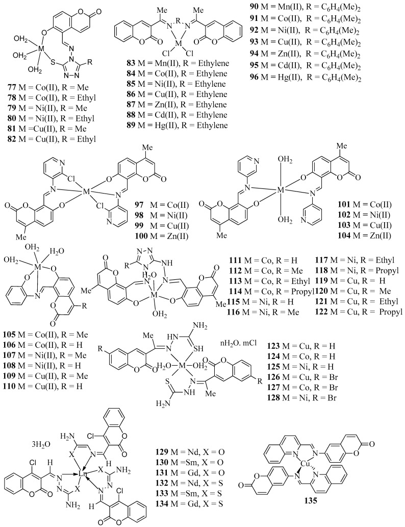
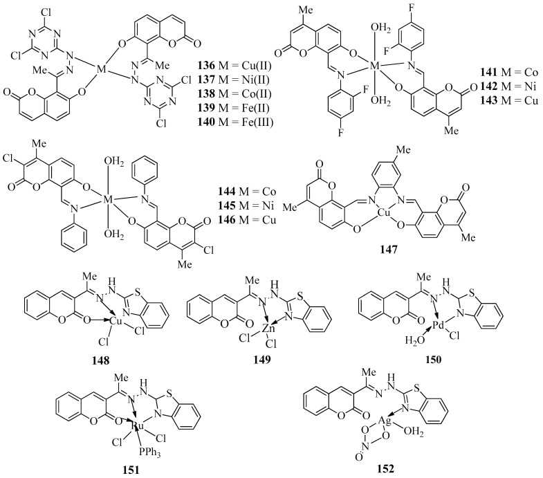
Patil et al, in 2011 reported the synthesis of a series of Co(II), Ni(II), and Cu(II) complexes (77–82) (Figure 5) with Schiff bases derived from 3-substituted-4-amino-5-mercapto-1,2,4-triazole and 5-formyl-6-hydroxy coumarin [43]. All synthesized metal complexes (77–82) were evaluated for antibacterial activities against four bacterial species, E. coli, S. aureus, P. aeruginosa, and S. typhi, and antifungal efficacy towards three fungi, namely A. niger, A. flavus, and Cladosporium through the minimum inhibitory concentration method. All the metal complexes (77–82) showed promising antimicrobial activities against some bacterial and fungal strains. Among the prepared metal complexes, Co(II) complex 77 showed maximum efficacy against P. aeruginosa with MIC 10 µg/mL and Ni(II) complex 80 exhibited the highest antifungal activity against Cladosporium with MIC 10 µg/mL.
Figure 5.
Structures of coumarin-derived imine–metal complexes (77–152).
The metal complexes (83–96) (Figure 5) with Schiff bases derived from the condensation of 3-acetylcoumarine with ethylenediamine and orthophenylenediameine were developed by Akkasali et al. [44]. All the developed compounds (83–96) were screened for their possible antimicrobial activity against E. coli, P. aeruginosa, A. niger, and C. albicans. The prepared metal complexes (83–96) showed moderate to high activity against the tested organisms. Among the complexes, copper complex 93 showed a maximum zone of inhibition against P. aeroginosa and nickel complexes 85 and 92 displayed higher antifungal activity against A. niger. In addition, nickel complex 92 also revealed good activity against C. albicans. However, all the developed compounds were less active than the standard drugs ciprofloxacin and fluconazole and the data presented in this review could be a helpful guide for medicinal chemists who are working in the area. Dhumwad and co-workers have designed the synthesis of metal complexes (97–104) (Figure 5) of Schiff bases formed through the condensation of 8-formyl-7-hydroxy-4-methyl-coumarin with 3-amino-pyridine and 3-amino-2-chloro-pyridine [45]. The synthesized metal complexes (97–104) were evaluated for their in vitro antibacterial potential against S. aureus and E. coli, and antifungal efficacy against two fungi, viz., A. niger and C. albicans. Among the metal complexes, copper complexes 99 and 103 are found to be most active against the tested bacteria with a MIC value of 1.56 µg/mL, which is almost equal to that of the standard drug, norfloxacin. Therefore, the prepared copper complexes 99 and 103 are found to be more effective bactericides than fungicides.
Patil et al. have structured and developed a new class of Co(II), Ni(II), and Cu(II) complexes (105–110) (Figure 5) with Schiff bases derived from 8-formyl-7-hydroxy-4-methylcoumarin or 5-formyl-6-hydroxycoumarin and o-aminophenol and assessed them for their in vitro antibacterial action against microorganisms such as E. coli, P. aeruginosa, S. typhi, A. flavus and Cladosporium, A. flavus, Cladosporium, and A. niger (Table 1) [46]. From the MIC studies, it has been found that the metal complexes were more active against both bacterial and fungal species than parent ligands. Among the metal complexes, cobalt complex 105 and nickel complex 107 revealed high activity against E. coli, P. aeruginosa, and S. typhi. Furthermore, cobalt complex 105 and nickel complex 107 also had good antifungal activity against A. flavus, Cladosporium, and A. niger. Patil et al. have also designed and synthesized a series of cobalt(II), nickel(II), and copper(II) complexes (111–122) (Figure 5) with newly derived biologically active ligands that are prepared from the condensation of 3-substituted-4-amino-5-hydrazino-1,2,4-triazole and 8-formyl-7-hydroxy-4-methylcoumarin [47]. All the metal complexes (111–122) were evaluated for their antibacterial efficacy against four bacterial strains, namely E. coli, S. aureas, S. pyogenes, and P. aeruginosa using Gentamycin as the standard. From the antibacterial studies, it was found that all the synthesized metal complexes (111–122) were more active compared with the corresponding parent Schiff bases. Metal complex 116 showed maximum inhibition against S. aureas and P. aeruginosa at 25 µg/mL. Furthermore, metal complex 118 showed maximum inhibition towards E. coli at 25 µg/mL. Also, metal complex 120 displayed highest inhibition against P. aeruginosa at 25 µg/mL, whereas metal complex 121 revealed maximum inhibition towards S. aureas and S. pyogenes at 25 µg/mL. The synthesized metal complexes 111–122 (Table 1) were also screened for their antifungal activities against A. Flavus, Cladosporium, and A. niger using the standard drug fluconazole. Metal complex 114 presented MIC 10 µg/mL against S. aureus and A. Niger, whereas metal complex 120 showed MIC 10 µg/mL against A. Flavus.
A series of Schiff base complexes of Cu(II), Co(II), and Ni(II) (123–128) (Figure 5) with two coumarin-3-yl thiosemicarbazone derivatives (1E)-1-(1-(2-oxo-2H-chromen-3-yl)ethylidene)thiosemicarbazide and (1E)-1-(1-(6-bromo-2-oxo-2H-chromen-3-yl)ethylidene)thiosemicarbazide were constructed and tested for their in vitro antibacterial potential towards both Gram-negative and Gram-positive bacterial species such as E. coli, P. aeruginosa, B. subtili, and S. aureus by Refat et al. [48]. From the in vitro antibacterial evaluation, it was found that all the synthesized metal complexes 123–128 showed more efficacy than their parent Schiff base ligands against both Gram-negative and Gram-positive bacterial species such as E. coli, P. aeruginosa, B. subtili, and S. aureus. Conversely, metal complexes 124 and 128 exhibited maximum inhibition zone against B. subtili. Similarly, metal complexes 124 and 127 exhibited maximum inhibition zone against S. aureus, whereas metal complex 127 displayed maximum inhibition zone against E. coli. Kapoor et al, in 2012 explored the microwave-assisted synthesis and biological evaluation of coumarin-based lanthanide complexes (129–134) (Figure 5) [49]. All complexes (129–134) were successfully synthesized using rare earth metals Nd(III), Sm(III), and Gm(III) with ligands 3-formyl-4-chlorocoumarin hydrazinecarboxamide and 3-formyl-4-chlorocoumarinhydrazine carbothioamide. All the lanthanide complexes 129–134 were evaluated for their antimicrobial activity against four bacteria (E. coli, P. aeruginosa, B. subtilis, and S. aureus), and three fungi (C. albicans, A. niger, and F. oxysporum). From the results, it has been found that complexes showed more activity compared with the paternal Schiff base ligands and were also compared against standard drugs ciprofloxacin for bacterial species and fluconazole for fungal species. All the aforementioned lanthanide complexes (129–134) showed MIC values in between 10–40 MIC (µg/mL). In particular, Lanthanide complex 130 displayed MIC 10 µg/mL against E. coli, S. aureus, C. albicans, A. niger, and F. oxysporum w, 15 µg/mL against P. aeruginosa, and 20 µg·mL−1 against B. subtilis. Conversely, complex 131 showed 84 ± 0.6% inhibition against F. oxysporum at 100 ppm, whereas complex 129 showed 78 ± 0.7% inhibition against C. albicans at 100 ppm (Table 1).
In the recent past, Modak et al. reported the synthesis of copper complex 135 (Figure 5) with imine ligand 6-((quinolin-2-ylmethylene)amino)-2H-chromen-2-one achieved from derivatization of natural compound coumarin [50]. The potential antibacterial effect of copper complex 135 was assessed for Flavobacterium psychrophilum (F. psychrophilum) isolated 10094 and efficacy was IC50 16.1 ± 0.9 µg/mL. However, MIC and MBC of the copper complex 135 were found to be lower (32 µg/mL) than the precursor (64 µg/mL). Novel classes of metal complexes (136–140) (Figure 5) that are derived from 7-hydroxy coumarin hydrazone of s-triazine derivatives were evaluated for their in vitro antibacterial potency against tested pathogens by the research group of Jani [51]. Antibacterial activity was tested using the agar diffusion method against E. coli and S. aureus and A. niger was used for antifungal activity. From the zone of inhibition data, it was concluded that metal complex 136 exhibited higher inhibition towards S. aureus, E. coli, Aspergillus niger, and S. pyogenes, whereas the other metal complexes 136–140 revealed moderate activity. All metal complexes (136–140) were inactive against P. klebsiella (Table 1).
In 2016, Patil and co-workers reported the synthesis of a series of Co(II), Ni(II), and Cu(II)complexes (141–146) (Figure 5) from the reaction 8-formyl-7-hydroxy-4-methylcoumarin/3-chloro-8-formyl-7-hydroxy-4-methylcoumarin with 2,4-difluoroaniline/o-toluidine [52]. Antimicrobial activities of metal complexes 141–146 were carried out through the disc diffusion method against two bacterial strains (P. auregenosa and Proteusmirabilis) with gentamicin as the standard and two fungal strains (A. niger and R. oryzae) with antifungal drug fluconazole (Table 1). Among different halogenated metal complexes, copper complex 146 exhibited more activity against A. niger. Nickel complex 145 displayed a maximum zone of inhibition against P. auregenosa, whereas copper and nickel complexes 143, 145, and 146 revealed maximum inhibition against Proteusmirabilis. Copper complex 147 (Figure 5) with a salen-type ligand derived from the condensation of 4-methyl-7-hydroxy-8-formylcoumarin with 3,4-diaminotoluene was screened for in vitro antibacterial activity by Sharma et al. through the broth dilution method against four bacterial strains: E. coli, S. aureus, P. aeruginosa, and K. pneumonia [53]. Compound 147 exhibited an MIC of 12.5-200 μg·mL−1 and appeared to be active against both Gram-positive and Gram-negative bacteria. Both experimental and theoretical calculations revealed that 147 has better antibacterial potential than the parent Schiff base ligand (Table 1).
A series of five new Cu (II), Zn (II), Pd (II), Ru (III), and Ag(I) complexes (148–152) (Figure 5) derived from the 3-acetylcoumarin-2-hydrazinobenzothiazole Schiff base have been synthesized and characterized by Atlam et al. [54]. All metal complexes 148–152 were screened for their antifungal effect towards two fungal strains, G. recinaceum (GR33) and O. latemarginatus (EM26). Palladium complex 150 acts as the best antifungal agent for GR33 with a 60% reduction, whereas the silver complex 152 has a 79% reduction for O. latemarginatus (EM26). The ligninolytic activity of the complexes on fungi was found using Poly R-dye decolorization ability. Results suggest that complexes showed decolorization at lower concentrations depending on the fungi used. From the observed results, it can be concluded that both palladium and silver complexes 152 and 150, respectively, were used for antifungal preservatives, whereas copper, zinc, and ruthenium complexes 148, 149, and 151, respectively, can be used for lignolytic activity. Observed results were confirmed using computational studies and molecular docking studies help to provide a better understanding of binding interactions (Table 1).
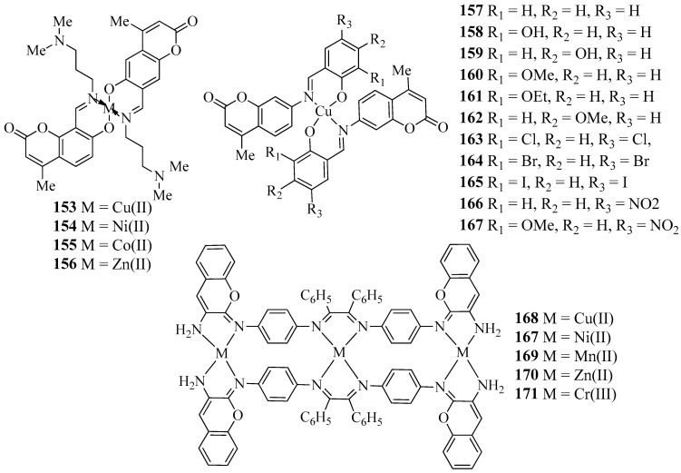
Sawant et al. reported the synthesis, characterization and antimicrobial activity of novel transition metal complexes of 4-methyl-7-hydroxy 8-formyl coumarin. The prepared Schiff base acts as a bidentate ligand for the complexation with Cu(II), Ni(II), Co(II), and Zn(II) ions. The Schiff base and their transition metal complexes (153–156) (Figure 6) were screened for their antimicrobial activity towards two strains, E. coli and A. niger, through the tube dilution method in order to examine the effect of metal ions upon chelation (Table 1). All the transition metal complexes 153–156 showed MIC values less than 20 µg/mL compared with the ligand, which has MIC values less than 200 µg/mL against both species. The results of antimicrobial activity indicate that the antimicrobial activity of the Schiff base was increased on complexation with metal ions [55]. In 2009, Creaven and co-workers reported the Cu(II) complexes (157–167) (Figure 6) of coumarin-derived Schiff bases. A few of the Cu(II) complexes were characterized through X-ray crystallography technique to determine the structures. Furthermore, all Cu(II) complexes 157–167 were screened for their anti-Candida activity. All Cu(II) complexes displayed excellent anti-Candida activity comparable to that of commercially available drugs such as ketoconazole and amphotericin B (Table 1) [56].
Figure 6.
Structures of coumarin-derived imine–metal complexes (153–171).
Recently, Chandrasekaran et al. described the synthesis, characterization, and antibacterial activity of new Cu (II), Ni (II), Mn (II), Zn (II), and Cr (III) complexes (168–171) (Figure 6) of Schiff bases derived from p-phenylenediamine, benzil, and 3-aminocoumarin. Structural analyses of all new transition metal complexes 168–171 were conducted through infrared, ultraviolet–visible, cyclic voltammetry, EPR, magnetic property, and thermogravimetric techniques. Furthermore, all transition metal complexes 168–171 were tested for their antibacterial activity against bacterial species such as K. pneumoniae, E. coli, and S. aureus by the disc diffusion method. The biological activity results concluded that the Schiff base transition metal complexes 168–171 have more efficiency compared with standard streptomycin drug and the transition metal complexes 168–171 arrested the growth of bacterium (Table 1) [57].
3.2. Anticancer Activity of Coumarin-Derived Imine–Metal Complexes
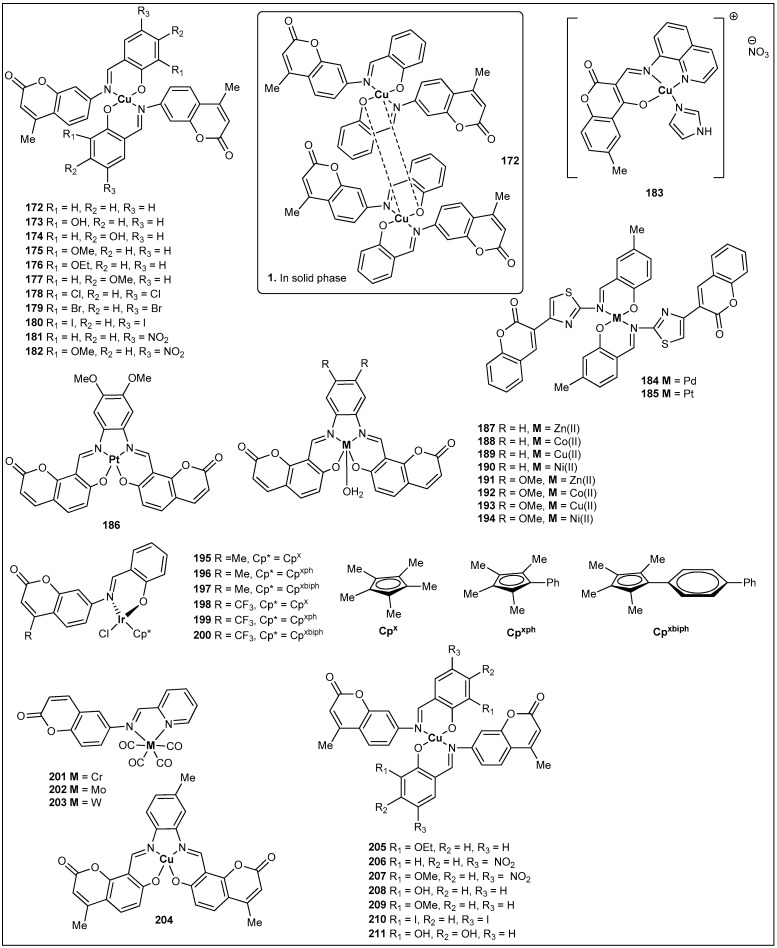
It is well known that coumarin derivatives present important cytotoxic activity against several cell lines [18]. It is also known that coordination of bioactive molecules to transition metals display enhanced inhibitory activity [58]. In this regard, Aazam and coworkers prepared the first coumarin-derived imine–metal complex that has been evaluated for its anticancer activity. This complex was synthesized from the reaction of 4-methyl-7-(salicylideneamino)coumarin with Cu(OAc)2. Interestingly, single crystal X-ray diffraction studies exhibited that the complex crystallized as a dimeric copper coumarin complex, 172, as depicted in Figure 7 [59]. In 2009, Creaven and coworkers corroborated this observation when they also observed the same dimeric copper complex [57]. Nonetheless, Creaven and coworkers went further and prepared a total of eleven coumarin copper complexes (172–182) (Figure 7) (Table 2). Although, other complexes were crystallized (e.g., 179), only complex 172 was observed as dimeric copper complex [60]. A year later, Creaven et al. reported the cytotoxicity activity of the eleven complexes against two human cell lines: human colon cancer cells (HT29) and human breast cancer cells (MCF-7) using the methylthiazolyldiphenyl-tetrazolium bromide assay (MTT) [60]. Unfortunately, none of the complexes were cytotoxic against colon cancer cells and only complexes 180 and 182 showed cytotoxicity against breast cancer cells, with IC50 values of 79.8 μM and 34.5 μM, respectively. Values that are comparable with the positive control mitoxantrone (IC50 value of 44 μM) (Table 2). In an attempt to try to increase the observed cytotoxicity, the authors reduced the coumarin shift bases to amines. Sadly, their respective copper complexes were not successfully synthesized [60].
Figure 7.
Structures of coumarin-derived imine–metal complexes (172–211).
Table 2.
Anticancer activity data of coumarin-derived imine–metal complexes.
| Compound | Cell Line | IC50 (µM) | Ref. | Compound No. | Cell Line | IC50 (µM) | Ref. |
|---|---|---|---|---|---|---|---|
| 172 | MCF-7 | no reported | [58] | 187 | HeLa | 86.9 ± 9.0 | [63] |
| 173 | MCF-7 | no reported | [58] | 188 | HeLa | 3.5 ± 1.2 | [63] |
| 174 | MCF-7 | no reported | [58] | 189 | HeLa | 52.5 ± 1.0 | [63] |
| 175 | MCF-7 | no reported | [58] | 190 | HeLa | 72.7 ± 8.1 | [63] |
| 176 | MCF-7 | no reported | [58] | 191 | HeLa | 90.7 ± 2.5 | [63] |
| 177 | MCF-7 | no reported | [58] | 192 | HeLa | 4.1 ± 0.9 | [63] |
| 178 | MCF-7 | no reported | [58] | 193 | HeLa | 83.5 ± 4.7 | [63] |
| 179 | MCF-7 | no reported | [58] | 194 | HeLa | 85.1 ± 1.6 | [63] |
| 180 | MCF-7 | 79.8 ± 5 | [58] | Cisplatin | HeLa | 11.6 ± 2.7 | [63] |
| 181 | MCF-7 | no reported | [58] | 195 | A549 | 30.9 ± 1.6 | [63] |
| 182 | MCF-7 | 34.5 ± 6 | [58] | 196 | HeLa | 9.9 ± 0.1 | [64] |
| Mitoxantrone | MCF-7 | 44 ± 3 | [58] | 197 | HeLa | 10.8 ± 0.1 | [64] |
| 183 | A549 | 4.6 ± 0.3 | [59] | 198 | A549 | 39.6 ± 1.0 | [64] |
| Cisplatin | A549 | 57.7 ± 0.9 | [59] | 199 | HeLa | 22.1 ± 0.5 | [64] |
| 184 | LNCap | 10.05 | [59] | 200 | A549 | 33.4 ± 0.7 | [64] |
| 185 | LNCap | 21.53 | [59] | Cisplatin | A549 | 21.3 ± 1.7 | [64] |
| 186 | HeLa | 61.4 ± 1.1 | [59] | Cisplatin | HeLa | 7.5 ± 0.2 | [64] |
In 2017, Tabassum et al. reported the synthesis of one coumarin-derived imine–metal complex, 183 (Figure 7), that has an appended imidazole (Figure 4) [59]. In vitro anticancer studies revealed that complex 183 has superior cytotoxicity towards the A549 adenocarcinoma cell line in comparison with the standard drug cisplatin (Table 2), which is a very promising discovery as A549 is a cisplatin-resistant cell line [61] (Table 2). The same year, Sahin and coworkers reported the synthesis, characterization, and anticancer activity of two coumarin-derived imine–metal complexes. Synthesis of complex 184 (Figure 7) used Na2PdCl4 to incorporate palladium to the ligand, whereas K2PtCl4 was used to install platinum into complex 185 (Figure 7) [62]. Both complexes were evaluated against three human cancer cell lines: prostate cancer cells (ATTC), colon carcinoma cells (LNCap), and breast cancer cells (MCF-7) using the MTT assay. The data from the in vitro antitumor activity showed that palladium complex 184 had the most prominent activity against the three screened cell lines, with an IC50 of 18.15 μg/mL for MCF-7, an IC50 of 10.05 μg/mL for LNCap, and an IC50 of 15.98 μg/mL for ATTC. Values that are 3- to 5-fold better than the ligand starting material [60].
In 2020, Hurtado et al. reported nine tetradentate coumarin Schiff-based metal complexes (186–194) (Figure 7) [63]. Two coumarin ligands and the five metals (Zn(OAc)2, Co(OAc)2, Cu(OAc)2, Ni(OAc)2, and K2PtCl4) were used to prepare the nine complexes (Figure 4). A cytotoxicity activity was carried out using an MTT assay in the carcinogenic cell line HeLa (human cervical cancer cells), and, to make the study more relevant, two noncarcinogenic cell lines, HFF-1 (human foreskin fibroblast cells) and HaCaT (human keratinocytes) were screened. Data from this study showed that both ligands and seven of the screened complexes displayed higher IC50 values than the standard drug cisplatin (IC50 = 11.6 μM). Fortunately, both cobalt complexes 188 and 192 exhibited lower IC50 values, or in other words the highest cytotoxicity towards HeLa cells. For instance, complex 188 showed an IC50 = 3.5 μM and complex 192 showed an IC50 = 4.1 μM (Table 2). Thus, they have promising anticarcinogenic potential [64]. The same year, Liu et al. published the synthesis, characterization, and anticancer activity of six coumarin-derived imine–iridium complexes (195–200) (Figure 7) [64]. The MTT assay was used to determine the anticancer activity of the former stated complexes. The studied human cancer cell lines were lung cancer cells (A549) and cervical cancer cells (HeLa) with cisplatin as the positive control. All complexes displayed IC50 values ranging from 9.9 μM to 40.7 μM, with complex 196 showing almost double (12 μM) the anticancer activity of cisplatin (21.3 μM) against lung cancer cells (Table 2) [64]. It is important to note that fluorinated complexes showed similar or lower antitumor activity.
3.3. Antioxidant Activity of Coumarin-Derived Imine–Metal Complexes
Following similar methodology as mentioned above, three new coumarin-derived imine–metal complexes were synthesized by Sinha et al. from the condensation of 6-aminocoumarin and pyridine-2-carboxaldehyde to make a ligand [65]. The ligand was reacted with Cr(CO)4, Mo(CO)4, and W(CO)4 to produce complexes (201–203) (Figure 7). The three complexes were tested for their antioxidant properties and their antioxidant activity (radical scavenging activity) was examined with reference to 1,1-diphenyl-2-picrylhydrazyl (DPPH), hydroxyl radical (OH·), superoxide(O2−), and nitroxyl radical (NO·). The data revealed that the complexes had a maximum percentage of inhibition at concentrations of 0.03 mg/mL, 0.3 mg/mL, and 0.01 mg/mL for [Cr(CO)4(L)], [Mo(CO)4(L)], and [W(CO)4(L)], respectively. Therefore, complex 203, having the heaviest element (W), shows the highest scavenging ability. In 2012, Halli and coworkers reported another series of coumarin-derived imine–metal complexes (41–46) (Figure 4) [38]. In addition to the antimicrobial activity (reported above), an antioxidant assay (DPPH) was investigated for all the synthesized complexes, and the tabulated data in Table 3 shows that the top radical scavengers were complexes 42 (Ni), 44 (Cd), and 46 (Hg) (Table 3) at a concentration of 100 μg/mL [66]. Similarly, Sharma et al. reported the synthesis, antibacterial, and antioxidant activity of complex 204 (Figure 7) [67]. Although complex 39 presented some scavenging ability (30%), it was lower in comparison with the standards gallic acid (90%) and quercetin (80%) at a concentration of 62.5 μg/mL. Finally, Creaven’s group has also studied a series of coumarin-derived imine–copper complexes (205–211) (Table 3) (Figure 7) [66]. Besides cytotoxicity studies (data not relevant) of those complexes, their antioxidant activity was studied in wild type cells (Saccharomyces cerevisiae). Interestingly, the survival rates for wild-type cells exposed to oxidative stress (with menadione) was excellent. For instance, wild-type cells treated with complexes 205–211 had a typical survival of around 90%, versus 15% for untreated cells. However, when the oxidative stress was induced with H2O2, none of the complexes were able to protect the cells, perhaps due to the lack of CAT enzymes in the selected wild-type cells [68]. In summary, it is safe to say that all the coumarin-derived imine–metal complexes present some scavenger activity, and that it is always superior to that of their respective coumarin ligands [68].
Table 3.
Antioxidant activity of selected coumarin-derived imine–metal complexes.
3.4. Anthelmintic Activity of Coumarin-Derived Imine–Metal Complexes
Drugs that are used to treat the infections of animals with parasitic worms are called anthelmintics. In 2015, Badami et al. reported the coumarin-derived imine Co(II), Ni(II), and Cu(II) complexes, 13–15 (Figure 4), for anthelmintic (Pheretima posthuma) activity study. On the basis of the obtained results, it can be considered that metal complexes are more active than their parent ligands. Predominantly, Cu(II) complex 15 exhibited excellent activity, compared with the standard drug, albendazole, at a 10 μg/mL concentration (Table 4) [33]. In the same year, Badami et al. also investigated anthelmintic activity of the Schiff bases metal complexes, 35–40 (Figure 4), on adult Indian earthworm, Pheretima posthuma. All the metal complexes 35–40 exhibited potent anthelmintic activity compared with the standard drug (albendazole) (Table 4). Specifically, metal complexes 37 and 40 displayed noticeable activity compared with the standard drug, albendazole, at a 10 µg/mL concentration [37].
Table 4.
Anthelmintic activity of the coumarin-derived imine–metal complexes.
| Compound | Conc.(μg/mL) | Time of Paralysis (min) |
Time of Death (min) |
Ref. |
|---|---|---|---|---|
| Albendazole | 10 | 3.48 ± 0.06 | 7.25 ± 0.14 | [33] |
| 13 | 10 | 7.40 ± 0.04 | 10.12 ± 0.03 | [33] |
| 14 | 10 | 9.13 ± 0.01 | 15.51 ± 0.00 | [33] |
| 15 | 10 | 5.39 ± 0.22 | 9.31 ± 0.01 | [33] |
| 13 | 2 | 12.14 ± 0.14 | 19.50 ± 0.02 | [33] |
| 14 | 2 | 16.10 ± 0.25 | 21.20 ± 0.09 | [33] |
| 15 | 2 | 10.27 ± 0.03 | 16.41 ± 0.06 | [33] |
| 35 | 10 | 4.30 ± 0.04 | 7.72 ± 0.03 | [37] |
| 36 | 10 | 3.43 ± 0.01 | 6.51 ± 0.20 | [37] |
| 37 | 10 | 3.29 ± 0.02 | 6.91 ± 0.01 | [37] |
| 38 | 10 | 54 ± 0.01 | 7.25 ± 0.01 | [37] |
| 39 | 10 | 3.25 ± 0.00 | 6.93 ± 0.10 | [37] |
| 40 | 10 | 3.20 ± 0.04 | 6.25 ± 0.12 | [37] |
| 35 | 2 | 8.24 ± 0.04 | 16.50 ± 0.09 | [37] |
| 36 | 2 | 7.20 ± 0.05 | 120.60 ± 0.09 | [37] |
| 37 | 2 | 7.17 ± 0.01 | 19.61 ± 0.06 | [37] |
| 38 | 2 | 7.10 ± 0.023 | 11.41 ± 0.05 | [37] |
| 39 | 2 | 6.10 ± 0.01 | 12.15 ± 0.09 | [37] |
| 40 | 2 | 5.14 ± 0.03 | 10.20 ± 0.09 | [37] |
3.5. Pesticidal and Nematicidal Activity of Coumarin-Derived Imine–Metal Complexes
Pests and nematodes inhabit numerous trophic levels and together disturb the entire ecological balance. They grow for centuries, causing diverse infections notoriously lethal to plants, humans, and animals. Due to this ever-present issue, it is vital to identify sustainable effective nematicides and insecticides. In 2011, Fahmi et al. reported pesticidal and nematocidal activities of lanthanide(III) complexes (129–131) (Figure 5) with 3-formyl-4-chlorocoumarin hydrazinecarbothioamide and 3-formyl-4-chlorocoumarin hydrazinecarboxamide against Tribolium castaneum and Meloidogyne incognita. All the metal complexes exhibited good pesticidal activity compared with their corresponding ligands. Sm(III) complex 130 was especially effective as evidenced through the comparative study of their percentage mortality data with other metal complexes (129 and 131). The nematocidal activity displays that metal complexes 129–131 were more active in lowering the hatching of eggs than the corresponding ligands [46].
3.6. Enzyme Activity of Coumarin-Derived Imine–Metal Complexes
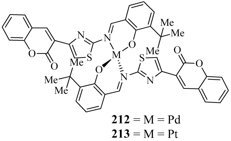
For the treatment of Alzheimer’s disease (AD), cholinesterase inhibition is the only valid target used. Hence, there is a continuous need for the development and synthesis of new cholinesterase inhibitor molecules. Recently, Özdemir and co-workers reported palladium and platinum complexes derived from combining coumarin and thiazole with 3-tertiary butyl salicylaldehyde, a new Schiff base [67]. Metal complexes 212 and 213 (Figure 8) were structurally characterized from various spectroscopic techniques. Additionally, the authors investigated the inhibitor potencies of metal complexes on three esterase enzymes, acetylcholinesterase (AChE), butyrylcholinesterase (BChE), and pancreatic cholesterol esterase activities (CEase). The anti-esterase activity results directed that metal complexes have good inhibition effects on the cholinesterase (AChE/BChE) and pancreatic cholesterol (CEase) enzymes. Particularly platinum complex 213 exhibited a high inhibition effect with lower IC50 values (12.0 μM for AChE, 23 μM for BChE, and 21.0 μM for CEase) compared with the reference drugs [67].
Figure 8.
Structures of coumarin-derived imine–metal complexes (212, 213).
4. Conclusions and Future Perspective
Searching through drug banks for coumarin-derived clinically approved agents has yielded several important drugs, including a treatment for HIV that combines several different compounds and includes calanolide A, a coumarin agent. Currently, a number of clinical trials are being conducted on several coumarin-based drugs alone or in combination with other drugs to treat common disorders, such as thrombosis, coagulation disorders, stroke (Ischemic), liver fibrosis, protein C deficiency, atrial fibrillation, etc. From these findings it can be seen that coumarin-based drugs have an important role in global health and well-being and are continuously researched to discover more clinical uses.
Coumarin scaffold has great potential in medicinal chemistry and is extensively used in drug design and development because of its vast biological properties. This scaffold is frequently used for designing small molecules with various biological activities. Metal complexes of Schiff bases with coumarin pharmacophores have played an important role in medicinal chemistry. Various coumarin-derived imine–metal complexes have been developed in the last three decades and most of them exhibited important pharmacological properties.
Author Contributions
Conceptualization, S.A.P. (Siddappa A. Patil), A.B. and S.A.P. (Shivaputra A. Patil); methodology, S.A.P. (Siddappa A. Patil), V.K., A.S., S.B.S. and M.R.F.; validation, A.B. and S.A.P. (Shivaputra A. Patil); data curation, S.A.P. (Siddappa A. Patil), V.K., A.S., S.B.S. and M.R.F.; writing—original draft preparation, S.A.P. (Siddappa A. Patil), A.B., S.A.P. (Shivaputra A. Patil), V.K., A.S., S.B.S. and M.R.F.; writing—review and editing, S.A.P. (Siddappa A. Patil), A.B. and S.A.P. (Shivaputra A. Patil), V.K., A.S., S.B.S. and M.R.F.; funding acquisition, S.A.P. (Siddappa A. Patil) and A.B. All authors have read and agreed to the published version of the manuscript.
Institutional Review Board Statement
Not applicable.
Informed Consent Statement
Not applicable.
Data Availability Statement
Not applicable.
Conflicts of Interest
The authors declare that they have no known competing financial interests or personal relationships that could have appeared to influence the work reported in this paper.
Funding Statement
The authors thank Jain University, India for financial support. The authors also acknowledge the ACSPRF-ND grant (58269-ND1) for financial support.
Footnotes
Publisher’s Note: MDPI stays neutral with regard to jurisdictional claims in published maps and institutional affiliations.
References
- 1.Stefanachi A., Leonetti F., Pisani L., Catto M., Carotti A. Coumarin: A natural, privileged and versatile scaffold for bioactive compounds. Molecules. 2018;23:250. doi: 10.3390/molecules23020250. [DOI] [PMC free article] [PubMed] [Google Scholar]
- 2.Vogel A. Darstellung von benzoesaure aus der tonka-boline und aus den meliIoten-oder steinklee-blumen. Ann. Phys. 1820;64:161–166. doi: 10.1002/andp.18200640205. [DOI] [Google Scholar]
- 3.Perkin W.H. On the artificial production of coumarin and formation of its homologues. J. Chem. Soc. 1868;21:53–63. doi: 10.1039/JS8682100053. [DOI] [Google Scholar]
- 4.Kontogiorgis C.A., Hadjipavlou-Litina D.J. Synthesis and antiinflammatory activity of coumarin derivatives. J. Med. Chem. 2005;48:6400–6408. doi: 10.1021/jm0580149. [DOI] [PubMed] [Google Scholar]
- 5.Silvan A.M., Abad M.J., Bermejo P., Sollhuber M., Villar A. Antiinflammatory activity of coumarins from Santolina oblonglifolia. J. Nat. Prod. 1996;59:1183–1185. doi: 10.1021/np960422f. [DOI] [PubMed] [Google Scholar]
- 6.Kontogiorgis C.A., Hadjipavlou-Litina D.J. Synthesis and biological evaluation of novel coumarin derivatives with a 7-azomethine linkage. Bioorg. Med. Chem. Lett. 2004;14:611–614. doi: 10.1016/j.bmcl.2003.11.060. [DOI] [PubMed] [Google Scholar]
- 7.Melagraki G., Afantitis A., Igglessi-Markopoulou O., Detsi A., Koufaki M., Kontogiorgis C., Hadjipavlou-Litina D.J. Synthesis and evaluation of the antioxidant and anti-inflammatory activity of novel coumarin-3-aminoamides and their alpha-lipoic acid adducts. Eur. J. Med. Chem. 2009;44:3020–3026. doi: 10.1016/j.ejmech.2008.12.027. [DOI] [PubMed] [Google Scholar]
- 8.Yoakim C., Bonneau P.R., Deziel R., Doyon L., Duan J., Guse I., Landry S., Malenfant E., Naud J., Ogilvie W.W., et al. Novel nevirapine-like inhibitors with improved activity against NNRTI-resistant HIV: 8-heteroarylthiomethyldipyridodiazepinone derivatives. Bioorg. Med. Chem. Lett. 2004;14:739–742. doi: 10.1016/j.bmcl.2003.11.049. [DOI] [PubMed] [Google Scholar]
- 9.Yu D., Suzuki M., Xie L., Morris-Natschke S.L., Lee K.H. Recent progress in the development of coumarin derivatives as potent anti-HIV agents. Med. Res. Rev. 2003;23:322–345. doi: 10.1002/med.10034. [DOI] [PubMed] [Google Scholar]
- 10.Kamal A., Adil S.F., Tamboli J.R., Jaki R., Siddardha B., Murthy U.S.N. Synthesis of coumarin linked naphthalimide conjugates as potential anticancer and antimicrobial agents. Lett. Drug. Des. Discov. 2009;6:201–209. doi: 10.2174/157018009787847855. [DOI] [Google Scholar]
- 11.Barot K.P., Jain S.V., Kremer L., Singh S., Ghate M.D. Recent advances and therapeutic journey of coumarins: Current status and perspectives. Med. Chem. Res. 2015;24:2771–2798. doi: 10.1007/s00044-015-1350-8. [DOI] [Google Scholar]
- 12.Annunziata F., Pinna C., Dallavalle S., Tamborini L., Pinto A. An overview of coumarin as a versatile and readily accessible scaffold with broad-ranging biological activities. Int. J. Mol. Sci. 2020;21:4618. doi: 10.3390/ijms21134618. [DOI] [PMC free article] [PubMed] [Google Scholar]
- 13.Pereira T.M., Franco D.P., Vitorio F., Kummerle A.E. Coumarin Compounds in Medicinal Chemistry: Some Important Examples from the Last Years. Curr. Top. Med. Chem. 2018;18:124–148. doi: 10.2174/1568026618666180329115523. [DOI] [PubMed] [Google Scholar]
- 14.May J.M., Owens T.W., Mandler M.D. The Antibiotic Novobiocin binds and activates the ATPase that powers lipopolysaccharide transport. J. Am. Chem. Soc. 2017;139:17221–17224. doi: 10.1021/jacs.7b07736. [DOI] [PMC free article] [PubMed] [Google Scholar]
- 15.Li S.M., Heide L. New aminocoumarin antibiotics from genetically engineered Streptomyces strains. Curr. Med. Chem. 2005;12:419–427. doi: 10.2174/0929867053363063. [DOI] [PubMed] [Google Scholar]
- 16.Eustáquio A.S., Gust B., Luft T., Li S.M., Chater K.F., Heide L. Clorobiocin biosynthesis in Streptomyces: Identification of the halogenase and generation of structural analogs. Chem. Biol. 2003;10:279–288. doi: 10.1016/S1074-5521(03)00051-6. [DOI] [PubMed] [Google Scholar]
- 17.Ghanghas P., Choudhary A., Kumar D., Poonia K. Coordination metal complexes with Schiff bases: Useful pharmacophores with comprehensive biological applications. Inorg. Chem. Commun. 2021;130:108710. doi: 10.1016/j.inoche.2021.108710. [DOI] [Google Scholar]
- 18.Patil S.A., Patil S.A., Fariyike T., Marichev K.O., Martinez H.M.H., Bugarin A. Medicinal applications of coumarins bearing azetidinone and thiazolidinone moieties. Future Med. Chem. 2021;13:1907–1934. doi: 10.4155/fmc-2021-0193. [DOI] [PubMed] [Google Scholar]
- 19.Delcourt M.-L., Reynaud C., Turcaud S., Favereau L., Crassous J., Micouin L., Benedetti E. 3D coumarin systems based on [2.2]paracyclophane: Synthesis, spectroscopic characterization, and chiroptical properties. J. Org. Chem. 2019;84:888–899. doi: 10.1021/acs.joc.8b02773. [DOI] [PubMed] [Google Scholar]
- 20.Halter O., Plenio H. Fluorescent dyes in organometallic chemistry: Coumarin-tagged NHC–metal complexes. Eur. J. Inorg. Chem. 2018;25:2935–2943. doi: 10.1002/ejic.201800395. [DOI] [Google Scholar]
- 21.Bagihalli G.B., Avaji P.G., Patil S.A., Badami P.S. Synthesis, spectral characterization, in vitro antibacterial, antifungal and cytotoxic activities of Co(II), Ni(II) and Cu(II) complexes with 1,2,4-triazole Schiff bases. Eur. J. Med. Chem. 2018;43:2639–2649. doi: 10.1016/j.ejmech.2008.02.013. [DOI] [PubMed] [Google Scholar]
- 22.Tumer M., Koksal H., Sener M.K., Serin S. Antimicrobial activity studies of the binuclear metal complexes derived from tridentate Schiff base ligands. Transit. Metal. Chem. 1999;24:414–420. doi: 10.1023/A:1006973823926. [DOI] [Google Scholar]
- 23.Mladenovic M., Vukovic N., Sukdolak S., Solujic S. Design of novel 4-hydroxy-chromene-2-one derivatives as antimicrobial agents. Molecules. 2010;15:4294–4308. doi: 10.3390/molecules15064294. [DOI] [PMC free article] [PubMed] [Google Scholar]
- 24.Vukovic N., Sukdolak S., Solujic S., Niciforovic N. An efficient synthesis and antioxidant properties of novel imino and amino derivatives of 4-hydroxy coumarin. Arch. Pharmacal Res. 2010;33:5–15. doi: 10.1007/s12272-010-2220-z. [DOI] [PubMed] [Google Scholar]
- 25.Gilani A.H., Shaheen F., Saeed S.A., Bibi S., Sadiq M., Faizi S. Hypotensive action of coumarin glycosides from Daucus carota. Phytomedicine. 2000;7:423–426. doi: 10.1016/S0944-7113(00)80064-1. [DOI] [PubMed] [Google Scholar]
- 26.Sandhya B., Giles D., Mathew V., Basavarajaswamy G., Abraham R. Synthesis, pharmacological evaluation and docking studies of coumarin derivatives. Eur. J. Med. Chem. 2011;46:4696–4701. doi: 10.1016/j.ejmech.2011.07.013. [DOI] [PubMed] [Google Scholar]
- 27.Cavettos G., Nano G.M., Palmisano G., Tagliapietra S. An asymmetric approach to coumarin anticoagulants via hetero-Diels–Alder cycloaddition. Tetrahedron Asymmetry. 2001;12:707–709. [Google Scholar]
- 28.Stanchev S., Momekov G., Jensen F., Manolov I. Synthesis, computational study and cytotoxic activity of new 4-hydroxycoumarin derivatives. Eur. J. Med. Chem. 2008;43:694–706. doi: 10.1016/j.ejmech.2007.05.005. [DOI] [PubMed] [Google Scholar]
- 29.Zabradnik M. The Production and Application of Fluorescent Brightening Agents. John Wiley & Sons; New York, NY, USA: 1992. [Google Scholar]
- 30.Wagner B.D. The use of coumarins as environmentally-sensitive fluorescent probes of heterogeneous inclusion systems. Molecules. 2009;14:210–237. doi: 10.3390/molecules14010210. [DOI] [PMC free article] [PubMed] [Google Scholar]
- 31.Abdel-Kader N.S., Moustafa H., El-Ansary A.L., Sherifa O.E., Farghaly A.M. A coumarin Schiff base and its Ag (I) and Cu (II) complexes: Synthesis, characterization, DFT calculations and biological applications. New J. Chem. 2021;45:7714–7730. doi: 10.1039/D0NJ05688J. [DOI] [Google Scholar]
- 32.Knittl E.T., Abou-Hussein A.A., Linert W. Syntheses, characterization, and biological activity of novel mono-and binuclear transition metal complexes with a hydrazone Schiff base derived from a coumarin derivative and oxalyldihydrazine. Monatsh. Chem. 2018;149:431–443. doi: 10.1007/s00706-017-2075-9. [DOI] [PMC free article] [PubMed] [Google Scholar]
- 33.Prabhakara C.T., Patil S.A., Kulkarni A.D., Naik V.H., Manjunatha M., Kinnala S.M., Badami P.S. Synthesis, spectral, thermal, fluorescence, antimicrobial, anthelmintic and DNA cleavage studies of mononuclear metal chelates of bi-dentate 2H-chromene-2-one Schiff base. J. Photochem. Photobiol. B Biol. 2015;148:322–332. doi: 10.1016/j.jphotobiol.2015.03.033. [DOI] [PubMed] [Google Scholar]
- 34.Raj K.M., Mruthyunjayaswamy B. Synthesis, spectroscopic characterization, electrochemistry and biological evaluation of some metal (II) complexes with ONO donor ligand containing benzo [b] thiophene and coumarin moieties. J. Mol. Struct. 2014;1074:572–582. [Google Scholar]
- 35.Elhusseiny A.F., Aazam E.S., Al-Amri H.M. Synthesis of new microbial pesticide metal complexes derived from coumarin-imine ligand. Spectrochim. Acta A Mol. Biomol. Spectrosc. 2014;128:852–863. doi: 10.1016/j.saa.2014.03.003. [DOI] [PubMed] [Google Scholar]
- 36.Abou-Hussein A., Linert W. Synthesis, spectroscopic studies and inhibitory activity against bactria and fungi of acyclic and macrocyclic transition metal complexes containing a triamine coumarine Schiff base ligand. Spectrochim. Acta A Mol. Biomol. Spectrosc. 2015;141:223–232. doi: 10.1016/j.saa.2015.01.063. [DOI] [PubMed] [Google Scholar]
- 37.Patil S.A., Prabhakara C.T., Halasangi B.M., Toragalmath S.S., Badami P.S. DNA cleavage, antibacterial, antifungal and anthelmintic studies of Co (II), Ni (II) and Cu (II) complexes of coumarin Schiff bases: Synthesis and spectral approach. Spectrochim. Acta A Mol. Biomol. Spectrosc. 2015;137:641–651. doi: 10.1016/j.saa.2014.08.028. [DOI] [PubMed] [Google Scholar]
- 38.Halli M., Sumathi R., Kinni M. Synthesis, spectroscopic characterization and biological evaluation studies of Schiff’s base derived from naphthofuran-2-carbohydrazide with 8-formyl-7-hydroxy-4-methyl coumarin and its metal complexes. Spectrochim. Acta A Mol. Biomol. Spectrosc. 2012;99:46–56. doi: 10.1016/j.saa.2012.08.089. [DOI] [PubMed] [Google Scholar]
- 39.Kalwania G., Bajroliya S. Synthesis, characterization and antimicrobial activities of 1, 2, 4-triazole-coumarin schiff bases and their Mn (II), Co (II) complexes. Asian, J. Chem. 2015;27:3956. doi: 10.14233/ajchem.2015.19010. [DOI] [Google Scholar]
- 40.Al-Bayati R., Mahdi F., Al-Amiery A. Synthesis, spectroscopic and antimicrobial studies of transition metal complexes of N-amino quinolone derivatives. Br. J. Pharmacol. Toxicol. 2011;2:5–11. [Google Scholar]
- 41.Nath P., Dhumwad S.D. Synthesis, characterization and antimicrobial studies of Co (II), Ni (II), Cu (II) and Zn (II) complexes derived from a Schiff base of 2-[(4-Methyl-2-oxo- 2H-chromen-7-yl) oxy] acetohydrazide with 3-formyl-2-hydroxy quinoline and 3-formyl-2-mercapto quinolone. J. Chem. Pharm. Res. 2012;4:851–865. [Google Scholar]
- 42.Patil S.A., Unki S.N., Badami P.S. In vitro antibacterial, antifungal, and DNA cleavage studies of coumarin Schiff bases and their metal complexes: Synthesis and spectral characterization. Med. Chem. Res. 2012;21:4017–4027. doi: 10.1007/s00044-011-9932-6. [DOI] [Google Scholar]
- 43.Patil S.A., Naik V.H., Kulkarni A.D., Unki S.N., Badami P.S. Synthesis, characterization, DNA cleavage, and in-vitro antimicrobial studies of Co(II), Ni(II), and Cu(II) complexes with Schiff bases of coumarin derivatives. J. Coord. Chem. 2011;64:2688–2697. doi: 10.1080/00958972.2011.594507. [DOI] [PubMed] [Google Scholar]
- 44.Akkasali R., Patil N., Angadi S.D. Synthesis, characterization and microbial activities of metal complexes with coumarine derivatives. Rasayan J. Chem. 2009;2:81–86. [Google Scholar]
- 45.Phaniband M.A., Dhumwad S.D., Pattan S.R. Synthesis, characterization, antimicrobial, and DNA cleavage studies of metal complexes of coumarin Schiff bases. Med. Chem. Res. 2011;20:493–502. doi: 10.1007/s00044-010-9343-0. [DOI] [Google Scholar]
- 46.Kulkarni A.D., Bagihalli G.B., Patil S.A., Badami P.S. Synthesis, characterization, electrochemical and in-vitro antimicrobial studies of Co(II), Ni(II), and Cu(II) complexes with Schiff bases of formyl coumarin derivatives. J. Coord. Chem. 2009;62:3060–3072. doi: 10.1080/00958970902914569. [DOI] [Google Scholar]
- 47.Bagihalli G.B., Badami P.S., Patil S.A. Synthesis, spectral characterization and in vitro biological studies of Co(II), Ni(II) and Cu(II) complexes with 1,2,4-triazole Schiff bases. J. Enzyme Inhib. Med. Chem. 2009;24:381–394. doi: 10.1080/14756360802187901. [DOI] [PubMed] [Google Scholar]
- 48.Refat M.S., El-Deen I.M., Anwer Z.M., El-Ghol S. Bivalent transition metal complexes of coumarin-3-yl thiosemicarbazone derivatives: Spectroscopic, antibacterial activity and thermogravimetric studies. J. Mol. Struct. 2009;920:149–162. doi: 10.1016/j.molstruc.2008.10.059. [DOI] [Google Scholar]
- 49.Kapoor P., Singh R.V., Fahmi N. Coordination chemistry of rare earth metal complexes with coumarin-based imines: Ecofriendly synthesis, characterization, antimicrobial, DNA cleavage, pesticidal, and nematicidal activity evaluations. J. Coord. Chem. 2012;65:262–277. doi: 10.1080/00958972.2011.649265. [DOI] [Google Scholar]
- 50.Aldabaldetrecu M., Parra M., Soto S., Arce P., Tello M., Guerrero J., Modak B. New Copper(I) Complex with a Coumarin as Ligand with Antibacterial Activity against Flavobacterium psychrophilum. Molecules. 2020;25:3183. doi: 10.3390/molecules25143183. [DOI] [PMC free article] [PubMed] [Google Scholar]
- 51.Jani G.R., Vyas K.B., Franco Z. Preparation and antimicrobial activity of s-triazine hydrazones of 7-hydroxy coumarin and their metal complexes. E-J. Chem. 2009;6:1228–1232. doi: 10.1155/2009/649654. [DOI] [Google Scholar]
- 52.Prabhakara C.T., Patil S.A., Toragalmath S.S., Kinnal S.M., Badami P.S. Synthesis, characterization and biological approach of metal chelates of some first row transition metal ions with halogenated bidentate coumarin Schiff bases containing N and O donor atoms. J. Photochem. Photobiol. B Biol. 2016;157:1–14. doi: 10.1016/j.jphotobiol.2016.02.004. [DOI] [PubMed] [Google Scholar]
- 53.Sharma V., Arora E.K., Cardoza S. Synthesis, antioxidant, antibacterial, and DFT study on a coumarin based salen-type Schiff base and its copper complex. Chem. Pap. 2016;70:1493–1502. doi: 10.1515/chempap-2016-0083. [DOI] [Google Scholar]
- 54.Elsayed S.A., El-Gharabawy H.M., Butler I.S., Atlam F.M. Novel metal complexes of 3-acetylcoumarin-2-hydrazinobenzothiazole Schiff base: Design, structural characterizations, DNA binding, DFT calculations, molecular docking and biological studies. Appl. Organomet. Chem. 2020;34:e5643. doi: 10.1002/aoc.5643. [DOI] [Google Scholar]
- 55.Pawar V., Chavan S.V., Yamgar R.S., Atram R.G., Thorat B.R., Bisht S., Sawant S.S. Synthesis and characterization of novel transition metal complexes of 4-methyl-7-hydroxy 8-formyl coumarin and their biological activities. Asian J. Res. Chem. 2011;4:1238–1242. [Google Scholar]
- 56.Creaven B.S., Devereux M., Karcz D., Kellett A., McCann M., Noble A., Walsh M. Copper(II) complexes of coumarin-derived Schiff bases and their anti-Candida activity. J. Inorg. Biochem. 2009;103:1196–1203. doi: 10.1016/j.jinorgbio.2009.05.017. [DOI] [PubMed] [Google Scholar]
- 57.Sivakumar K., Chandrasekaran V. Synthesis, characterization, DNA cleavage and biological activities of Schiff base derived from p-phenylenediamine, benzil and 3-aminocoumarin and their transition metal compound. J. Crit. Rev. 2020;7:2769–2778. [Google Scholar]
- 58.Patil S.A., Hoagland P., Patil S.A., Bugarin A. N-heterocyclic carbene-metal complexes as bio-organometallic antimicrobial and anticancer drugs, an update (2015–2020) Future Med. Chem. 2020;12:2239–2275. doi: 10.4155/fmc-2020-0175. [DOI] [PubMed] [Google Scholar]
- 59.Aazam E., Husseiny A.E.L., Hitchcock P., Alshehri J. Copper coumarin complex: Synthesis and crystal structure of [{Cu(4-methyl-7-(salicylideneamino)coumarin)2}2] Cent. Eur. J. Chem. 2008;6:319–323. doi: 10.2478/s11532-008-0018-3. [DOI] [Google Scholar]
- 60.Creaven B.S., Czeglédi E., Devereux M., Enyedy É.A., Foltyn-Arfa Kia A., Karcz D., Kellett A., McClean S., Nagy N.V., Noble A., et al. Biological activity and coordination modes of copper(II) complexes of Schiff base-derived coumarin ligands. Dalton Trans. 2010;39:10854. doi: 10.1039/c0dt00068j. [DOI] [PubMed] [Google Scholar]
- 61.Usman M., Zaki M., Khan R.A., Alsalme A., Ahmad M., Tabassum S. Coumarin centered copper(II) complex with appended-imidazole as cancer chemotherapeutic agents against lung cancer: Molecular insight via DFT-based vibrational analysis. RSC Adv. 2017;7:36056–36071. doi: 10.1039/C7RA05874H. [DOI] [Google Scholar]
- 62.Şahin Ö., Özdemir Ü.Ö., Seferoğlu N., Genc Z.K., Kaya K., Aydıner B., Tekin S., Seferoğlu Z. New Platinum (II) and palladium (II) complexes of coumarin-thiazole Schiff base with a fluorescent chemosensor properties: Synthesis, spectroscopic characterization, X-ray structure determination, in vitro anticancer activity on various human carcinoma cell lines and computational studies. J. Photochem. Photobiol. B Biol. 2018;178:428–439. doi: 10.1016/j.jphotobiol.2017.11.030. [DOI] [PubMed] [Google Scholar]
- 63.Mestizo P.D., Narváez D.M., Pinzón-Ulloa J.A., Bello D.T.D., Franco-Ulloa S., Macías M.A., Groot H., Miscione G.P., Suescun L., Hurtado J.J. Novel complexes with ONNO tetradentate coumarin Schiff-base donor ligands: X-ray structures, DFT calculations, molecular dynamics and potential anticarcinogenic activity. Biometals. 2021;34:119–140. doi: 10.1007/s10534-020-00268-8. [DOI] [PubMed] [Google Scholar]
- 64.Liu C., Liu X., Ge X. Fluorescent iridium (III) coumarin-salicylaldehyde Schiff base compounds as lysosome-targeted antitumor agents. Dalton Trans. 2020;49:5988–5998. doi: 10.1039/D0DT00627K. [DOI] [PubMed] [Google Scholar]
- 65.Datta P., Mukhopadhyay A.P., Manna P., Tiekink E.R.T., Chandra Sil P., Sinha C. Structure, photophysics, electrochemistry, DFT calculation, and in-vitro antioxidant activity of coumarin Schiff base complexes of group 6 metal carbonyls. J. Inorg. Biochem. 2011;105:577–588. doi: 10.1016/j.jinorgbio.2010.04.013. [DOI] [PubMed] [Google Scholar]
- 66.MacLean L., Karcz D., Jenkins H., McClean S., Devereux M., Howe O., Pereira M.D., May N.V., Enyedy É.A., Creaven B.S. Copper(II) complexes of coumarin-derived Schiff base ligands: Pro- or antioxidant activity in MCF-7 cells? J. Inorg. Biochem. 2019;197:110702. doi: 10.1016/j.jinorgbio.2019.110702. [DOI] [PubMed] [Google Scholar]
- 67.Şahin Ö., Özdemir Ü.Ö., Seferoğlu N., Adem S., Seferoğlu Z. Synthesis, characterization, molecular docking and in vitro screening of new metal complexes with coumarin Schiff base as anticholine esterase and antipancreatic cholesterol esterase agents. J. Biomol. Struct. Dyn. 2022;40:4460–4474. doi: 10.1080/07391102.2020.1858163. [DOI] [PubMed] [Google Scholar]
- 68.Pradhan D.A., Koshal A.K., Chauhan N. Coumarin based Schiff base: Synthesis and their antioxidant and antimicrobial activity. IOSR J. Pharm. 2019;9:1–5. [Google Scholar]
Associated Data
This section collects any data citations, data availability statements, or supplementary materials included in this article.
Data Availability Statement
Not applicable.


