Abstract
Objectives
Fetal growth restriction (FGR) may alter brain development permanently, resulting in lifelong structural and functional changes. However, in studies addressing this research question, FGR singletons have been compared primarily to matched appropriately grown singletons, a design which is inherently biased by differences in genetic and maternal factors. To overcome these limitations, we conducted a within‐pair comparison of neonatal structural cerebral ultrasound measurements in monochorionic twin pairs with selective FGR (sFGR).
Methods
Structural cerebral measurements on neonatal cerebral ultrasound were compared between the smaller and larger twins of monochorionic twin pairs with sFGR, defined as a birth‐weight discordance (BWD) ≥ 20%, born in our center between 2010 and 2020. Measurements from each twin pair were also compared with those of an appropriately grown singleton, matched according to sex and gestational age at birth.
Results
Included were 58 twin pairs with sFGR, with a median gestational age at birth of 31.7 (interquartile range, 29.9–33.8) weeks and a median birth weight of 1155 g for the smaller twin and 1725 g for the larger twin (median BWD, 32%). Compared with both the larger twin and the singleton, the smaller twin had significantly smaller cerebral structures (corpus callosum, vermis, cerebellum), less white/deep gray matter and smaller intracranial surface area and volume. Intracranial‐volume discordance and BWD correlated significantly (R 2 = 0.228, P < 0.0001). The median intracranial‐volume discordance was smaller than the median BWD (19% vs 32%, P < 0.0001). After correction for intracranial volume, only one of the observed differences (biparietal diameter) remained significant for the smaller twin vs both the larger twin and the singleton.
Conclusions
In monochorionic twins with sFGR, neonatal cerebral ultrasound reveals an overall, proportional restriction in brain growth, with smaller cerebral structures, less white/deep gray matter and smaller overall brain‐size parameters in the smaller twin. There was a positive linear relationship between BWD and intracranial‐volume discordance, with intracranial‐volume discordance being smaller than BWD. © 2021 The Authors. Ultrasound in Obstetrics & Gynecology published by John Wiley & Sons Ltd on behalf of International Society of Ultrasound in Obstetrics and Gynecology.
Keywords: brain development, monochorionic twins, neonatal cerebral ultrasound, selective FGR
CONTRIBUTION —
What are the novel findings of this work?
This is an extensive overview of structural cerebral measurements on neonatal cerebral ultrasound in monochorionic twin pairs with selective fetal growth restriction and a matched, appropriately grown singleton. It shows that the smaller twin presents with an overall restriction in brain growth, with smaller cerebral structures (corpus callosum, vermis, cerebellum), less white/deep gray matter and smaller overall brain‐size parameters.
What are the clinical implications of this work?
Our results reinforce the hypothesis that FGR has significant implications for brain development.
Introduction
Approximately 10% of all pregnancies are affected by fetal growth restriction (FGR), characterized by the failure of the fetus to reach its growth potential 1 . FGR in singletons is multifactorial in origin, by way of maternal, fetal or placental determinants, and is responsible for a large proportion of both perinatal morbidity and mortality 2 . It is hypothesized that FGR can alter permanently fetal development, including brain development, resulting in lifelong structural and functional changes.
The hemodynamic adaptation of the brain to suboptimal growth conditions can be detected antenatally as ‘brain sparing’, a redistribution of blood flow to the brain indicated by a decreased cerebroplacental ratio (CPR) 3 . Despite this supposedly protective mechanism, deficits in brain structures are prevalent in FGR singletons, and include reduced intracranial volume, corpus callosal size and cerebellar diameter 4 , 5 . These structural deficits are known to have significant consequences for brain function in childhood, being associated with, for example, lower cognitive test scores and impaired motor skills 6 .
So far, in studies on the impact of FGR on brain structure and function, FGR singletons have been compared primarily to matched appropriate‐for‐gestational‐age singletons 4 , 7 . However, this study design is inherently biased by differences in genetic and maternal factors, which potentially influence outcomes and thereby limit comparability. These limitations are not present when research is performed in an identical‐twin model with discordance in fetal growth 8 .
Monochorionic (MC) twins have a single placenta that can be shared unequally, resulting in an unbalanced nutrient and oxygen supply and a subsequent discordant growth pattern, known as selective FGR (sFGR) 9 . These twins allow comparison of a growth‐restricted twin with its genetically identical appropriately grown cotwin, with identical maternal characteristics. To date, no study has evaluated cerebral ultrasound (cUS) parameters in this specific twin population. The aim of this study was to conduct a within‐pair comparison of neonatal structural cUS measurements in MC twin pairs with sFGR.
Methods
This study was approved and the requirement for written informed consent was waived by the ethics committee of the Leiden University Medical Center (LUMC), as it was a retrospective analysis of clinically indicated ultrasound examinations (protocol G21.011). All consecutive MC twin pairs with sFGR, defined as a birth‐weight discordance (BWD) ≥ 20%, born in our center (the national referral center for complicated MC twin pregnancies) between 2010 and 2020 were eligible for inclusion. BWD was calculated as 10 : (birth weight of larger twin − birth weight of smaller twin)/birth weight of larger twin × 100. Cases with twin–twin transfusion syndrome and those with twin anemia–polycythemia sequence were excluded due to the likely additional effect of these complications on brain development 11 , 12 . We also excluded MC triplet pregnancies and cases with twin reversed arterial perfusion and/or other congenital abnormalities 12 . Structural measurements could not be performed when no cUS was available for either one or both of the neonates. Each twin pair was matched to one appropriate‐for‐gestational‐age singleton without cerebral injury, to account for differences between twins and singletons. The singletons were selected from our neonatology patient database and were born in the same time period as the included twins. Per twin pair, a singleton was selected with the same sex and gestational age at birth. In order to minimize factors that might influence cerebral outcome for this group, singletons with asphyxia, congenital abnormality or infection, and singletons born after alloimmunization (with or without fetal therapy) during pregnancy were not included.
Clinical characteristics
The following maternal and obstetric baseline characteristics were retrieved from patient files: maternal age, gravidity, parity, Gratacós classification for sFGR 13 (Type I defined as positive end‐diastolic flow, Type II defined as persistent absent or reversed end‐diastolic flow and Type III defined as intermittent absent or reversed end‐diastolic flow), presence of brain sparing (defined as CPR < 1 for at least 2 weeks, with CPR calculated as the pulsatility index of the middle cerebral artery divided by the pulsatility index of the umbilical artery), and, in this case, gestational age at start and duration of brain sparing 14 , monoamnionicity and mode of delivery. The following neonatal baseline characteristics were retrieved: gestational age at birth, sex, BWD, birth weight (in g) and whether neonates were born small‐for‐gestational age (defined as birth weight < 10th centile) 15 . Placental share was calculated and expressed as a percentage of the total placental area, based on the margins of the twin‐specific dyes after standard colored dye injection of MC twin placentae 16 . The percentages were calculated using Image J version 1.57 17 .
cUS measurements
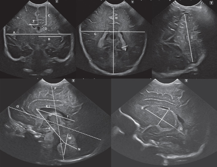
Before 2015, cUS was performed using an Aloka α ultrasound system (Hitachi Medical Systems Holding AG, Zug, Switzerland). From 2015 onwards, a Canon Aplio 400 or Aplio i700 system (Canon Medical Systems B.V., The Netherlands) was used. Between 1 and 3 days after birth, cUS was performed by the attending neonatologist, all of whom had extensive experience with this imaging modality as it is part of standard care in LUMC. Head circumference at birth and corresponding z‐score were documented 18 . Cerebral measurements were performed offline using retrieved images from the first available cUS examination after birth (Clinical Assistant, RVC B.V., The Netherlands). The resistance index of the anterior cerebral artery was recorded and calculated as: (peak systolic velocity − end‐diastolic velocity)/peak systolic velocity. The following structural measurements were performed by a single researcher with expertise in neonatal neuroimaging (S.G.G.) to avoid interobserver variability 4 : anterior horn width, ventricular index (VI), ventriculoatrial width (VAW), thalamo‐occipital distance (TOD), interhemispheric fissure width, corpus callosal length, corpus callosal height, callosum–fastigium length, vermis height, vermis width, transverse cerebellar diameter, frontal white matter height, deep gray matter width, deep gray matter surface area, biparietal diameter, intracranial fronto‐occipital diameter (FOD), intracranial height, axial intracranial surface area and intracranial volume 19 (Table S1 and Figure 1). Intracranial‐volume discordance was calculated as: (intracranial volume of larger twin − intracranial volume of smaller twin)/intracranial volume of larger twin × 100. The researcher was not blinded to the group (smaller twin, larger twin or singleton).
Figure 1.

Ultrasound images giving overview of neonatal cerebral measurements: A and G, biparietal diameter; B, width of deep gray matter; C, height of frontal white matter; D, anterior horn width; E, ventricular index; F, ventriculoatrial width; G and H, used in calculation of intracranial surface area; I, transverse cerebellar diameter; J, corpus callosal length; K, corpus callosal height; L, callosum–fastigium length; M, vermis height; N, vermis width; O, intracranial fronto‐occipital diameter; P, intracranial height; Q, thalamo‐occipital distance; R and S, used in calculation of deep gray matter surface area.
Measurements were compared between the smaller and larger twin, the smaller twin and the matched singleton, and the larger twin and the singleton. In order to examine whether certain structures were affected to a greater extent than others, the analyses were also corrected for intracranial volume 19 . Both uncorrected and corrected measurements are presented herein, because having a smaller brain in itself might have consequences for future neurodevelopment. To evaluate reliability, measurements were repeated by the same researcher in a random sample of 18 neonates (10% of the population), and the intraclass correlation coefficient (ICC) was calculated for every measurement. ICC values < 0.50 were indicative of poor reliability and values between 0.50–0.75 indicated moderate reliability 20 .
Brain lesions seen on cUS
We recorded the presence of brain lesions, including pseudocysts, germinolytic cysts, subependymal or choroid plexus cysts, lenticulostriate vasculopathy, intraventricular hemorrhage (IVH) Grade 1–4 21 , periventricular leukomalacia (PVL) Grade 1–4 22 , ventricular dilatation > 97th percentile 23 and parenchymal hemorrhage. Severe cerebral injury was defined as IVH ≥ Grade 3, cystic PVL (c‐PVL) ≥ Grade 2; ventricular dilatation > 97th percentile, arterial or venous infarction or porencephalic or parenchymal cysts.
Brain maturation
Brain maturation in the twin pairs was assessed by two other researchers (L.S.d.V. and S.J.S.) with expertise in neonatal neuroimaging. These researchers did not perform any structural measurements and were blinded to the group (smaller or larger twin) and gestational age at birth. Maturation was scored in three planes according to the appearance and increasing complexity of the principal sulci, as described by Murphy et al. 24 . Overall maturity was determined on the first cUS examination after birth and based on the comparison of actual gestational age at birth with the maturation score for at least two out of three planes, and was categorized as either normal, 2–4 weeks behind or > 4 weeks behind.
Statistical analysis
Statistical analyses were performed using IBM Statistics Version 25.0 (SPSS, IBM Corp, Armonk, NY, USA). Data are presented as median (interquartile range (IQR)), n/N (%) or n (%). Given the nature of the study population (twin pairs), the analyses took into account that observations between cotwins are not independent, by using the Wilcoxon signed‐rank test (non‐parametric test for related samples) and generalized estimating equations (GEE). To test for association between sFGR and the structural cerebral measurements, the Wilcoxon signed‐rank test was used. A GEE was used to test for association between sFGR and the structural cerebral measurements, corrected for intracranial volume. A GEE was also used to test for association between sFGR and the presence of brain lesions. As the GEE cannot be used when an outcome event does not occur in one of the groups, under these circumstances an adjustment to the data was applied, in which one unaffected twin was considered as an affected twin for both groups (smaller/larger twin) for the purpose of the statistical analysis; this results in more conservative P‐values.
Intracranial‐volume discordance was tested for correlation with BWD and placental‐share discordance and plotted against BWD and placental‐share discordance for each type of sFGR, using sRStudio Version 2021.9.2.382 (RStudio, PBC, Boston, MA, USA). The ICC of each structural measurement was calculated in a two‐way mixed‐effects model based on a single measurement.
P < 0.05 was considered statistically significant. For every structural measurement, three comparisons were performed, i.e. smaller twin vs larger twin, smaller twin vs singleton and larger twin vs singleton. Therefore, a Bonferroni adjustment was applied to correct for multiple testing, resulting in a significance level set at P < 0.017 (i.e. 0.05/3) for the structural measurements.
Results
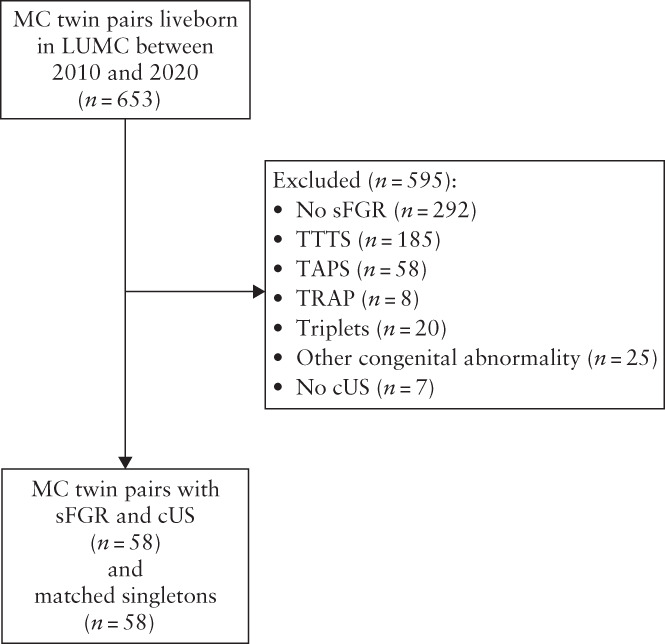
Of the 653 liveborn MC twin pairs delivered at the LUMC between 2010 and 2020, pairs which did not have sFGR (n = 292) or which met the aforementioned exclusion criteria (n = 296) were excluded. Of the remaining pairs, seven did not have a cUS available for either one or both of the twins. Thus, 58 twin pairs with sFGR and an available cUS were included in the analyses (Figure 2). Hence, 58 appropriate‐for‐gestational‐age singletons without cerebral injury and matched for sex and gestational age at birth were included as well.
Figure 2.

Flowchart summarizing inclusion of neonates in the study. cUS, cerebral ultrasound; LUMC, Leiden University Medical Center; MC, monochorionic; sFGR, selective fetal growth restriction; TAPS, twin anemia–polycythemia sequence; TRAP, twin reversed arterial perfusion; TTTS, twin–twin transfusion syndrome.
Clinical characteristics
Baseline maternal, obstetric and neonatal characteristics are presented in Table 1. As expected, antenatal brain sparing was observed primarily in the smaller twin (76.8% (43/56)), for a median of 7 (IQR, 4–9) weeks, as a sign of hemodynamic adaptation of the brain to suboptimal growth conditions. Brain sparing was observed in only 1.8% (1/56) of larger twins, with a duration of 4 weeks. Of the 58 twin pregnancies, 39.7% (23/58) were classified as Gratacós Type I, 17.2% (10/58) as Type II and 43.1% (25/58) as Type III. The median gestational age at birth was 31.7 (IQR, 29.9–33.8) weeks and nearly 80% of twins were delivered by Cesarean section. The median BWD was 31.5% (IQR, 26.7–38.1%), with the smaller twin weighing 1155 (IQR, 886–1433) g and the larger twin weighing 1725 (IQR, 1386–2145) g. In line with the difference in birth weight, the proportion of neonates born small‐for‐gestational age was 94.8% (55/58) for the smaller twin and 13.8% (8/58) for the larger twin. Conforming to the pathophysiology of sFGR, the smaller twin had a smaller placental share compared with that of the larger twin: 30.0% (IQR, 25.3–34.7%) vs 70.0% (IQR, 65.3–74.7%). For the matched singletons, the median gestational age at birth was 31.7 (IQR, 29.9–33.8) weeks and the birth weight was 1758 (IQR, 1528–2164) g respectively.
Table 1.
Baseline maternal, obstetric and neonatal characteristics for twin pregnancies with selective fetal growth restriction (sFGR) and matched singletons*
| Characteristic | sFGR twins (n = 116; 58 pregnancies) | Matched singleton (n = 58) | ||
|---|---|---|---|---|
| All | Smaller twin (n = 58) | Larger twin (n = 58) | ||
| Maternal age (years) | 31 (28–34) | — | — | — |
| Gravidity | 1 (1–2) | — | — | — |
| Parity | 0 (0–1) | — | — | — |
| Gratacós type | ||||
| Type I | 23/58 (39.7) | — | — | — |
| Type II | 10/58 (17.2) | — | — | — |
| Type III | 25/58 (43.1) | — | — | — |
| Brain sparing | — | 43/56 (76.8) | 1/56 (1.8) | — |
| GA at start (weeks) | — | 19.6 (17.4–21.4) | 15.9 | — |
| Duration (weeks) | — | 7 (4–9) | 4 | — |
| Monoamniotic | 6/58 (10.3) | — | — | — |
| GA at birth (weeks) | 31.7 (29.9–33.8) | — | — | 31.7 (29.9–33.8) |
| Female neonate | 52/116 (44.8) | — | — | 26/58 (44.8) |
| Cesarean delivery | 92/116 (79.3) | — | — | — |
| BWD (%) | 31.5 (26.7–38.1) | — | — | — |
| Birth weight (g) | — | 1155 (886–1433) | 1725 (1386–2145) | 1758 (1528–2164) |
| Small‐for‐gestational age | — | 55/58 (94.8) | 8/58 (13.8) | 0 (0.0) |
| Placental share (%) | — | 30.0 (25.3–34.7) | 70.0 (65.3–74.7) | — |
Data are presented as median (interquartile range), n/N (%) or absolute values.
Matched for gestational age and sex.
BWD, birth‐weight discordance; GA, gestational age.
cUS measurements
Structural cUS measurements are summarized in Table 2. As expected, based on the difference in birth weight, head circumference at birth and corresponding z‐score were lower for the smaller twin compared with both the larger twin and the singleton: the median (IQR) was 27.1 (25.0–29.3) cm with z‐score of –1.3 (–1.9 to –0.1) for the smaller twin, 29.0 (27.5–30.0) cm with z‐score of 0.5 (–0.5 to 1.2) for the larger twin and 29.0 (27.5–30.0) cm with z‐score of 0.1 (–0.5 to 0.9) for the singleton (P < 0.0001 for both).
Table 2.
Neonatal cerebral ultrasound (cUS) parameters in twins with selective fetal growth restriction (sFGR) and matched singletons*
| Parameter | Smaller twin (n = 58) | Larger twin (n = 58) | P (small vs large) | Matched singleton (n = 58) | P (small vs singleton) | P (large vs singleton) |
|---|---|---|---|---|---|---|
| GA at cUS (weeks) | 31.9 (29.9–34.0) | 31.9 (29.9–34.0) | 31.7 (30.0–34.0) | 0.615 | 0.608 | |
| Postnatal age at cUS (days) | 2 (1–2) | 2 (1–2) | 2 (1–3) | 0.063 | 0.060 | |
| Head circumference (cm) | 27.1 (25.0–29.3) | 29.0 (27.5–30.0) | < 0.0001 | 29.0 (27.5–30.0) | < 0.0001 | 0.435 |
| Head circumference z‐score | –1.3 (–1.9 to –0.1) | 0.5 (–0.5 to 1.2) | < 0.0001 | 0.1 (–0.5 to 0.9) | < 0.0001 | 0.481 |
| RI‐ACA | 0.7 (0.6–0.8) | 0.8 (0.7–0.8) | 0.062 | 0.7 (0.6–0.8) | 0.441 | 0.177 |
| Ventricular | ||||||
| AHW (mm) | ||||||
| Right | 0.7 (0.3–1.4) | 0.6 (0.3–1.0) | 0.136 | 0.6 (0.0–1.1) | 0.048 | 0.820 |
| Left | 0.8 (0.3–1.4) | 0.6 (0.3–1.4) | 0.797 | 0.6 (0.0–1.3) | 0.382 | 0.593 |
| VI (mm) | ||||||
| Right | 9.6 (8.6–10.9) | 9.5 (8.8–10.7) | 0.991 | 9.8 (8.8–10.8) | 0.462 | 0.341 |
| Left | 9.5 (8.8–10.5) | 9.6 (8.8–10.4) | 0.486 | 9.9 (9.2–10.5) | 0.241 | 0.188 |
| VAW (mm) | ||||||
| Right | 6.1 (5.2–7.4) | 6.4 (5.5–7.6) | 0.809 | 6.4 (5.6–7.5) | 0.554 | 0.874 |
| Left | 6.2 (5.2–7.4) | 6.8 (6.1–7.8) | 0.036 | 7.0 (5.9–8.0) | 0.105 | 0.863 |
| TOD (mm) | ||||||
| Right | 15.6 (13.5–18.4) | 16.0 (12.7–18.0) | 0.385 | 12.8 (10.7–15.9) | < 0.0001† | 0.007† |
| Left | 16.0 (14.1–18.1) | 16.2 (13.7–18.9) | 0.750 | 14.6 (11.8–18.7) | 0.078 | 0.109 |
| IFW (mm) | 0 (0–0) | 0 (0–0) | 0.347 | 0 (0–0) | 0.386 | 0.875 |
| Brain structures | ||||||
| Corpus callosum (mm) | ||||||
| Length | 37.6 (35.6–41.1) | 39.8 (37.7–43.1) | < 0.0001 | 40.8 (38.4–42.0) | 0.001 | 0.461 |
| Height | 2.1 (1.8–2.4) | 2.3 (2.0–2.6) | 0.003 | 1.8 (1.5–2.0) | < 0.0001† | < 0.0001† |
| Callosum–fastigium length (mm) | 42.0 (39.8–45.0) | 43.2 (41.6–46.0) | < 0.0001 | 43.4 (42.1–45.1) | 0.014 | 0.585 |
| Vermis (mm) | ||||||
| Height | 18.3 (16.6–20.3) | 19.2 (18.1–21.1) | < 0.0001 | 18.7 (17.2–19.8) | 0.364 | 0.003† |
| Width | 11.7 (10.3–13.1) | 12.0 (10.2–14.2) | < 0.0001 | 11.1 (10.2–12.4) | 0.215† | 0.132 |
| TCD (cm) | 3.5 (3.1–4.0) | 3.8 (3.5–4.3) | < 0.0001 | 3.8 (3.5–4.1) | < 0.0001 | 0.851 |
| White/deep gray matter | ||||||
| Frontal white matter height (mm) | ||||||
| Right | 18.5 (16.9–20.2) | 19.4 (18.1–20.6) | 0.002† | 19.9 (18.3–21.0) | 0.006 | 0.429 |
| Left | 18.8 (16.8–20.1) | 19.4 (17.7–21.0) | < 0.0001 | 19.8 (18.4–20.7) | 0.001 | 0.530 |
| Deep gray matter width (mm) | ||||||
| Right | 22.4 (20.8–24.9) | 24.0 (22.4–27.2) | < 0.0001 | 24.0 (23.0–26.2) | < 0.0001 | 0.993 |
| Left | 22.8 (21.1–24.7) | 24.3 (22.3–26.7) | < 0.0001 | 24.4 (22.5–25.8) | < 0.0001 | 0.969 |
| Deep gray matter surface area (mm2) | ||||||
| Right | 379 (330–460) | 436 (393–499) | < 0.0001 | 417 (372–466) | 0.003 | 0.001 |
| Left | 378 (331–452) | 447 (403–486) | < 0.0001 | 418 (385–448) | 0.106 | < 0.0001† |
| Overall brain size | ||||||
| Biparietal diameter (cm) | 6.6 (6.1–7.0) | 7.0 (6.5–7.7) | < 0.0001† | 7.2 (6.8–7.5) | < 0.0001† | 0.706 |
| Intracranial | ||||||
| Surface area (cm2) | 34.9 (30.9–43.3) | 41.1 (37.1–47.5) | < 0.0001 | 40.5 (36.3–44.1) | < 0.0001 | 0.088 |
| FOD (cm) | 8.3 (7.5–9.0) | 8.7 (8.3–9.2) | < 0.0001 | 8.7 (8.3–9.1) | < 0.0001† | 0.718 |
| Height (cm) | 6.7 (6.3–7.3) | 7.1 (6.8–7.7) | < 0.0001 | 7.3 (6.9–7.6) | < 0.0001 | 0.619 |
| Axial surface area (cm2) | 42.6 (37.2–49.1) | 49.3 (43.4–55.3) | < 0.0001 | 49.3 (45.5–52.6) | < 0.0001 | 0.794 |
| Volume (cm3) | 191 (155–240) | 231 (199–283) | < 0.0001 | 245 (210–266) | < 0.0001 | 0.730 |
Data are presented as median (interquartile range).
Matched for gestational age and sex.
Significant after correction for intracranial‐volume discordance (Table S3).
AHW, anterior horn width; FOD, fronto‐occipital diameter; GA, gestational age; IFW, interhemispheric fissure width; RI‐ACA, resistance index in anterior cerebral artery; TCD, transverse cerebellar diameter; TOD, thalamo‐occipital distance; VAW, ventriculoatrial width; VI, ventricular index.
The structural measurements could be divided into four categories: ventricular, brain structures, white/deep gray matter and overall brain‐size parameters. Ventricular parameters did not differ between groups, except for the right TOD which was smaller for the singleton (12.8 (IQR, 10.7–15.9) mm) in comparison with both the smaller (15.6 (IQR, 13.5–18.4) mm, P < 0.0001) and the larger (16.0 (IQR, 12.7–18.0) mm, P = 0.007) twin. This difference was independent of intracranial volume (Table S2).
All other structural measurements (brain structures, white/deep gray matter and overall brain‐size parameters) were significantly smaller for the smaller twin compared with the larger twin, in line with the difference in head circumference. Thus, there was overall restriction in brain growth of the smaller twin. When corrected for intracranial volume, only the right frontal white matter height (P = 0.003) and biparietal diameter (P < 0.0001) remained significantly different.
The measurements of brain structures, white/deep gray matter and overall brain‐size parameters that differed between the smaller and larger twin also differed between the smaller twin and the singleton (indicating comparability of the larger twin and the singleton), except for the vermis height (P = 0.364) and width (P = 0.215) and left deep gray matter surface area (P = 0.106). When corrected for intracranial volume, the differences that persisted were in the corpus callosal height (P < 0.0001), biparietal diameter (P < 0.0001) and FOD (P = 0.014). Vermis width became significantly different after correction (P = 0.007).
Measurements that were significantly different between the larger twin and the singleton (thereby indicative of possible incomparability of these groups) were in two brain structures (corpus callosal height (P < 0.0001) and vermis height (P = 0.003)) and both right and left deep gray matter surface area (P = 0.001 and P < 0.0001, respectively). Three of these differences between the larger twin and singleton persisted after correction for intracranial volume: corpus callosal height (P < 0.0001), vermis height (P = 0.005) and left deep gray matter surface area (P < 0.0001).
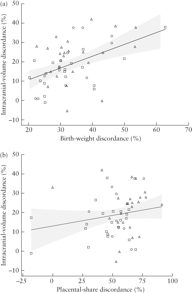
Intracranial‐volume discordance and BWD correlated significantly, with a Pearson's correlation coefficient of 0.477 (R 2 = 0.228, P < 0.0001) (Figure 3a). The median intracranial‐volume discordance was smaller than the median BWD (19.3% vs 31.5%, P < 0.0001). Intracranial‐volume discordance and placental‐share discordance did not correlate significantly (Pearson's correlation coefficient of 0.198 (R 2 = 0.039, P = 0.144)) (Figure 3b). There was no significant difference in intracranial‐volume discordance between the three sFGR types (P = 0.080).
Figure 3.

Scatterplots, with regression lines and 95% CIs, in 58 twin pregnancies with selective fetal growth restriction (sFGR), depicting the association of intracranial‐volume discordance on neonatal cerebral ultrasound (cUS) with: (a) birth‐weight discordance (R
2 linear = 0.228); and (b) placental‐share discordance (R
2 linear = 0.039). Regression lines are based on the group as a whole. Intracranial‐volume discordance was missing in two cases due to insufficient cUS quality. Cases were categorized according to Gratacós type as: Type I ( ), Type II (
 ) or Type III (
 ).
).
The majority of the structural measurements had an ICC indicative of good‐to‐excellent reliability, the exception being the left VI, right VAW and vermis width, which had moderate reliability (Table S3).
Brain lesions seen on cUS
The brain lesions observed on neonatal cUS are presented in Table 3. If PVL was present, the smaller twin presented more often with PVL Grade 1 (transient periventricular densities > 7 days after birth) than did the larger twin (100% (10/10) vs 86% (12/14), P < 0.0001). Severe cerebral injury was present in 7% (4/58) of larger twins and 0% (0/58) of smaller twins (P = 0.065). These four twins presented as follows. The first had c‐PVL Grade 3, 15 days after birth (gestational age at birth, 28.9 weeks; birth weight, 1262 g; severe respiratory morbidity and patent ductus arteriosus; died 15 days after birth, following redirection of care because of severity of cerebral injury). The second had a periventricular hemorrhagic infarction with ventricular dilatation 3 days after birth (gestational age at birth, 28.4 weeks; birth weight, 1210 g; severe respiratory and cardiovascular morbidity; died 6 days after birth, following redirection of care because of severity of cerebral injury). The third had a periventricular hemorrhagic infarction 3 days after birth (gestational age at birth, 30.4 weeks; birth weight, 1740 g; severe respiratory morbidity). The fourth had c‐PVL Grade 3 and IVH Grade 2, 7 days after birth (gestational age at birth, 29.6 weeks; birth weight, 1450 g; severe respiratory morbidity). The first case was from a pregnancy classified antenatally as sFGR Type II and the other three cases were from pregnancies classified as Type III.
Table 3.
Neonatal brain lesions seen on cerebral ultrasound in twins with selective fetal growth restriction
| Lesion | Smaller twin (n = 58) | Larger twin (n = 58) | P |
|---|---|---|---|
| Pseudocyst | 9/58 (16) | 3/58 (5) | 0.065 |
| Lenticulostriate vasculopathy | 1/58 (2) | 2/58 (3) | 0.571 |
| Intraventricular hemorrhage | 6/58 (10) | 6/58 (10) | 1.000 |
| Grade 1 | 5/6 (83) | 2/6 (33) | 0.519 |
| Grade 2 | 1/6 (17) | 2/6 (33) | 0.683 |
| Grade 3 | 0/6 (0) | 0/6 (0) | 1.000 |
| Grade 4 (venous infarction) | 0/6 (0) | 2/6 (33) | 0.190 |
| Periventricular leukomalacia | 10/53 (19) | 14/53 (26) | 0.333 |
| Grade 1 | 10/10 (100) | 12/14 (86) | < 0.0001 |
| Grade 2 | 0/10 (0) | 0/14 (0) | 1.000 |
| Grade 3 | 0/10 (0) | 2/14 (14) | 0.482 |
| Grade 4 | 0/10 (0) | 0/14 (0) | 1.000 |
| Ventricular dilatation > 97th centile | 0/58 (0) | 1/58 (2) | 0.323 |
| Parenchymal hemorrhage | 0/58 (0) | 1/58 (2) | 0.323 |
| Severe cerebral injury | 0/58 (0) | 4/58 (7) | 0.065 |
Data are presented as n/N (%).
Brain maturation
Overall brain maturation was categorized as 2–4 weeks behind in 9% (5/55) of smaller twins as opposed to 16% (9/57) of larger twins (P = 0.281) and it was > 4 weeks behind in 2% (1/55) of smaller twins as opposed to 2% (1/58) of larger twins (P = 0.979). Maturation could not be scored in three cases due to insufficient quality of the cUS. In two twin pairs (both born at a gestational age of 31 weeks), the maturation of both the smaller and the larger twin was behind.
Discussion
This is the first study evaluating neonatal cerebral measurements in a unique identical‐twin population affected by sFGR, controlling for genetic and maternal factors. Our study shows that the smaller twin presents with an overall restriction in brain growth, with smaller cerebral structures (corpus callosum, vermis, cerebellum), less white/deep gray matter and smaller overall brain‐size parameters. The measurements were different primarily for the smaller twin in comparison with both the larger twin and the matched singleton, demonstrating that the larger twin has similar outcomes to those of the matched singleton. After correction for intracranial volume, only one of the observed differences (biparietal diameter) remained significant for the smaller twin vs both the larger twin and the singleton, indicating a global, proportional decrease in brain growth. Furthermore, there was a positive linear relationship between BWD and intracranial‐volume discordance. The intracranial volume discordance appeared to be smaller than the BWD, indicating a certain degree of brain preservation.
The observed structural deficits have been linked previously to functional consequences in singletons. Small head circumference can be considered a strong predictor of adverse neurodevelopmental outcome, with increased rates of cerebral palsy and impaired cognitive and motor development 6 , 25 . Smaller corpus callosal size in preterm infants has been associated with speech and language difficulties, motor delay, cerebral palsy and a lower full‐scale intelligence quotient in adolescence 26 , 27 , 28 . Moreover, a study in monozygotic twins has shown that even subtle differences in birth weight within the normal range in full‐term twins is associated with alterations in brain structure which persist into adolescence and can be correlated with neurodevelopmental outcome 29 . It is important to realize that any neurodevelopmental consequences of (s)FGR are influenced by the time of onset and severity, as well as the gestational age at birth. Preterm birth is generally considered to exacerbate any deficits.
Our results are similar to those of a pilot study on cUS measurements in singletons, reporting that FGR neonates with antenatal brain sparing have a smaller corpus callosum and cerebellum 4 . In our study, no additional effect of brain sparing was found. However, only 77% of the smaller twins presented with antenatal brain sparing. Brain sparing can be considered a marker of the severity of FGR, as there is redistribution of blood to vital organs in response to unfavorable intrauterine circumstances, and has been linked to adverse neurobehavioral outcome 14 . The term ‘brain sparing’ is therefore a misnomer, as it appears to be an indication of a type of FGR with an even greater impairment of brain growth 4 . This is also illustrated by the fact that the sole parameter that remained significantly different between the smaller twin and both the larger twin and the singleton after correction for intracranial volume was the biparietal diameter, indicative of smaller head growth and associated with adverse neurodevelopmental outcome 30 .
With regard to cerebral injury, a systematic literature review reported an incidence of approximately 8% in MC twins with sFGR, with a particularly increased risk for the larger twin 31 . We found a similar incidence of severe cerebral injury in larger twins (7% (4/58)). Each of these four larger twins was born between 28 and 30 weeks of gestation, were from pregnancies with sFGR Type II or III, presented with severe respiratory morbidity, did not have cerebral abnormalities in utero and developed cerebral injury 3–15 days after birth. It has been suggested that the larger twin is already at increased risk of cerebral injury in utero, due to fetofetal shifts in blood volume through large anastomoses, resulting in hypoxic injury 13 , 32 , 33 . Hence, one would expect the injury to be visible antenatally or within 1 or 2 days after birth. As this was not the case, our data are more in line with the hypothesis that (iatrogenic) prematurity plays an additional role in the development of cerebral injury in the larger twin 31 . As yet, there is no consensus on the timing of delivery for sFGR twins. The benefits of prolonging the pregnancy, with the risk of fetal demise of the smaller twin and subsequent demise or neurological damage of the larger twin, must be weighed against the risks of prematurity 34 .
Our study is limited by its retrospective design. As cUS was performed for the detection of cerebral injury, the quality was not always optimal to conduct all measurements or to score maturation. Moreover, it would have been desirable to include a control group of uncomplicated MC twin pregnancies, as these can also present with brain injury and may show cerebral growth alterations without any known antenatal complications. Lastly, we were unable to find differences in cerebral maturation on cUS, possibly because we used a scoring system that uses a number of rough markers of maturation. A more detailed, validated scoring system for cUS scans is currently unavailable. Nevertheless, our results are strengthened by the unique population of identical twins discordant in birth weight, inherently controlling for sex, gestational age at birth and genetic and maternal factors. By including matched singletons, we were able to investigate changes that are specific to MC twins and may also be present in the larger twin.
More research is necessary to investigate the effects of the observed structural differences on brain function. A systematic review published by our group concluded that the smaller twin of MC pairs with sFGR is at increased risk of neurodevelopmental impairment 35 . However, this was based on just five articles with varying degrees of validity. Long‐term follow‐up with neurodevelopmental testing is needed to provide more conclusive evidence. Ideally, magnetic resonance imaging at term should be performed to achieve a better understanding of alterations in brain growth, maturation and connectivity. Longitudinal neuroimaging beyond the neonatal period should be used to assess whether these alterations are permanent or whether there is catch‐up growth over time.
To conclude, sFGR in MC twins is associated with an overall restriction in brain growth on neonatal cUS. The severity of BWD and intracranial‐volume discordance are positively correlated, suggesting that the BWD translates into a discordance in brain size as well (this size discordance being smaller than BWD). Our results reinforce the hypothesis that FGR has significant implications for brain development.
Supporting information
Table S1 Definitions of the structural measurements on neonatal cerebral ultrasound
Table S2 Neonatal cerebral ultrasound parameters in twins with selective fetal growth restriction and matched singletons, corrected for intracranial volume
Table S3 Intraclass correlation coefficients for the neonatal cerebral ultrasound parameters
DATA AVAILABILITY STATEMENT
The data that support the findings of this study are available on request from the corresponding author. The data are not publicly available due to privacy or ethical restrictions.
References
- 1. Colella M, Frerot A, Novais ARB, Baud O. Neonatal and Long‐Term Consequences of Fetal Growth Restriction. Curr Pediatr Rev 2018; 14: 212–218. [DOI] [PMC free article] [PubMed] [Google Scholar]
- 2. Cetin I, Alvino G. Intrauterine growth restriction: implications for placental metabolism and transport. A review. Placenta 2009; 30 Suppl A: S77–82. [DOI] [PubMed] [Google Scholar]
- 3. Figueras F, Gratacos E. Update on the diagnosis and classification of fetal growth restriction and proposal of a stage‐based management protocol. Fetal Diagn Ther 2014; 36: 86–98. [DOI] [PubMed] [Google Scholar]
- 4. Pharande P, Krishnamurthy M, Whiteley G, Sasi A, Malhotra A. Ultrasound Measurements of Intracranial Structures in Growth‐Restricted Neonates with Fetal Blood Flow Redistribution: A Pilot Observational Study. Neonatology 2020; 117: 446–452. [DOI] [PubMed] [Google Scholar]
- 5. Miller SL, Huppi PS, Mallard C. The consequences of fetal growth restriction on brain structure and neurodevelopmental outcome. J Physiol 2016; 594: 807–823. [DOI] [PMC free article] [PubMed] [Google Scholar]
- 6. Baschat AA. Neurodevelopment after fetal growth restriction. Fetal Diagn Ther 2014; 36: 136–142. [DOI] [PubMed] [Google Scholar]
- 7. Brembilla G, Righini A, Scelsa B, Lista G, Balestriero M, Cesari E, Castoldi FM, Di Stasi M, Ciardi C, Ligato E, Taricco E, Cetin I. Neuroimaging and neurodevelopmental outcome after early fetal growth restriction: NEUROPROJECT‐FGR. Pediatr Res 2021; 90: 869–875. [DOI] [PubMed] [Google Scholar]
- 8. Groene SG, Todtenhaupt P, van Zwet EW, van Pel M, Berkhout RJM, Haak MC, Roest AAW, Lopriore E, van Klink JMM, Heijmans BT. TwinLIFE: The Twin Longitudinal Investigation of FEtal Discordance. Twin Res Hum Genet 2019; 22: 617–622. [DOI] [PubMed] [Google Scholar]
- 9. Bennasar M, Eixarch E, Martinez JM, Gratacos E. Selective intrauterine growth restriction in monochorionic diamniotic twin pregnancies. Semin Fetal Neonatal Med 2017; 22: 376–382. [DOI] [PubMed] [Google Scholar]
- 10. Khalil A, Beune I, Hecher K, Wynia K, Ganzevoort W, Reed K, Lewi L, Oepkes D, Gratacos E, Thilaganathan B, Gordijn SJ. Consensus definition and essential reporting parameters of selective fetal growth restriction in twin pregnancy: a Delphi procedure. Ultrasound Obstet Gynecol 2019; 53: 47–54. [DOI] [PubMed] [Google Scholar]
- 11. Tollenaar LSA, Lopriore E, Middeldorp JM, Haak MC, Klumper FJ, Oepkes D, Slaghekke F. Improved prediction of twin anemia‐polycythemia sequence by delta middle cerebral artery peak systolic velocity: new antenatal classification system. Ultrasound Obstet Gynecol 2019; 53: 788–793. [DOI] [PMC free article] [PubMed] [Google Scholar]
- 12. Sueters M, Oepkes D. Diagnosis of twin‐to‐twin transfusion syndrome, selective fetal growth restriction, twin anaemia‐polycythaemia sequence, and twin reversed arterial perfusion sequence. Best Pract Res Clin Obstet Gynaecol 2014; 28: 215–226. [DOI] [PubMed] [Google Scholar]
- 13. Gratacos E, Lewi L, Munoz B, Acosta‐Rojas R, Hernandez‐Andrade E, Martinez JM, Carreras E, Deprest J. A classification system for selective intrauterine growth restriction in monochorionic pregnancies according to umbilical artery Doppler flow in the smaller twin. Ultrasound Obstet Gynecol 2007; 30: 28–34. [DOI] [PubMed] [Google Scholar]
- 14. Figueras F, Cruz‐Martinez R, Sanz‐Cortes M, Arranz A, Illa M, Botet F, Costas‐Moragas C, Gratacos E. Neurobehavioral outcomes in preterm, growth‐restricted infants with and without prenatal advanced signs of brain‐sparing. Ultrasound Obstet Gynecol 2011; 38: 288–294. [DOI] [PubMed] [Google Scholar]
- 15. Hoftiezer L, Hof MHP, Dijs‐Elsinga J, Hogeveen M, Hukkelhoven C, van Lingen RA. From population reference to national standard: new and improved birthweight charts. Am J Obstet Gynecol 2019; 220: 383.e1–17. [DOI] [PubMed] [Google Scholar]
- 16. Lopriore E, Slaghekke F, Middeldorp JM, Klumper FJ, van Lith JM, Walther FJ, Oepkes D. Accurate and simple evaluation of vascular anastomoses in monochorionic placenta using colored dye. J Vis Exp 2011; 55: e3208. [DOI] [PMC free article] [PubMed] [Google Scholar]
- 17.Image J. https://imagej.net/software/imagej/#publication.
- 18. Chou JH, Roumiantsev S, Singh R. PediTools Electronic Growth Chart Calculators: Applications in Clinical Care, Research, and Quality Improvement. J Med Internet Res 2020; 22: e16204. [DOI] [PMC free article] [PubMed] [Google Scholar]
- 19. Graca AM, Cardoso KR, da Costa JM, Cowan FM. Cerebral volume at term age: comparison between preterm and term‐born infants using cranial ultrasound. Early Hum Dev 2013; 89: 643–648. [DOI] [PubMed] [Google Scholar]
- 20. Koo TK, Li MY. A Guideline of Selecting and Reporting Intraclass Correlation Coefficients for Reliability Research. J Chiropr Med 2016; 15: 155–163. [DOI] [PMC free article] [PubMed] [Google Scholar]
- 21. Volpe JJ. Intraventricular hemorrhage and brain injury in the premature infant. Diagnosis, prognosis, and prevention. Clin Perinatol 1989; 16: 387–411. [PubMed] [Google Scholar]
- 22. de Vries LS, Eken P, Dubowitz LM. The spectrum of leukomalacia using cranial ultrasound. Behav Brain Res 1992; 49: 1–6. [DOI] [PubMed] [Google Scholar]
- 23. Levene MI. Measurement of the growth of the lateral ventricles in preterm infants with real‐time ultrasound. Arch Dis Child 1981; 56: 900–904. [DOI] [PMC free article] [PubMed] [Google Scholar]
- 24. Murphy NP, Rennie J, Cooke RW. Cranial ultrasound assessment of gestational age in low birthweight infants. Arch Dis Child 1989; 64: 569–572. [DOI] [PMC free article] [PubMed] [Google Scholar]
- 25. Gale CR, O'Callaghan FJ, Bredow M, Martyn CN, Avon Longitudinal Study of Parents and Children Study Team. The influence of head growth in fetal life, infancy, and childhood on intelligence at the ages of 4 and 8 years. Pediatrics 2006; 118: 1486–1492. [DOI] [PubMed] [Google Scholar]
- 26. Nosarti C, Rushe TM, Woodruff PW, Stewart AL, Rifkin L, Murray RM. Corpus callosum size and very preterm birth: relationship to neuropsychological outcome. Brain 2004; 127: 2080–2089. [DOI] [PubMed] [Google Scholar]
- 27. Anderson NG, Laurent I, Woodward LJ, Inder TE. Detection of impaired growth of the corpus callosum in premature infants. Pediatrics 2006; 118: 951–960. [DOI] [PubMed] [Google Scholar]
- 28. Klebermass‐Schrehof K, Aumuller S, Goeral K, Vergesslich‐Rothschild K, Fuiko R, Brandstetter S, Berger A, Jilma B, Haiden N. Biometry of the corpus callosum assessed by 3D ultrasound and its correlation to neurodevelopmental outcome in very low birth weight infants. J Perinatol 2017; 37: 448–453. [DOI] [PubMed] [Google Scholar]
- 29. Raznahan A, Greenstein D, Lee NR, Clasen LS, Giedd JN. Prenatal growth in humans and postnatal brain maturation into late adolescence. Proc Natl Acad Sci U S A 2012; 109: 11366–11371. [DOI] [PMC free article] [PubMed] [Google Scholar]
- 30. Hasegawa Y, Aoki S, Kurasawa K, Takahashi T, Hirahara F. Association of biparietal diameter growth rate with neurodevelopment in infants with fetal growth restriction. Taiwan J Obstet Gynecol 2015; 54: 371–375. [DOI] [PubMed] [Google Scholar]
- 31. Inklaar MJ, van Klink JM, Stolk TT, van Zwet EW, Oepkes D, Lopriore E. Cerebral injury in monochorionic twins with selective intrauterine growth restriction: a systematic review. Prenat Diagn 2014; 34: 205–213. [DOI] [PubMed] [Google Scholar]
- 32. Valsky DV, Eixarch E, Martinez JM, Crispi F, Gratacos E. Selective intrauterine growth restriction in monochorionic twins: pathophysiology, diagnostic approach and management dilemmas. Semin Fetal Neonatal Med 2010; 15: 342–348. [DOI] [PubMed] [Google Scholar]
- 33. Groene SG, Tollenaar LSA, Slaghekke F, Middeldorp JM, Haak M, Oepkes D, Lopriore E. Placental characteristics in monochorionic twins with selective intrauterine growth restriction in relation to the umbilical artery Doppler classification. Placenta 2018; 71: 1–5. [DOI] [PubMed] [Google Scholar]
- 34. Hillman SC, Morris RK, Kilby MD. Co‐twin prognosis after single fetal death: a systematic review and meta‐analysis. Obstet Gynecol 2011; 118: 928–940. [DOI] [PubMed] [Google Scholar]
- 35. Groene SG, Tollenaar LSA, Oepkes D, Lopriore E, van Klink JMM. The Impact of Selective Fetal Growth Restriction or Birth Weight Discordance on Long‐Term Neurodevelopment in Monochorionic Twins: A Systematic Literature Review. J Clin Med 2019; 8: 944. [DOI] [PMC free article] [PubMed] [Google Scholar]
Associated Data
This section collects any data citations, data availability statements, or supplementary materials included in this article.
Supplementary Materials
Table S1 Definitions of the structural measurements on neonatal cerebral ultrasound
Table S2 Neonatal cerebral ultrasound parameters in twins with selective fetal growth restriction and matched singletons, corrected for intracranial volume
Table S3 Intraclass correlation coefficients for the neonatal cerebral ultrasound parameters
Data Availability Statement
The data that support the findings of this study are available on request from the corresponding author. The data are not publicly available due to privacy or ethical restrictions.


