Abstract
Purpose
We have previously studied clinical and allergological aspects of sick building syndrome (SBS) cases with ocular disorders and found that SBS is suggested to be partially induced by an allergic response. We analyzed the cytokine production profiles of conjunctival and peripheral blood lymphocytes in patients with SBS with ocular manifestations to further evaluate the pathophysiology of SBS from an immunological standpoint.
Methods
We obtained conjunctival samples and peripheral blood mononuclear cells (PBMC) from 15 cases of SBS with ocular findings, 49 cases of allergic conjunctival diseases (ACD) (allergic conjunctivitis (AC), atopic keratoconjunctivitis (AKC), and vernal keratoconjunctivitis (VKC)), and normal controls. Frequencies of cytokine-producing T cells were analyzed by flow cytometry based on an intracellular cytokine staining method.
Results
Although no significant difference was observed in the percentage of interferon (IFN)-γ-producing CD4+ T cells in PBMC between patients with SBS and controls, the percentage of interleukin (IL)-4-producing PBMC CD4+ T cells in patients with SBS was significantly higher than that in controls. The percentage of IL-4-producing CD4+ T cells in the conjunctiva in patients with SBS was significantly higher than that in controls, whereas it was significantly lower than that in AKC and VKC. A significant correlation was observed between the percentage of IL-4-producing CD4+ T cells in the conjunctiva and clinical score.
Conclusion
These results suggest that SBS may be a kind of allergic disorder and that IL-4 plays a role in the development of allergic disorders in SBS ocular lesions.
Keywords: sick building syndrome, allergic conjunctival disease, interferon-γ, interleukin-4, lymphocyte
Introduction
Symptoms associated with poor quality of the indoor environment include various non-specific symptoms affecting the eyes, nose, throat, and skin and general symptoms like headache and tiredness, sometimes denoted as sick building syndrome (SBS).1 Irritation and dryness of the eyes together with a blocked or runny nose and dry throat are especially considered an important part of the oculonasal mucosal symptoms of SBS.2 Symptoms of SBS occur only in specific buildings or dwellings and disappear when the person is out of the environment. We have previously studied clinical and allergological aspects of SBS cases with ocular disorders with special reference to allergic conjunctival diseases (ACD); allergic conjunctivitis (AC), atopic keratoconjunctivitis (AKC) and vernal keratoconjunctivitis (VKC), from the standpoint of analyzing their similarities and differences, especially with respect to local immunological features, and reported that conjunctival lesions were observed in all cases and corneal abnormalities were found in two-thirds of cases of SBS.3 Mean serum total IgE level in SBS was significantly higher than that in AC; however, it was significantly lower than that in AKC and VKC.3 Eosinophil count in peripheral blood and the number of positive allergens in multiple allergen simultaneous test (MAST) were significantly lower in SBS than in AKC and VKC. Significant elevation of tear interleukin (IL)-4 was observed in SBS and ACD.3 We have previously evaluated tear cytokine levels in ACD and reported that tear IL-4 was significantly elevated only in AKC, while tear IL-5 level in patients with diseases was associated with proliferative disorders, such as VKC and AKC with proliferative change, meaning that type 2 helper T cell (Th2) cytokines play an important pathophysiological role in severe ocular allergic conditions, whereas, in contrast, Th1 cytokine (interferon (IFN)-γ and IL-2) levels did not significantly differ among different types of ACD.4 However, in contrast to ACD, elevation of other cytokines in tears was not observed in SBS.3 From these results, it is considered that SBS may be partially induced by an allergic response; however, the clinical and local immunological features of SBS suggest that SBS may belong to a different entity from ACD.3 There remains a need for studies evaluating the pathophysiology of the ocular disorders found in SBS cases based on inflammatory cells on the ocular surface to highlight the responses observed in human cases.
A significantly elevated level of tear IL-4 detected by enzyme-linked immunosorbent assay (ELISA) has been reported in patients with SBS with ocular complications in our previous study.3 IL-4 has been shown to induce the production of extracellular matrix components by fibroblasts,5 and the major source of IL-4 is thought to be Th2. However, it is still unclear whether such increased cytokine production is caused by increased numbers of cytokine-producing cells or enhanced ability of cytokine production. Analysis of cytokine production has been approached by measuring each cytokine by ELISA or mRNA levels. Flow cytometric analysis of cytokine-producing cells has been reported in several disorders including ocular allergic disease.6–8 Therefore, we applied this method to detect intracellular cytokines in individual cells and determined the percentage of IL-4- or IFN-γ-producing CD4+ T cells in the conjunctiva and peripheral blood from patients with SBS with ocular complications in comparison with ACD, to evaluate the relationship of these parameters with clinical severity in this study.
Materials and Methods
Study Population
This was a consecutive case series study of 15 patients (4 male and 11 female) who attended Yokohama City University Medical Center with SBS symptoms, especially ocular irritation in a specific building. They were asked to complete a self-reporting questionnaire survey as previously proposed by Lu et al,9 and written informed consent was obtained from all patients. SBS was diagnosed according to SBS symptoms, defined as one or more selected symptoms specified in the questionnaire for at least 1–3 days a week while at work in the office in the previous month, but which improved or disappeared after work or on days without work. The diagnosis of SBS was made according to the classification and diagnostic criteria proposed by Miyajima et al,10 in brief: 1) The cause of the onset of a disease relates to a move, a new house or building, the reconstruction of a house or building, and/or the use of a new or different daily toiletry. 2) Symptoms appear within a particular room and/or a particular house/building. 3) When the patient leaves the house/building, symptoms improve or disappear. 4) The detection of indoor environmental pollution is critical evidence. When patients met all of the criteria 1–3, they were diagnosed as having SBS.10 The symptoms of SBS were identified individually as several groups including the eyes, as described previously.9 Those with a past history of medical treatment for systemic diseases, such as hypertension, cardiac disease, or hyperthyroidism, were excluded from the study population.
For comparative evaluation with SBS, 49 patients with ACD who also attended Yokohama City University Medical Center were included in this study. ACD was classified into AC, AKC, or VKC according to the guidelines for the diagnosis and treatment of conjunctivitis and past reports.11,12 The ACD patients consisted of 14 with AC (6 male and 8 female), 14 with AKC (6 male and 8 female), and 21 with VKC (17 male and 4 female). Thirteen normal (non-allergic) subjects (5 male and 8 female) were subjected to immunological testing for comparison. While AC is in the mild spectrum without proliferative change, AKC, VKC and GPC belong to the severe spectrum of ACD with proliferative lesions such as giant papillae or corneal plaques.11,12 This study was performed in line with the principles of the Declaration of Helsinki. Approval was granted by the Review Committee of the Clinical Research Committee of Yokohama City University Medical Center. Informed consent was obtained from all individual participants included in the study.
Clinical Grading
Clinical evaluation of ocular findings was carried out according to the ocular clinical grading system reported previously.12 Ocular findings of slit-lamp examination were recorded on the patients’ first visit to our outpatient clinic. Ten objective ocular clinical findings of conjunctival, limbal, and corneal lesions were graded on a 4-point scale (0 = none, 1 = mild, 2 = moderate, and 3 = severe; left and right eyes separately in each case). The total score of 10 findings, with a maximum of 30, taking the score of the more severe side in bilateral cases, was used as the clinical score (Table 1).
Table 1.
Criteria for Clinical Evaluation of Allergic Ocular Findings
| Palpebral Conjunctiva | Hyperemia | Severe | Impossible to distinguish individual blood vessels |
| Moderate | Dilatation of many vessels | ||
| Mild | Dilatation of several vessels | ||
| None | No manifestations | ||
| Edema | Severe | Diffuse marked edema | |
| Moderate | Diffuse mild edema | ||
| Mild | Localized edema | ||
| None | No manifestations | ||
| Follicles | Severe | ≥20 follicles | |
| Moderate | 10–19 follicles | ||
| Mild | 1–9 follicles | ||
| None | No manifestations | ||
| Papillae | Severe | Diameter ≥0.6 mm | |
| Moderate | Diameter 0.3–0.5 mm | ||
| Mild | Diameter 0.1–0.2 mm | ||
| None | No manifestations | ||
| Giant papillae | Severe | Elevated papillae in 1/2 or more of upper palpebral conjunctiva | |
| Moderate | Elevated papillae in less than 1/2 of upper palpebral conjunctiva | ||
| Mild | Flat giant papillae | ||
| None | No manifestations | ||
| Bulbar conjunctiva | Hyperemia | Severe | Vasodilatation of all vessels |
| Moderate | Dilatation of many vessels | ||
| Mild | Dilatation of several vessels | ||
| None | No manifestations | ||
| Chemosis | Severe | Cyst-like chemosis of entire conjunctiva | |
| Moderate | Diffuse thin chemosis | ||
| Mild | Partial conjunctival swelling | ||
| None | No manifestations | ||
| Limbus | Limbal edema | Severe | In ≥2/3 of circumference |
| Moderate | In 1/3 to 2/3 of circumference | ||
| Mild | In less than 1/3 of circumference | ||
| None | No manifestations | ||
| Trantas’ dots | Severe | ≥9 dots | |
| Moderate | 5–8 dots | ||
| Mild | 1–4 dots | ||
| None | No manifestations | ||
| Cornea | Epithelial lesions | Severe | Shield ulcer or epithelial erosion |
| Moderate | Superficial punctate keratitis with filamentary debris | ||
| Mild | Superficial punctate keratitis | ||
| None | No manifestations |
Note: In cases having giant papillae, papillae and giant papillae should be graded simultaneously.
Monoclonal Antibodies
Phycoerythrin (PE)-conjugated anti-human IFN-γ monoclonal antibody (mAb) (mouse IgG1) and PE-conjugated anti-human IL-4 mAb (mouse lgG1) were purchased from Becton Dickinson (BD) Biosciences (San Diego, CA). PE-labeled isotype-matched immunoglobulin (Ig) (BD Biosciences) was used as the negative control. Fluorescein isothiocyanate (FITC)- or PE-CyTM5-conjugated anti-human CD3 mAb and PE-CyTM5-conjugated anti-human CD8 mAb were purchased from BD Biosciences. Fluorescein isothiocyanate (FITC)-labeled rabbit anti-mouse Ig was purchased from Dako (Glostrup, Denmark).
Cell Preparation and Culture
Conjunctival cell specimens from the upper palpebral conjunctiva were collected using a Cytobrush13 (Cytobrush Small, Medscand, Malmo, Sweden) under topical anesthesia with xylocaine eye drops. These specimens were kept in 2 mL of Gibco Roswell Park Memorial Institute (RPMI)-1640 medium (Thermo Fisher Scientific Inc., Göteborg, Sweden) on ice. These cells were then prepared for intracellular cytokine production assay according to the method described previously.7 Six milliliters of heparinized venous blood was also obtained. Peripheral blood mononuclear cells (PBMC) were isolated by density gradient centrifugation (Nycomed Diagnostics, Oslo, Norway) and washed three times with RPMI-1640 medium. PBMC (4 × 10⁶ cells/mL) were cultured in 24-well culture plates under the conditions described above.
Immunofluorescence Staining and Flow Cytometric Analysis
After stimulation with phorbol myristate acetate (PMA) and calcium ionophore, the cells were washed in phosphate-buffer saline (PBS) with 1% fetal calf serum (FCS) (staining buffer) and stained with FITC-conjugated anti-human CD3 mAb and PE-CyTM5-conjugated anti-human CD8 mAb for 30 minutes. The cells were washed twice with staining buffer and then fixed in cold PBS containing 2% paraformaldehyde for 15 minutes. After two washes in PBS, cells were washed in 0.1% saponin/PBS for permeabilization of the cell surface and resuspended in 100 μL of 0.1% saponin/PBS containing PE-conjugated anti-human IFN-γ Ab (1 μg) or PE-conjugated anti-human IL-4 mAb (1 μg) for 30 minutes on ice. The cells were washed once in 0.1% saponin/PBS and once in staining buffer. The cells were resuspended in 500 μL staining buffer. Cells were analyzed on a FACScanTM flow-cytometer (BD Biosciences) using CELL Quest software (BD Biosciences). Negative isotype controls were used to verify the staining specificity of the antibodies used. Analysis gates were set on lymphocytes according to forward- and side-scatter properties. We analyzed the frequency of cytokine-producing T cells in the conjunctiva in the gates set on lymphocytes according to forward- and side-scatter properties. In this study, we analyzed the frequencies of IFN-γ- or IL-4-producing T cells in this gate as reported in our previous study.6 CD3 and CD8 expression on T cells were investigated because CD4 expression disappeared after 24 h with PMA and calcium ionophore. We calculated the frequency of CD4+ T cells as (CD3+ T cells [%] - {CD3+CD8+ T cells [%]}).6
Statistical Analysis
Data are presented as mean ± standard error of the mean (SEM). One-way ANOVA with Tukey–Kramer test was conducted to identify differences among patient groups. Spearman’s rank correlation coefficient was calculated to evaluate the correlation between cytokine levels and clinical scores. GraphPad Prism9J (MDF Co. Ltd., Tokyo, Japan) was used for statistical analysis. A P-value of <0.05 was accepted as statistically significant.
Results
Percentage of IFN-γ- or IL-4-Producing CD4+ T Cells in PBMC
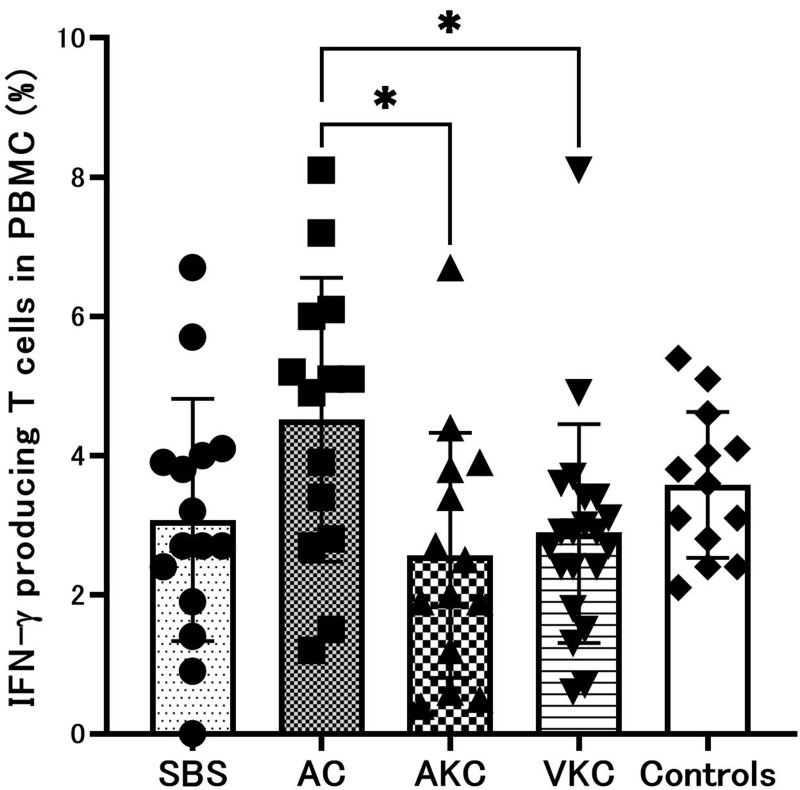
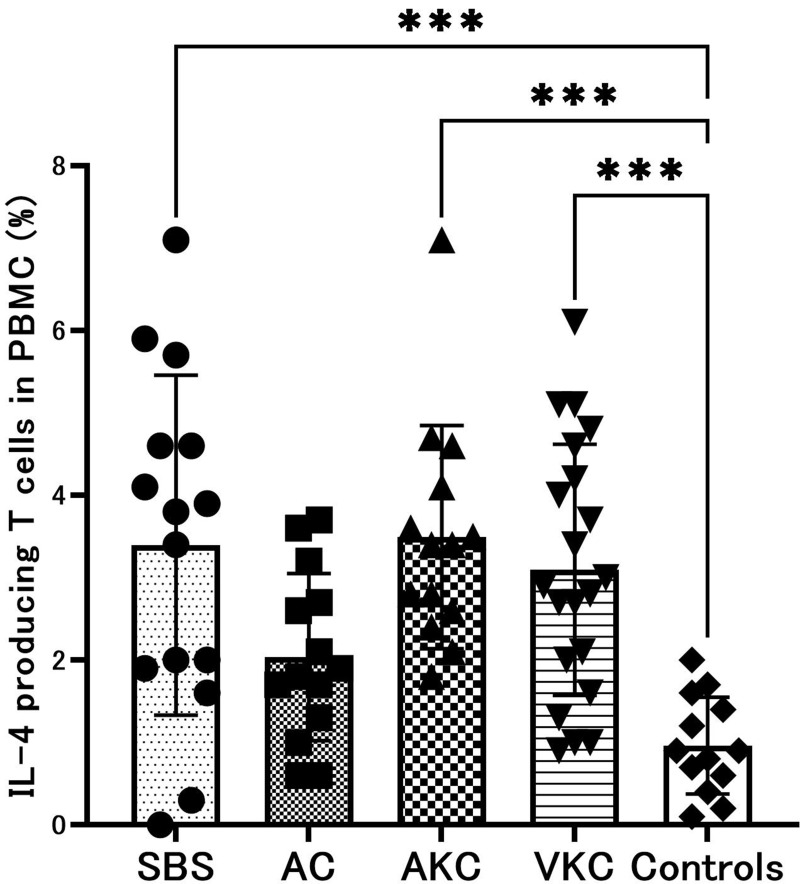
The percentage of peripheral blood CD4+ T cells in ACD and SBS did not show a significant difference to that in healthy controls (data not shown). We compared the percentages of IFN-γ- or IL-4-producing CD4+ T cells in PBMC between patients with SBS or ACD and healthy controls. The percentage of IFN-γ-producing CD4+ T cells in PBMC did not show a significant difference among patients with SBS and ACD and normal controls except for between AC and AKC (P < 0.05) and between AC and VKC (P < 0.05) (Figure 1). The percentage of IL-4-producing CD4+ T cells in PBMC from patients with SBS (P < 0.01), AKC (P < 0.01), or VKC (P < 0.01) was significantly higher than that in PBMC from normal controls (Figure 1), whereas no significant difference was observed in the percentage of IL-4-producing CD4+ T cells among patients with SBS and different types of ACD (Figure 2).
Figure 1.
Percentage of IFN-γ-producing CD4+ T cells in PBMC. The results are expressed in percent as mean ± SEM (*P < 0.05).
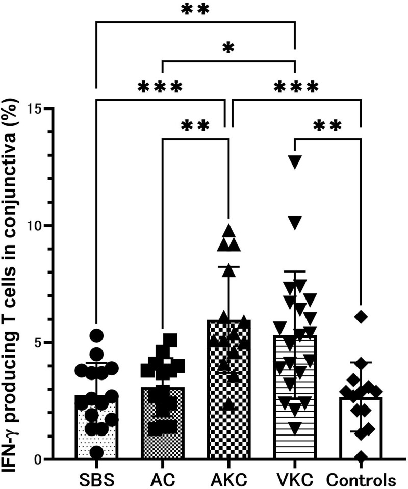
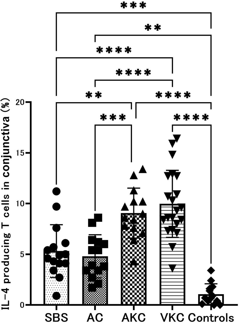
Abbreviations: PBMC, peripheral blood mononuclear cells; SBS, sick building syndrome; AC, allergic conjunctivitis; AKC, atopic keratoconjunctivitis; VKC, vernal keratoconjunctivitis.
Figure 2.
Percentage of IL-4-producing CD4+ T cells in PBMC. The results are expressed in percent as mean ± SEM (***P < 0.001).
Percentage of IFN-γ- or IL-4-Producing CD4+ T Cells in Conjunctiva
The percentage of T cells detected in gated conjunctival lymphocytes was comparable among patients with SBS, ACD and normal controls, and was approximately 70% (data not shown). The percentages of conjunctival IFN-γ-producing CD4+ T cells in patients with AKC (P < 0.001) and VKC (P < 0.01) were significantly higher than that in patients with SBS (Figure 3). The percentages of conjunctival IFN-γ-producing CD4+ T cells in patients with AKC (P < 0.01) and VKC (P < 0.05) were significantly higher than that in patients with AC (Figure 3). The percentages of conjunctival IFN-γ-producing CD4+ T cells in patients with AKC (P < 0.001) and VKC (P < 0.01) were significantly higher than that in normal controls (Figure 3). There was no significant difference in the percentage of INF-γ-producing CD4+ T cells between patients with VKC and AKC (Figure 3). The percentage of conjunctival IL-4-producing CD4+ T cells in patients with SBS was significantly higher than that in normal controls (P < 0.001), whereas it was significantly lower than those in patients with AKC (P < 0.01) and VKC (P < 0.0001) (Figure 4). The percentage of conjunctival IL-4-producing CD4+ T cells in patients with AC was significantly higher than that in normal controls (P < 0.01), whereas it was significantly lower than those in patients with AKC (P < 0.001) and VKC (P < 0.0001) (Figure 4). No significant difference was observed in the percentage of conjunctival IL-4-producing CD4+ T cells between SBS and AC, and between AKC and VKC (Figure 4).
Figure 3.
Percentage of IFN-γ-producing CD4+ T cells in conjunctiva. The results are expressed in percent as mean ± SEM (*P < 0.05; **P < 0.01; ***P < 0.001).
Figure 4.
Percentage of IL-4-producing CD4+ T cells in conjunctiva. The results are expressed in percent as mean ± SEM (**P < 0.01; ***P < 0.001; ****P<0.0001).
Correlation Between Percentage of IFN-γ- or IL-4-Producing CD4+ T Cells in PBMC or Conjunctiva and Clinical Score in Patients with SBS
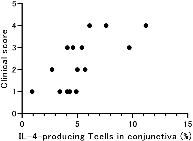
The correlation coefficients between the percentage of IFN-γ- or IL-4-producing T cells in PBMC or conjunctiva and clinical score in patients with SBS are shown in Table 2. A significant correlation was observed only between the percentage of IL-4-producing T cells in the conjunctiva and clinical score (P = 0.0025) (Figure 5). A relatively large coefficient was observed between PBMC IL-4-producing T cell percentage and clinical score (r = 0.48), but this did not reach statistical significance.
Table 2.
Correlation Between Percentage of IFN-γ- or IL-4-Producing CD4+ T Cells in PBMC or Conjunctiva and Clinical Score in Patients with SBS
| Cells | Correlation Coefficient |
|---|---|
| IFN-γ-producing CD4+ T cells in PBMC | −0.056 |
| IL-4-producing CD4+ T cells in PBMC | 0.48 (P= 0.072) |
| IFN-γ-producing CD4+ T cells in conjunctiva | 0.082 |
| IL-4-producing CD4+ T cells in conjunctiva | 0.72 (P = 0.0025) |
Abbreviations: PBMC, peripheral blood mononuclear cells; SBS, sick building syndrome; IFN-γ, interferon-γ; IL-4, interleukin-4.
Figure 5.
Correlation between percentage of IL-4-producing CD4+ T cells in conjunctiva and clinical score in patients with SBS. Correlation coefficient between percentage of IL-4-producing CD4+ T cells in conjunctiva and clinical score was significant (P = 0.0025, Spearman’s rank test).
Discussion
We found that the percentage of IFN-γ-producing CD4+ T cells in PBMC did not show a significant difference between patients with SBS and normal controls (Figure 1); in contrast, the percentage of IL-4-producing CD4+ T cells in PBMC from patients with SBS was significantly higher than that in PBMC from normal controls (Figure 2). These results indicate that the systemic cytokine production profile observed in CD4+ T cells of patients with SBS had different characteristics to that of normal controls, and suggest that increased frequency of IL-4-producing CD4+ T cells may be one reason for increased type 2 cytokine production in patients with SBS. There have been few studies on the immunological features of SBS, whereas a previous study reported that eosinophil count in peripheral blood was a predictor of SBS.14 This might support the systemic immunological finding of elevated percentage of IL-4-producing CD4+ T cells in PBMC. However, further evaluation is necessary regarding the systemic allergological aspects of SBS.
In this study, the significantly higher percentage of conjunctival IL-4-producing CD4+ T cells in patients with SBS compared with normal controls (Figure 4) was similar to the finding of our previous report that significant elevation of tear IL-4 was observed in patients with SBS compared to that in normal controls.3 The percentage of IFN-γ-producing CD4+ T cells in the conjunctiva in SBS did not show a significant elevation compared with that in normal controls (Figure 3). We reported the absence of a significant change in IFN-γ level in tears of SBS patients compared with that in normal controls, and this seems to support our present results.3 The result that the nasal lavage fluid level of IFN-γ was significantly lower and the IL-4/IFN-γ ratio was significantly higher in allergic than in control children led to the hypothesis that deficient release of Th1 cytokines, such as IFN-γ, plays an important role in the pathogenesis of allergic inflammation.15 Regardless of whether defective IFN-γ secretion is primary or a consequence of suppression by other cytokines, it would enhance the release of Th2 cytokines in allergic subjects, which in turn would facilitate the development of allergic inflammation, since SBS and AC, which are on the mild spectrum of ACD, showed a similar tendency regarding the percentage of IFN-γ-producing CD4+ conjunctival T cells in this study (Figure 3).
It has been revealed that the clinical severity of SBS correlated significantly with the percentage of IL-4-producing T cells in the conjunctiva, whereas the percentage of IFN-γ-producing conjunctival T cells did not correlate with clinical score in this study. This seems to be consistent with the tendency towards a switch from a predominantly type 2 response in the cytokine pattern that is observed in allergic disorders;16 however, there have been few reports on the levels of IL-4 and IFN-γ with regard to the clinical ocular severity of ACD. Leonardi et al reported that IL-4 tear levels were increased in VKC and AKC compared with controls, but only IFN-γ was significantly correlated with corneal involvement.17 They suggested that Th1 cells are locally activated and IFN-γ has a role in the pro-inflammatory phase in the active phase of chronic allergic eye diseases via IFN-γ-secreting cells, such as conjunctival fibroblasts, other than mononuclear cells.18,19 As mentioned above, in the present study, we carried out intracellular cytokine assays in CD4+ T cells in the conjunctiva and PBMC; therefore, this may explain the discordance between our results and those of Leonardi et al17–19 Thus, from the results that both type 1 and type 2 cytokines are produced in CD4+ T lymphocytes in SBS, with some difference in the percentage observed in the severe end of the spectrum of ACD, the possibility that IFN-γ plays a role in the development of allergic disorders in SBS ocular lesions cannot be excluded, regardless of our results.
There are several limitations of this study as follows: Firstly, we focused on the features of local cytokine production in SBS in comparison with ACD, and other biomarkers, such as chemokines, eosinophil cationic protein, matrix metalloproteinases, and periostin, etc., were not evaluated in this study. Lee et al reported that the expression of platelet-derived growth factor receptor alpha gene (PDGFRA) and mouse double minute 2 (MDM2) transcripts was significantly higher in peripheral blood lymphocytes obtained from residents in new buildings than in seven control individuals, and their results suggest that the elevated levels of PDGFRA and MDM2 may be associated with the formaldehyde-induced pathophysiology that was shown to be closely related to SBS in a recent study.20 Multifactorial mechanisms should be evaluated in future studies including PDGFRA and MDM2 as potential biomarkers of this disease. Secondly, this study was designed from the standpoint of ophthalmological aspects of SBS; therefore, the study population was small, and measurement of environmental parameters using calibrated instruments for each patient was not done because of the restriction of laboratory equipment and considerable diversity in the specific location of each patient. The causal elements might vary in each case, such as volatile organic compounds (VOC), odors, dust and bioaerosols. A VOC exposure study might be meaningful for direct clinical observation of ocular findings, but this type of experimental equipment is still restricted in Japan at present.21
Acknowledgments
This work was supported by a Grant-in-Aid for Encouragement of Scientists (21K09709) from the Ministry of Education, Science, Sports, and Culture of Japan. We thank Dr W Gray for editing this manuscript.
Disclosure
The authors report no conflict of interest for this work.
References
- 1.Burge PS. Sick building syndrome. Occup Environ Med. 2004;61(2):185–190. doi: 10.1136/oem.2003.008813 [DOI] [PMC free article] [PubMed] [Google Scholar]
- 2.Editorial. Sick building syndrome. Lancet. 1991;338(8781):1493–1494. doi: 10.1016/0140-6736(91)92305-L [DOI] [PubMed] [Google Scholar]
- 3.Saeki Y, Kadonosono K, Uchio E. Clinical and allergological analysis of ocular manifestations of sick building syndrome. Clin Ophthalmol. 2017;11:517–522. doi: 10.2147/OPTH.S124500 [DOI] [PMC free article] [PubMed] [Google Scholar]
- 4.Uchio E, Ono SY, Ikezawa Z, Ohno S. Tear levels of interferon-gamma, interleukin (IL)-2, IL-4 and IL-5 in patients with vernal keratoconjunctivitis, atopic keratoconjunctivitis and allergic conjunctivitis. Clin Exp Allergy. 2000;30(1):103–109. doi: 10.1046/j.1365-2222.2000.00699.x [DOI] [PubMed] [Google Scholar]
- 5.Postlethwaite AE, Holness MA, Katai H, Raghow R. Human fibroblasts synthesize elevated levels of extracellular matrix proteins in response to interleukin 4. J Clin Invest. 1992;90(4):1479–1485. doi: 10.1172/JCI116015 [DOI] [PMC free article] [PubMed] [Google Scholar]
- 6.Matsuura N, Uchio E, Nakazawa M, et al. Predominance of infiltrating IL-4-producing T cells in conjunctiva of patients with allergic conjunctival disease. Curr Eye Res. 2004;29(4–5):235–243. doi: 10.1080/02713680490516738 [DOI] [PubMed] [Google Scholar]
- 7.Sugi-Ikai N, Nakazawa M, Nakamura S, Ohno S, Minami M. Increased frequencies of interleukin-2- and interferon-gamma-producing T cells in patients with active Behçet’s disease. Invest Ophthalmol Vis Sci. 1998;39(6):996–1004. [PubMed] [Google Scholar]
- 8.Tsuji-Yamada J, Nakazawa M, Minami M, Saeki T. Increased frequency of interleukin 4 producing CD4+ and CD8+ cells in peripheral blood from patients with systemic sclerosis. J Rheumatol. 2001;28(6):1252–1258. [PubMed] [Google Scholar]
- 9.Lu CY, Lin JM, Chen YY, Chen YC. Building-related symptoms among office employees associated with indoor carbon dioxide and total volatile organic compounds. Int J Environ Res Public Health. 2015;12(6):5833–5845. doi: 10.3390/ijerph120605833 [DOI] [PMC free article] [PubMed] [Google Scholar]
- 10.Miyajima E, Tsunoda M, Sugiura Y, et al. The diagnosis of sick house syndrome: the contribution of diagnostic criteria and determination of chemicals in an indoor environment. Tokai J Exp Clin Med. 2015;40(2):69–75. [PubMed] [Google Scholar]
- 11.BenEzra D. Guidelines on the diagnosis and treatment of conjunctivitis. Ocul Immunol Inflamm. 1994;2(suppl):17–26. [Google Scholar]
- 12.Uchio E, Kimura R, Migita H, Kozawa M, Kadonosono K. Demographic aspects of allergic conjunctival diseases and evaluation of new criteria for clinical assessment of ocular allergy. Graefes Arch Clin Exp Ophthalmol. 2008;246(2):291–296. doi: 10.1007/s00417-007-0697-z [DOI] [PubMed] [Google Scholar]
- 13.Del Prete G, Maggi E, Parronchi P, et al. IL-4 is an essential factor for the IgE synthesis induced in vitro by human T cell clones and their supernatants. J Immunol. 1988;140(12):4193–4198. [PubMed] [Google Scholar]
- 14.Sahlberg B, Norbäck D, Wieslander G, Gislason T, Janson C. Onset of mucosal, dermal, and general symptoms in relation to biomarkers and exposures in the dwelling: a cohort study from 1992 to 2002. Indoor Air. 2012;22(4):331–338. doi: 10.1111/j.1600-0668.2012.00766.x [DOI] [PubMed] [Google Scholar]
- 15.Benson M, Strannegård IL, Wennergren G, Strannegård O. Low levels of interferon-gamma in nasal fluid accompany raised levels of T-helper 2 cytokines in children with ongoing allergic rhinitis. Pediatr Allergy Immunol. 2000;11(1):20–28. doi: 10.1034/j.1399-3038.2000.00051.x [DOI] [PubMed] [Google Scholar]
- 16.Glück J, Rogala B, Mazur B. Intracellular production of IL-2, IL-4 and IFN-gamma by peripheral blood CD3+ cells in intermittent allergic rhinitis. Inflamm Res. 2005;54(2):91–95. doi: 10.1007/s00011-004-1328-3 [DOI] [PubMed] [Google Scholar]
- 17.Leonardi A, Fregona IA, Plebani M, Secchi AG, Calder VL. Th1- and Th2-type cytokines in chronic ocular allergy. Graefes Arch Clin Exp Ophthalmol. 2006;244(10):1240–1245. doi: 10.1007/s00417-006-0285-7 [DOI] [PubMed] [Google Scholar]
- 18.Leonardi A, Jose PJ, Zhan H, Calder VL. Tear and mucus eotaxin-1 and eotaxin-2 in allergic keratoconjunctivitis. Ophthalmology. 2003;110:487–492. doi: 10.1016/S0161-6420(02)01767-0 [DOI] [PubMed] [Google Scholar]
- 19.Leonardi A, Cortivo R, Fregona IA, Plebani M, Secchi AG, Abatangelo G. The effects of Th2 cytokines on collagen, MMP-1 and TIMP-1 expression in conjunctival fibroblasts. Invest Ophthalmol Vis Sci. 2003;44(1):183–189. doi: 10.1167/iovs.02-0420 [DOI] [PubMed] [Google Scholar]
- 20.Lee MH, Lee BH, Shin HS, Lee MO. Elevated levels of PDGF receptor and MDM2 as potential biomarkers for formaldehyde intoxication. Toxicol Res. 2008;24(1):45–49. doi: 10.5487/TR.2008.24.1.045 [DOI] [PMC free article] [PubMed] [Google Scholar]
- 21.Brouwer DH, De Pater NA, Zomer C, Lurvink MW, van Hemmen JJ. An experimental study to investigate the feasibility to classify paints according to neurotoxicological risks: occupational air requirement (OAR) and indoor use of alkyd paints. Ann Occup Hyg. 2005;49(5):443–451. doi: 10.1093/annhyg/mei007 [DOI] [PubMed] [Google Scholar]


