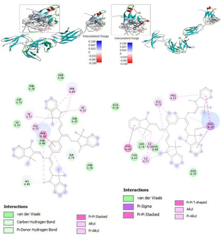
Figure 3.
Docking of Bis-PMSs 5 and 6 to the IL-6 receptor (IL-6R) containing α and β subunits. Upper panels show a 3D model of IL-6R; the binding site (cavity) is localized in IL-6Rβ. Electron density in the binding site is shown as a color scale from blue (low) to red (high). Lower panels show the interaction of Bis-PMSs 5 and 6 with the amino acid residues of a homology model of IL-6R. Bis-PMS 5 and 6 docked to the IL-6R, −9.5 and, −8.1 kcal/mol, respectively.

