Abstract
Objective
Subtypes in type 1 diabetes pathogenesis have been implicated based on the first-appearing autoantibody (primary autoantibody). We set out to describe the glucose metabolism in preclinical diabetes in relation to the primary autoantibody in children with HLA-conferred disease susceptibility.
Design and methods
Dysglycemic markers are defined as a 10% increase in HbA1c in a 3–12 months interval or HbA1c ≥5.9% (41 mmol/mol) in two consecutive samples, impaired fasting glucose or impaired glucose tolerance, or a random plasma glucose value ≥7.8 mmol/L. A primary autoantibody could be detected in 295 children who later developed at least 1 additional biochemical autoantibody. These children were divided into three groups: insulin autoantibody (IAA) multiple (n = 143), GAD antibody (GADA) multiple (n = 126) and islet antigen 2 antibody (IA-2A) multiple (n = 26). Another 229 children seroconverted to positivity only for a single biochemical autoantibody and were grouped as IAA only (n = 87), GADA only (n = 114) and IA-2A only (n = 28).
Results
No consistent differences were observed in selected autoantibody groups during the preclinical period. At diagnosis, children with IAA only showed the highest HbA1c (P < 0.001 between groups) and the highest random plasma glucose (P = 0.005 between groups). Children with IA-2A only progressed to type 1 diabetes as frequently as those with IA-2A multiple (46% vs 54%, P = 0.297) whereas those with IAA only or GADA only progressed less often than children with IAA multiple or GADA multiple (22% vs 62% (P < 0.001) and 7% vs 43% (P < 0.001)), respectively.
Conclusions
The phenotype of preclinical diabetes defined by the primary autoantibody is not associated with any discernible differences in glucose metabolism before the clinical disease manifestation.
Keywords: type 1 diabetes, islet autoantibodies, preclinical type 1 diabetes, glucose metabolism, dysglycemia
Introduction
Type 1 diabetes is one of the most common chronic diseases in childhood. The incidence rate has been rising throughout the Western world (1). The incidence is highest in Finland, in 2006 as high as 64.3 per 100,000 children under the age of 15 years (2). The presentation of type 1 diabetes is usually acute with the presence of typical symptoms. Diabetic ketoacidosis is present in approximately 20–30% of newly diagnosed patients but significantly less in children followed in prospective studies (3). The risk of type 1 diabetes can be estimated based on the HLA genotype (4) and a series of single nucleotide polymorphisms outside the HLA region (5). The presence of ≥2 islet autoantibodies is a strong predictive factor with a risk of 70% for clinical diabetes over the subsequent 10 years (6). Abnormalities in the glucose metabolism appear approximately 2 years before the clinical presentation (7). Metabolic parameters assessed based on HbA1c (8), oral glucose tolerance test (OGTT) (9), random plasma glucose (9), C-peptide (10) and intravenous glucose tolerance test (11) can predict the timing of the diagnosis. For secondary prevention studies, the ability to predict the time and risk of the disease may facilitate the design of shorter and lessexpensive trials by using glucose markers as inclusion criteria (7).
Despite a series of intervention studies aimed at slowing down or preventing type 1 diabetes, long-term positive effects have not been achieved (12). A possible explanation for the lack of success is heterogeneity in the disease process (13). The islet autoantibodies have been shown to correlate with the time from seroconversion to clinical disease and also with the age at diagnosis. Insulin autoantibodies (IAA) are typically present at a younger age than GAD antibodies (GADA) (14), and children with islet antigen 2 antibodies (IA-2A) as their first-emerging autoantibody are characterized by rapid progression to overt disease (15). IAA and IA-2A are associated with the HLA DR4-DQ8 haplotype, whereas GADA positivity is linked to the HLA DR3-DQ2 haplotype (16, 17, 18, 19, 20).
In the current study, we set out to examine the association between the first-emerging, that is, the primary autoantibody (IAA, GADA or IA-2A) with metabolic parameters based on HbA1c, OGTT and random plasma glucose in order to recognize differences in glucose metabolism between subtypes of type 1 diabetes defined by the primary autoantibody.
Materials and methods
Study design
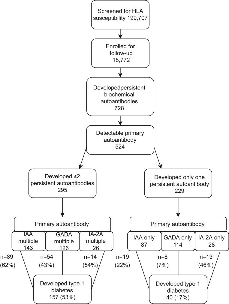
The Type 1 Diabetes Prediction and Prevention (DIPP) study is a Finnish birth cohort study in which children with HLA-conferred susceptibility to type 1 diabetes are observed from birth. The recruitment started in November 1994 and still continues in three university hospitals (Oulu, Tampere and Turku). Screening for genetic risk is performed from cord blood. Families with an infant carrying eligible HLA genotypes associated with an increased risk of type 1 diabetes were invited for prospective follow-up at 3–12-month intervals until the age of 15 years or development of clinical disease. Islet autoantibodies were analyzed at each visit. If a child seroconverted to positivity for islet cell antibodies (ICA) and at least one biochemical autoantibody (IAA, GADA and/or IA-2A), then the monitoring of HbA1c, OGTT and random plasma glucose was started. HbA1c and random plasma glucose were analyzed at every 3-month visit, and OGTT was performed once a year. The diagnosis of type 1 diabetes was based on typical symptoms and high plasma glucose or two diabetic OGTT tests in asymptomatic subjects as the World Health Organization (WHO) recommends (21). The study population included in the current report comprises 524 children with a detectable first-appearing autoantibody (IAA, GADA or IA-2A) and is summarized in Fig. 1. Children presenting with ≥2 biochemical autoantibodies during the whole follow-up and children with only one biochemical autoantibody have been analyzed separately. Children who developed ≥2 biochemical autoantibodies during follow-up are referred to as IAA multiple, GADA multiple and IA-2A multiple. Subjects with only a single biochemical autoantibody during follow-up are referred to as IAA only, GADA only and IA-2A only. Data collected by August 31, 2014, was included in this analysis, updated from previously reported (8, 9). The DIPP study has been approved by the Ethics Committees in the participating universities and hospital districts (Turku University Ethics Committee, 10/1994 §228). All families taking part in the study have provided written informed consent.
Figure 1.
Flow-chart showing children with detectable first-appearing islet autoantibodies from the Type 1 Diabetes Prediction and Prevention (DIPP) study. The children presenting with ≥2 biochemical autoantibodies during the whole follow-up and those with only one biochemical autoantibody have been analyzed separately. Data collected by August 31, 2014, was included in this analysis.
Immunological screening
Participants who seroconverted to positivity for any of the diabetes-associated islet autoantibodies, that is, ICA, IAA, GADA or IA-2A, were rescheduled for follow-up visits at 3-month intervals. ICA has been analyzed in the DIPP study, but these antibodies were not considered in the current study. Seroconversion was defined as the time when the first biochemical autoantibody was detected for the first time and multiple (when two or more) (≥2) autoantibodies were detected for the first time. A positive result required a positive autoantibody also in the subsequent sample (persistent positivity). Autoantibody analyses were performed as described previously (14). The positive cut-off limits have been reported previously (9). In the Diabetes Autoantibody Standardization workshop in 2005, the following sensitivities and specificities were recorded: IAA 58% and 96%, GADA 82% and 96% and to IA-2A 72% and 100%, respectively.
Genetic screening
HLA-conferred susceptibility to type 1 diabetes was analyzed using cord blood as described earlier (22). According to various HLA DRB1–DQA1–DQB1 haplotype combinations, six risk groups were identified: high, moderately increased, slightly increased, neutral, slightly decreased and strongly decreased (22). For the current study, risk groups of neutral, slightly decreased risk and strongly decreased risk were combined due to low number of children, and the analyses were performed with four risk groups.
HbA1c assays
In the Oulu University Hospital, an immunoassay-based method was applied throughout the study. In the Tampere University Hospital, fast protein liquid chromatography (FPLC) was used until June 9, 1999, when the local laboratory changed to an immunoassay-based method. In the Turku University Hospital, the FPLC method was used until August 5, 1996. Thereafter, a method based on high performance liquid chromatography (HPLC) was applied, and this method was eventually changed to an immunoassay-based method on September 1, 2013. Assays and devices have been described in detail previously (8).
Oral glucose tolerance tests and glucose assays
The oral glucose tolerance test (1.75 g/kg body weight, up to a maximum of 75 g) was performed with a standard protocol after overnight fasting. Samples were taken in the fasting state and at 120 min. In the Oulu University Hospital, capillary samples were used, whereas in Tampere and Turku, tests were based on venous samples. In the Oulu University Hospital, a glucose dehydrogenase-based method was used until May 2000, and thereafter, a glucose oxidase method was used. In Tampere and Turku, the hexokinase method was applied during the whole study.
Random plasma glucose assays
Venous plasma samples were obtained in all study sites. In Oulu, the dehydrogenase method was used until May 2000, whereafter, the enzymatic glucose hexokinase method was utilized for the analysis of glucose. In Tampere, the hexokinase-based method was used until September 2006, and thereafter, a glucose dehydrogenase method was applied. In Turku, the glucose dehydrogenase method was used during the whole follow-up.
Definition of dysglycemia
The definitions of dysglycemia used in the current study have been described earlier (8, 9). We previously proposed predictive HbA1c values based on a 10% rise in HbA1c levels during an interval of 3–12 month or HbA1c ≥5.9% (41 mmol/mol) in two consecutive samples. An increase of 10% from the baseline value has been suggested also in other studies (23). In the OGTT, the cut-offs recommended by WHO for aberrant values have been applied and categorized as normal, impaired fasting glucose (IFG), impaired glucose tolerance (IGT) and diabetic. For random plasma glucose, a cut-off of ≥7.8 mmol/L was used, the value being the same as for IGT on OGTTs.
Statistical analyses
A total of six different parameters of glucose metabolism were analyzed: (1) a 10% increase in HbA1c during a 3–12-month interval, (2) two consecutive HbA1c values ≥5.9% (41 mmol/mol), (3) IFG, (4) IGT, (5) both IFG and IGT and (6) random plasma glucose value ≥7.8 mmol/L.
The chi-square test was applied to investigate the observed proportions and tested glucose parameters between primary autoantibody groups. The variance analysis (ANOVA) with Tukey’s HSD test was used to analyze differences in age at seroconversion, age at ≥2 biochemical autoantibodies, time from seroconversion to dysglycemia and time from dysglycemia to diagnosis (in progressors only) between the groups (IAA multiple, GADA multiple, IA-2A multiple, IAA only, GADA only and IA-2A only) and separately between progressors and non-progressors.
The linear mixed model (LMM) with random intercept and first-order autoregressive (AR1) covariance structure for repeated measurements was used to analyze HbA1c, 0 min and 120 min glucose levels in OGTT and random plasma glucose concentrations over time between the autoantibody groups and separately between progressors and non-progressors. The random intercepts and repeated measurements were nested within subjects and subjects within the hospital. The group-by-time interaction was included in the model to test differences between group means at each time point. Sex, age at sampling and HLA risk were included in the LMM model as fixed variables. For time-to-event analyses, Kaplan–Meier survival curves were used and statistical significance was tested with the log-rank method. All analyses were performed using IBM SPSS Statistic 24.0.0.1 for Windows and StatsDirect statistical software version 3.0.181. Figures were drawn using OriginPro 2015.
Results
Between November 1994 and August 2014 altogether, 18,772 infants with increased genetic risk were enrolled for regular follow-up in the DIPP study. During the follow-up, 728 children developed at least one persistent biochemical islet autoantibody. A total of 204 children seroconverted to positivity for at least two autoantibodies and accordingly the first-appearing autoantibody could not be defined. Therefore, these children were excluded from further analyzes. The first-appearing autoantibody (IAA, GADA or IA-2A) could be detected in 524 individuals, out of whom 295 (56%) developed at least 1 additional biochemical autoantibody (IAA multiple, GADA multiple or IA-2A multiple) during the follow-up and 157 (53%) of these children developed type 1 diabetes. A total of 229 children with a detectable first-appearing autoantibody developed no additional biochemical autoantibodies (IAA only, GADA only or IA-2A only), and among these, 40 (17%) developed type 1 diabetes. Children in the IAA multiple and GADA multiple groups developed type 1 diabetes with substantially higher frequency than those with IAA only (62% vs 22%, P < 0.001) and GADA only (43% vs 7%, P < 0.001). In the IA-2A multiple and IA-2A only groups, progression rate was similar during the follow-up (54% vs 46%, P = 0.297), see Fig. 1. The proportions of HLA-risk genotypes in each study group are presented in Table 1.
Table 1.
Characteristics of children with detectable first-appearing islet autoantibodies from the Type 1 Diabetes Prediction and Prevention (DIPP) study. The groups of children who developed ≥2 biochemical autoantibodies during follow-up are referred to as IAA multiple, GADA multiple and IA-2A multiple. The groups of children with only a single biochemical autoantibody during follow-up are referred to as IAA only, GADA only, IA-2A only. Data collected by August 31, 2014, was included in this analysis.
| IAA multiplea | GADA multiplea | IA-2A multiplea | P between groups | IAA onlyb | GADA onlyb | IA-2A onlyb | P between groups | |
|---|---|---|---|---|---|---|---|---|
| Progressors | N = 89 | N = 54 | N = 14 | N = 19 | N = 8 | N = 13 | ||
| Gender, boys, n (%) | 57 (64) | 26 (48) | 10 (71) | 0.107 | 12 (63) | 3 (38) | 9 (69) | 0.328 |
| HLA risk, n (%) | 0.667 | 0.117 | ||||||
| Neutral or decreased | 1 (1) | 1 (1) | 0 (0) | 0 (0) | 0 (0) | 0 (0) | ||
| Low | 15 (17) | 8 (15) | 1 (7) | 4 (21) | 1 (13) | 0 (0) | ||
| Moderate | 45 (51) | 29 (54) | 11 (79) | 7 (39) | 6 (75) | 10 (77) | ||
| High | 28 (31) | 19 (35) | 2 (14) | 8 (42) | 1 (13) | 3 (23) | ||
| Mean number of HbA1c tests | 7.5 | 8.6 | 6.9 | 2.7 | 2.9 | 5.8 | ||
| Mean number of OGTTs | 3.2 | 3.6 | 2.4 | 1.0 | 1.1 | 2.9 | ||
| Mean number of random plasma glucose tests | 6.1 | 8.0 | 7.7 | 4.0 | 6.5 | 4.1 | ||
| Two consecutive HbA1c values ≥5.9% (41 mmol/mol), n (%) | 26 (29) | 18 (33) | 7 (50) | 0.549 | 4 (21) | 0 (0) | 5 (38) | 0.230 |
| Any dysglycemiac, n (%) | 63 (71) | 40 (74) | 11 (79) | 0.796 | 7 (37) | 1 (13) | 7 (54) | 0.164 |
| Time from seroconversion to multiple autoantibodies, mean (s.d.) | 0.6 (0.5) | 1.1 (1.2) | 1.2 (1.1) | 0.001 | NA | NA | NA | |
| Time from seroconversion to detection of any dysglycemiac mean (s.d.) | 3.1 (2.5) | 3.7 (2.9) | 2.2 (1.6) | 0.189 | 2.4 (1.9) | 1.4 (NA) | 1.4 (0.8) | 0.480 |
| Time from multiple autoantibodies to detection of any dysglycemia, mean (s.d.) | 2.3 (2.3) | 2.2 (2.4) | 1.0 (1.3) | 0.186 | NA | NA | NA | |
| Time from detection of dysglycemia to type 1 diabetes, mean (s.d.) | 1.8 (2.7) | 2.2 (2.4) | 1.7 (1.8) | 0.728 | 2.2 (1.5) | 3.9 (NA) | 1.7 (2.1) | 0.543 |
| Time from seroconversion to type 1 diabetes, mean (s.d.) | 4.2 (3.1) | 4.8 (3.0) | 4.1 (2.4) | 0.485 | 2.3 (2.5) | 3.6 (3.8) | 2.9 (2.3) | 0.510 |
| Age (years) at type 1 diabetes diagnosis, mean (s.d.) | 5.9 (3.5) | 9.1 (4.1) | 7.6 (4.1) | <0.001 | 4.6 (3.7) | 9.4 (3.7) | 7.4 (3.3) | 0.008 |
| Non-progressors | N=54 | N=72 | N=12 | N=68 | N=106 | N=15 | ||
| Gender, boys, n (%) | 37 (66) | 39 (52) | 8 (69) | 0.240 | 35 (54) | 67 (61) | 11 (71) | 0.164 |
| HLA riskd, n (%) | 0.091 | 0.374 | ||||||
| Neutral or decreased | 0 (0) | 1 (1) | 0 (1) | 6 (9) | 11 (10) | 0 (0) | ||
| Low | 8 (15) | 15 (21) | 2 (17) | 19 (28) | 24 (23) | 3 (20) | ||
| Moderate | 40 (74) | 34 (47) | 8 (67) | 35 (51) | 49 (46) | 10 (67) | ||
| High | 6 (11) | 22 (31) | 2 (17) | 8 (12) | 22 (21) | 1 (7) | ||
| Mean number of HbA1c tests | 11.8 | 15.3 | 11.8 | 7.3 | 7.8 | 6.4 | ||
| Mean number of OGTTs | 2.7 | 3.9 | 3.0 | 0.7 | 1.2 | 1.7 | ||
| Mean number of random plasma glucose tests | 9.0 | 9.7 | 9.1 | 7.2 | 7.1 | 5.3 | ||
| Two consecutive HbA1c values ≥5.9% (41 mmol/mol), n (%) | 7 (13) | 6 (8) | 1 (8) | 0.565 | 5 (7) | 9 (8) | 0 (0) | 0.453 |
| Any dysglycemiac, n (%) | 29 (54) | 33 (46) | 4 (33) | 0.392 | 27 (40) | 37 (35) | 6 (40) | 0.790 |
| Time from seroconversion to multiple autoantibodies, mean (s.d.) | 1.4 (2.0) | 1.8 (2.2) | 1.1 (0.9) | 0.479 | NA | NA | NA | |
| Time from seroconversion to detection of any dysglycemiac mean (s.d.) | 4.4 (3.5) | 3.3 (2.3) | 5.4 (4.2) | 0.309 | 3.6 (3.1) | 4.5 (3.4) | 2.2 (1.0) | 0.204 |
| Time from multiple autoantibodies to detection of any dysglycemia, mean (s.d.) | 2.3 (2.5) | 1.2 (1.9) | 3.6 (4.4) | 0.052 | NA | NA | NA | |
| Age (years) at last visit, mean (s.d.) | 8.1 (4.5) | 12.4 (4.3) | 8.2 (5.3) | <0.001 | 9.2 (4.4) | 11.3 (4.3) | 9.0 (5.2) | 0.008 |
aThe first observed autoantibody was IAA, GADA or IA-2A with no other autoantibodies in the same sample followed by at least one more biochemical autoantibody; bThe first observed autoantibody was IAA, GADA or IA-2A with no other autoantibodies in the same sample. No other biochemical autoantibodies were detected during follow-up; cDysglycemia was defined as 10% rise in HbA1c during 3–12 months, two consecutive HbA1c values ≥5.9% (41 mmol/mol), IFG, IGT or random plasma glucose ≥7.8 mmol/L; dOne non-progressor child in IA-2A only group had a rare HLA genotype not possible to define.
A total of 4598 HbA1c samples were taken (3110 in children with ≥2 biochemical autoantibodies and 1565 in children with a single biochemical autoantibody), OGTTs were performed 1248 times (983 and 265) and random plasma glucose samples were taken 3881 times (2374 and 1507).
HbA1c, OGTT and random plasma glucose
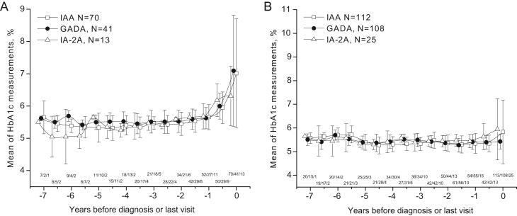
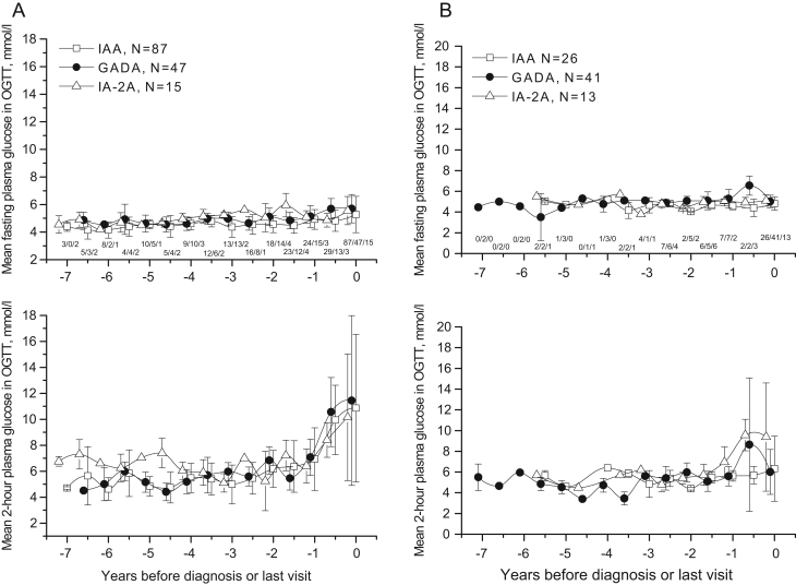
Measured metabolic parameters (HbA1c, OGTT data and random plasma glucose) were compared between the different primary autoantibody groups during the follow-up. In the groups of progressors, mean adjusted HbA1c values varied from 4.7% (28 mmol/mol) to 8.2% (66 mmol/mol) increasing toward diagnosis (Fig. 2A). No consistent significant differences were observed between the autoantibody groups during the preclinical period. At the time of diagnosis, IAA only showed slightly higher adjusted HbA1c values (mean 8.2% (66 mmol/mol (95% CI 7.6–8.8% (60–73 mmol/mol)) compared to most other groups: IAA multiple (mean 7.1% (54 mmol/mol (95% CI 6.7–7.5% (50–59 mmol/mol)), GADA multiple (mean 7.0% (53 mmol/mol (95% CI 6.6–7.4% (49–57 mmol/mol)), IA-2A multiple (mean 7.3% (56 mmol/mol (95% CI 6.6–8.0% (49–64 mmol/mol)), GADA only (mean 7.8% (62 mmol/mol (95% CI 6.8–8.8% (51–73 mmol/mol)) and IA-2A only (mean 6.7% (50 mmol/mol (95% CI 6.0–7.4% (42–57 mmol/mol)). In non-progressors, no consistent differences were observed (Fig. 2B). HbA1c values at the last visit showed minimal variation between primary autoantibody groups, with mean values between 5.2 and 5.4% (33–36 mmol/mol). Neither among the progressors (Fig. 3A) nor among the non-progressors (Fig. 3B) we observed any significant differences between the three primary autoantibody groups in fasting plasma glucose concentrations in OGTTs during the preclinical period or at the time of diagnosis. Fasting plasma glucose values during the follow-up varied from 4.0 to 5.4 mmol/L in progressors and 4.1 to 5.4 mmol/L in the non-progressor groups. The 2-h glucose values varied from 3.4 mmol/L to 10.8 mmol/L in the progressor groups, increasing toward diagnosis (Fig. 3A). Still, no statistically significant differences between the primary autoantibody groups could be observed at any of the time points during the preclinical period or at the time of diagnosis. At diagnosis, the mean plasma glucose values were as follows: IAA multiple (mean 10.8 mmol/L (95% CI 9.4–12.2)), GADA multiple (mean 10.0 mmol/L (95% CI 8.5–11.5)), IA-2A multiple (mean 9.3 mmol/L (95% CI 6.9–11.6)), IAA only (mean 11.0 mmol/L (95% CI 8.1–14.0)), GADA only (mean 9.2 mmol/L (95% CI 6.0–12.3)), IA-2A only (mean 11.6 mmol/L (95% CI 9.1–14.1)). In non-progressors, no differences were seen (Fig. 3B).
Figure 2.
Adjusted mean HbA1c during the follow-up in children with different primary autoantibodies who progressed to type 1 diabetes (A) or remained disease-free (B). Children developing at least one additional autoantibody and children who remained positive for a single autoantibody only have been combined. The last point is the diagnosis of type 1 diabetes (A) or the last visit (B) (IAA, squares; GADA, dots; IA-2A, triangles). Whiskers show 95% CIs of the adjusted mean.
Figure 3.
Adjusted mean plasma glucose in OGTT (fasting and 2-h plasma glucose) during the follow-up in children with different primary autoantibodies who progressed to type 1 diabetes (A) or remained disease-free (B). Children developing at least one additional autoantibody and children who remained positive for a single autoantibody only have been combined. The last point is the diagnosis of type 1 diabetes (A) or the last visit (B) (IAA, squares; GADA, dots; IA-2A, triangles). Whiskers show 95% CIs of the adjusted mean.
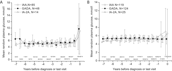
There was higher variation in the random plasma glucose values between the autoantibody groups than that seen in HbA1c values. In progressors, random plasma glucose varied from 4.4 mmol/L to 13.2 mmol/L, also increasing toward diagnosis (Fig. 4A). Again no consistent differences could be observed between the autoantibody groups during the preclinical period. At the time of diagnosis, mean random plasma glucose was highest in the group with IAA only (mean 13.2 mmol/L (95% CI 9.9–16.6)) when compared to other groups: IAA multiple (mean 9.1 mmol/L (95% CI 6.7–11.6)), GADA multiple (mean 9.8 mmol/L (95% CI 7.3–12.3)), IA-2A multiple (mean 8.2 mmol/L (95% CI 4.7–11.7)), GADA only (mean 9.2 mmol/l (95% CI 4.2–14.2)), IA-2A only (mean 9.5 mmol/L (95% CI 5.5–13.4)). In non-progressors, no differences were observed (Fig. 4B).
Figure 4.
Adjusted mean random plasma glucose during the follow-up in children with different primary autoantibodies who progressed to type 1 diabetes (A) or remained disease-free (B). Children developing at least one additional autoantibody, and children who remained positive for a single autoantibody only have been combined. The last point is the diagnosis of type 1 diabetes (A) or the last visit (B) (IAA, squares; GADA, dots; IA-2A, triangles). Whiskers show 95% CIs of the adjusted mean.
Time from seroconversion to dysglycemia and type 1 diabetes
The overall rate and overlap of different dysglycemia markers are presented in Supplementary Table 1 (see section on supplementary materials given at the end of this article). Detection of dysglycemia (a 10% rise in HbA1c, ≥5.9% (41 mmol/mol) in two consecutive samples, IFG, IGT or random plasma glucose ≥7.8 mmol/L) was common especially in children in the multiple autoantibody groups who progressed to type 1 diabetes, varying from 73 to 79% (Table 1). In progressors who developed only a single autoantibody, dysglycemia was observed in 13–54% of children. In the non-progressors, the occurrence of dysglycemia varied from 33 to 54% (Table 1). The previously identified more specific marker of HbA1c ≥5.9% (41 mmol/mol) in two consecutive samples was identified in up to 50% in the progressor groups but only up to 13% in the non-progressor groups (Table 1).
We calculated the time from the detection of the primary islet autoantibody to the development of the first-appearing dysglycemic marker and the time from dysglycemia to diagnosis of type 1 diabetes, as summarized in Table 1. No significant differences in the time from seroconversion to any of the markers were observed among the analyzed groups (Table 1) in either progressors or non-progressors. In Kaplan–Meier survival curves presenting time from seroconversion to various dysglycemia criteria, children with IA-2A as the first-appearing islet autoantibody showed a trend for faster progression than IAA and GADA groups (Supplementary Figs 1 and 2). In progressors, children with IA-2A as the primary autoantibody showed the shortest time from seroconversion to any dysglycemia (P = 0.008) and from seroconversion to IFG or IGT (P = 0.006), Supplementary Fig. 2.
Discussion
The prediction of type 1 diabetes has improved significantly during the past years along with increasing knowledge of islet autoantibodies and glucose metabolism. However, interventions aimed at slowing down or preventing type 1 diabetes have proven ineffective in most cases, possibly due to heterogeneity in the disease process. In the current study, we set out to characterize the glucose metabolism with HbA1c, OGTT and random plasma glucose in groups with IAA, GADA or IA-2A as the primary islet autoantibody. We confirm the previous finding that young age at disease presentation is associated with IAA as the primary islet autoantibody. We also found that IA-2A predisposes to a high risk of progression to type 1 diabetes even when being the only detectable autoantibody. No differences in glucose metabolism during the preclinical stage of type 1 diabetes could be observed between the three groups with different first-appearing biochemical islet autoantibodies, whether or not developing additional autoantibodies, indicating similar pathogenesis from the appearance of the first autoantibody to overt disease. Occurrence of dysglycemia was common in children later progressing to type 1 diabetes, with the highest specificity observed with HbA1c ≥5.9% (41 mmol/mol) in two consecutive samples as reported before (8, 9, 24).
Our cohort of 295 children with multiple biochemical autoantibodies and 229 children with only one biochemical autoantibody, where the first-appearing autoantibody can be defined, is unique worldwide. Due to the design of the DIPP study, including follow-up since birth and screening for islet autoantibodies, we were able to identify the initiation of the autoimmunity process and divide children into groups of IAA, GADA or IA-2A as the first-emerging islet autoantibody. Previously one analysis based on the DIPP study has been performed according to the first-appearing islet autoantibody status, suggesting a shorter time from seroconversion to clinical disease in children seroconverting first to IA-2A (15). In our study, we report a high progression rate (46%) also in children with IA-2A as a single biochemical islet autoantibody. The high number of autoantibody-positive children in the DIPP study among whom HbA1c, OGTT and random glucose were repeatedly measured during long follow-up allowed us to reliably describe the glucose metabolism in preclinical type 1 diabetes according to the first-appearing autoantibody specificities.
Heterogeneity in the pathogenic process triggering the start of autoimmunity and leading to progression to clinical disease has been implicated previously (13). A series of intervention trials with various approaches have been performed before the appearance of autoantibodies (primary prevention), after the detection of autoantibodies (secondary prevention) and after the diagnosis of clinical disease (tertiary prevention) with little or no success (12). In histological studies of pancreases from patients with disease presentation under 15 years of age and samples obtained less than 1 year after diagnosis, the proportion of inflammation in islets, that is, insulitis was 68% compared to 39% in older patients suggesting a more aggressive disease process in younger individuals (25). Different proportions of circulating lymphocyte subpopulations, for example, CD8 T cells, have as well been observed (26). In some patients, inflammation is also present in the exocrine pancreas (27), referring to the possibility of an infectious agent being involved (28).
An association between specific pathomechanisms and the primary islet autoantibody has been implicated. The T-cell response to proinsulin epitopes restricted to HLA DQ2/DQ8 heterozygous individuals suggests one specific route for the initiation of the autoimmune process (16). It has also been shown that IAA as the first autoantibody is associated with the DR4-DQ8 haplotype and GADA with the DR3-DQ2 haplotype (15, 29, 30). In the current study, we set out to analyze whether the first-emerging islet autoantibody correlates with glucose metabolism and the rate of disease progression. However, we could not observe any associations between the primary islet autoantibody and absolute values of glucose metabolism, numbers of dysglycemic markers or time from seroconversion to detected dysglycemia or overt type 1 diabetes during the preclinical period. This indicates that the pathogenic process leading to glucose intolerance is rather similar in children presenting with different primary islet autoantibodies. The only differences were observed at the time of diagnosis when the children with IAA only tended to have higher values than the other participants. Children with IAA as the first-appearing autoantibody were younger at disease onset which might explain the observed difference.
The size of the study population is after all a limitation of our work. Even with a large number of children with ≥2 biochemical autoantibodies (n = 295) and children with only one biochemical autoantibody (n = 229), the size of the IA-2A groups wase small (n = 26 and n = 28, respectively). With this sample size, the LMM analysis of the studied parameters provides relatively reliable results, but for the comparison of dysglycemic markers within each primary autoantibody group, the size represents a limitation. Our study population came from three clinical centers and was compiled over a time period of 20 years, with some changes in the analytical methods over time, which may introduce some additional variability. However, these potential confounding factors affect all groups to the same extent. The methods applied and the study centers were taken into account in the statistical analyses. This process has been explained in detail in our previous reports in which the criteria for dysglycemia were established (8, 9). Autoantibodies to zinc transporter 8 (ZnT8A) were not included in the current analysis, which could to some extent affect the true primary islet autoantibody status. However, ZnT8A usually develops at a rather late stage of preclinical type 1 diabetes and therefore the effect should be small (31).
It should be accepted that in the evolution of type 1 diabetes, as in other autoimmune diseases, multiple immune pathways are involved. It seems that any treatment aimed at changing the disease course needs to be performed with agents affecting several pathways. Primary autoantibodies can be a sign of specific phenotypes, although according to our current study, glucose metabolism in preclinical diabetes seems quite similar in all primary islet autoantibody groups. More studies are needed for a better understanding of underlying pathomechanisms in type 1 diabetes in order to find effective individualized prevention.
In conclusion, the first-appearing islet autoantibody does not affect the development of dysglycemia assessed by HbA1c, OGTT and random plasma glucose values during preclinical type 1 diabetes. However, IA-2A seems to predispose to a high risk of progression to clinical type 1 diabetes even as a single biochemical islet autoantibody. According to our study, children positive for IA-2A should be monitored and treated in a manner similar to that applied for children with ≥2 islet autoantibodies.
Supplementary Material
Declaration of interest
The authors declare that there is no conflict of interest that could be perceived as prejudicing the impartiality of the research reported.
Funding
This work was supported by the following grants. International: JDRF International (grants 4-1998-274, 4-1999-731, 4-2001-435); European Union (grant BMH4-CT98-3314); Novo Nordisk Foundation. Finland: Academy of Finland (Centre of Excellence in Molecular Systems Immunology and Physiology Research 2012-2017, Decision No. 250114); TEKES National Technology Agency of Finland; Special Research Funds for University Hospitals in Finland; Finnish Office for Health Technology Assessment; Diabetes Research Foundation, Finland; Sigrid Juselius Foundation; Emil Aaltonen Foundation; Jalmari and Rauha Ahokas Foundation; Signe and Ane Gyllenberg Foundation; the Research Foundation of Orion Corporation; Foundation for Pediatric Research; Alma and KA Snellman Foundation; Päivikki and Sakari Sohlberg Foundation, the Finnish Medical Foundation and Finnish Cultural Foundation, Northern Ostrobothnia Regional Fund.
Author contribution statement
O H had full access to all of the data in the study and takes responsibility for the integrity of the data and the accuracy of the data analysis. Study concept and design: O H, J I, O S, M K, R V. Acquisition of data: O H, S A, J I, M K, R V. Analysis and interpretation of data: O H, T P, M K, R V. Drafting of the manuscript: O H, T P, M K, R V. Critical revision of the manuscript for important intellectual content: O H, T P, S A, J I, O S, M K, R V. Statistical analysis: T P. Administrative, technical, or material support: O H, T P, J I, M K, R V. Study supervision: R V. All authors read and approved the final version.
Acknowledgements
The author thank the dedicated personnel of the DIPP study in Oulu, Tampere and Turku; and the study children and their families for their essential contribution.
References
- 1.DIAMOND Project Group. Incidence and trends of childhood type 1 diabetes worldwide 1990–1999. Diabetic Medicine 200623857–866. ( 10.1111/j.1464-5491.2006.01925.x) [DOI] [PubMed] [Google Scholar]
- 2.Harjutsalo V, Sund R, Knip M, Groop PH. Incidence of type 1 diabetes in Finland. JAMA 2013310427–428. ( 10.1001/jama.2013.8399) [DOI] [PubMed] [Google Scholar]
- 3.Steck AK, Larsson HE, Liu X, Veijola R, Toppari J, Hagopian WA, Haller MJ, Ahmed S, Akolkar B, Lernmark Ået al. Residual beta-cell function in diabetes children followed and diagnosed in the TEDDY study compared to community controls. Pediatric Diabetes 201718794–802. ( 10.1111/pedi.12485) [DOI] [PMC free article] [PubMed] [Google Scholar]
- 4.Noble JA, Valdes AM. Genetics of the HLA region in the prediction of type 1 diabetes. Current Diabetes Reports 201111533–542. ( 10.1007/s11892-011-0223-x) [DOI] [PMC free article] [PubMed] [Google Scholar]
- 5.Concannon P, Rich SS, Nepom GT. Genetics of type 1A diabetes. New England Journal of Medicine 20093601646–1654. ( 10.1056/NEJMra0808284) [DOI] [PubMed] [Google Scholar]
- 6.Ziegler AG, Rewers M, Simell O, Simell T, Lempainen J, Steck A, Winkler C, Ilonen J, Veijola R, Knip Met al. Seroconversion to multiple islet autoantibodies and risk of progression to diabetes in children. JAMA 20133092473–2479. ( 10.1001/jama.2013.6285) [DOI] [PMC free article] [PubMed] [Google Scholar]
- 7.Insel RA, Dunne JL, Atkinson MA, Chiang JL, Dabelea D, Gottlieb PA, Greenbaum CJ, Herold KC, Krischer JP, Lernmark Ået al. Staging presymptomatic type 1 diabetes: a scientific statement of JDRF, the endocrine society, and the American Diabetes Association. Diabetes Care 2015381964–1974. ( 10.2337/dc15-1419) [DOI] [PMC free article] [PubMed] [Google Scholar]
- 8.Helminen O, Aspholm S, Pokka T, Hautakangas MR, Haatanen N, Lempainen J, Ilonen J, Simell O, Knip M, Veijola R. HbA1c predicts time to diagnosis of type 1 diabetes in children at risk. Diabetes 2015641719–1727. ( 10.2337/db14-0497) [DOI] [PubMed] [Google Scholar]
- 9.Helminen O, Aspholm S, Pokka T, Ilonen J, Simell O, Veijola R, Knip M. OGTT and random plasma glucose in the prediction of type 1 diabetes and time to diagnosis. Diabetologia 2015581787–1796. ( 10.1007/s00125-015-3621-9) [DOI] [PubMed] [Google Scholar]
- 10.Sosenko JM, Palmer JP, Rafkin LE, Krischer JP, Cuthbertson D, Greenbaum CJ, Eisenbarth G, Skyler JS. & Diabetes Prevention Trial-Type 1 Study Group. Trends of earlier and later responses of C-peptide to oral glucose challenges with progression to type 1 diabetes in diabetes prevention trial-type 1 participants. Diabetes Care 201033620–625. ( 10.2337/dc09-1770) [DOI] [PMC free article] [PubMed] [Google Scholar]
- 11.Koskinen MK, Helminen O, Matomaki J, Aspholm S, Mykkanen J, Makinen M, Simell V, Vaha-Makila M, Simell T, Ilonen Jet al. Reduced beta-cell function in early preclinical type 1 diabetes. European Journal of Endocrinology 2016174251–259. ( 10.1530/EJE-15-0674) [DOI] [PMC free article] [PubMed] [Google Scholar]
- 12.Skyler JS.Prevention and reversal of type 1 diabetes – past challenges and future opportunities. Diabetes Care 201538997–1007. ( 10.2337/dc15-0349) [DOI] [PubMed] [Google Scholar]
- 13.Atkinson MA, von Herrath M, Powers AC, Clare-Salzler M. Current concepts on the pathogenesis of type 1 diabetes – considerations for attempts to prevent and reverse the disease. Diabetes Care 201538979–988. ( 10.2337/dc15-0144) [DOI] [PMC free article] [PubMed] [Google Scholar]
- 14.Siljander HT, Simell S, Hekkala A, Lahde J, Simell T, Vahasalo P, Veijola R, Ilonen J, Simell O, Knip M. Predictive characteristics of diabetes-associated autoantibodies among children with HLA-conferred disease susceptibility in the general population. Diabetes 2009582835–2842. ( 10.2337/db08-1305) [DOI] [PMC free article] [PubMed] [Google Scholar]
- 15.Ilonen J, Hammais A, Laine AP, Lempainen J, Vaarala O, Veijola R, Simell O, Knip M. Patterns of beta-cell autoantibody appearance and genetic associations during the first years of life. Diabetes 2013623636–3640. ( 10.2337/db13-0300) [DOI] [PMC free article] [PubMed] [Google Scholar]
- 16.Pathiraja V, Kuehlich JP, Campbell PD, Krishnamurthy B, Loudovaris T, Coates PT, Brodnicki TC, O'Connell PJ, Kedzierska K, Rodda Cet al. Proinsulin-specific, HLA-DQ8, and HLA-DQ8-transdimer-restricted CD4+ T cells infiltrate islets in type 1 diabetes. Diabetes 201564172–182. ( 10.2337/db14-0858) [DOI] [PubMed] [Google Scholar]
- 17.Hagopian WA, Sanjeevi CB, Kockum I, Landin-Olsson M, Karlsen AE, Sundkvist G, Dahlquist G, Palmer J, Lernmark A. Glutamate decarboxylase-, insulin-, and islet cell-antibodies and HLA typing to detect diabetes in a general population-based study of Swedish children. Journal of Clinical Investigation 1995951505–1511. ( 10.1172/JCI117822) [DOI] [PMC free article] [PubMed] [Google Scholar]
- 18.Genovese S, Bonfanti R, Bazzigaluppi E, Lampasona V, Benazzi E, Bosi E, Chiumello G, Bonifacio E. Association of IA-2 autoantibodies with HLA DR4 phenotypes in IDDM. Diabetologia 1996391223–1226. ( 10.1007/BF02658510) [DOI] [PubMed] [Google Scholar]
- 19.Ziegler AG, Standl E, Albert E, Mehnert H. HLA-associated insulin autoantibody formation in newly diagnosed type I diabetic patients. Diabetes 1991401146–1149. ( 10.2337/diab.40.9.1146) [DOI] [PubMed] [Google Scholar]
- 20.Sabbah E, Savola K, Kulmala P, Reijonen H, Veijola R, Vahasalo P, Karjalainen J, Ilonen J, Akerblom HK, Knip M. Disease-associated autoantibodies and HLA-DQB1 genotypes in children with newly diagnosed insulin-dependent diabetes mellitus (IDDM). the childhood diabetes in finland study group. Clinical and Experimental Immunology 199911678–83. ( 10.1046/j.1365-2249.1999.00863.x) [DOI] [PMC free article] [PubMed] [Google Scholar]
- 21.Alberti KG, Zimmet PZ. Definition, diagnosis and classification of diabetes mellitus and its complications. Part 1: diagnosis and classification of diabetes mellitus provisional report of a WHO consultation. Diabetic Medicine 199815539–553. () [DOI] [PubMed] [Google Scholar]
- 22.Ilonen J, Kiviniemi M, Lempainen J, Simell O, Toppari J, Veijola R, Knip M. & Finnish Pediatric Diabetes Register. Finnish Pediatric Diabetes Register: genetic susceptibility to type 1 diabetes in childhood – estimation of HLA class II associated disease risk and class II effect in various phases of islet autoimmunity. Pediatric Diabetes 201617 (Supplement 22) 8–16. ( 10.1111/pedi.12327) [DOI] [PubMed] [Google Scholar]
- 23.Krischer JPType 1 Diabetes TrialNet Study Group. The use of intermediate endpoints in the design of type 1 diabetes prevention trials. Diabetologia 2013561919–1924. ( 10.1007/s00125-013-2960-7) [DOI] [PMC free article] [PubMed] [Google Scholar]
- 24.Helminen O, Pokka T, Tossavainen P, Ilonen J, Knip M, Veijola R. Continuous glucose monitoring and HbA1c in the evaluation of glucose metabolism in children at high risk for type 1 diabetes mellitus. Diabetes Research and Clinical Practice 201612089–96. ( 10.1016/j.diabres.2016.07.027) [DOI] [PubMed] [Google Scholar]
- 25.In't Veld P.Insulitis in human type 1 diabetes: the quest for an elusive lesion. Islets 20113131–138. ( 10.4161/isl.3.4.15728) [DOI] [PMC free article] [PubMed] [Google Scholar]
- 26.Skowera A, Ladell K, McLaren JE, Dolton G, Matthews KK, Gostick E, Kronenberg-Versteeg D, Eichmann M, Knight RR, Heck Set al. Beta-cell-specific CD8 T cell phenotype in type 1 diabetes reflects chronic autoantigen exposure. Diabetes 201564916–925. ( 10.2337/db14-0332) [DOI] [PMC free article] [PubMed] [Google Scholar]
- 27.Rodriguez-Calvo T, Ekwall O, Amirian N, Zapardiel-Gonzalo J, von Herrath MG. Increased immune cell infiltration of the exocrine pancreas: a possible contribution to the pathogenesis of type 1 diabetes. Diabetes 2014633880–3890. ( 10.2337/db14-0549) [DOI] [PMC free article] [PubMed] [Google Scholar]
- 28.Vaarala O, Atkinson MA, Neu J. The ‘perfect storm’ for type 1 diabetes: the complex interplay between intestinal microbiota, gut permeability, and mucosal immunity. Diabetes 2008572555–2562. ( 10.2337/db08-0331) [DOI] [PMC free article] [PubMed] [Google Scholar]
- 29.Krischer JP, Lynch KF, Schatz DA, Ilonen J, Lernmark Å, Hagopian WA, Rewers MJ, She JX, Simell OG, Toppari Jet al. The 6 year incidence of diabetes-associated autoantibodies in genetically at-risk children: the TEDDY study. Diabetologia 201558980–987. ( 10.1007/s00125-015-3514-y) [DOI] [PMC free article] [PubMed] [Google Scholar]
- 30.Giannopoulou EZ, Winkler C, Chmiel R, Matzke C, Scholz M, Beyerlein A, Achenbach P, Bonifacio E, Ziegler AG. Islet autoantibody phenotypes and incidence in children at increased risk for type 1 diabetes. Diabetologia 2015582317–2323. ( 10.1007/s00125-015-3672-y) [DOI] [PubMed] [Google Scholar]
- 31.Salonen KM, Ryhanen S, Harkonen T, Ilonen J, Knip M. & Finnish Pediatric Diabetes Register. Autoantibodies against zinc transporter 8 are related to age, metabolic state and HLA DR genotype in children with newly diagnosed type 1 diabetes. Diabetes/Metabolism Research and Reviews 201329646–654. ( 10.1002/dmrr.2440) [DOI] [PubMed] [Google Scholar]
Associated Data
This section collects any data citations, data availability statements, or supplementary materials included in this article.


 This work is licensed under a


