



Abstract
Background
Electrocardiogram (ECG) is one of the most common noninvasive diagnostic tools that can provide useful information regarding a patient’s health status. Deep learning (DL) is an area of intense exploration that leads the way in most attempts to create powerful diagnostic models based on physiological signals.
Objective
This study aimed to provide a systematic review of DL methods applied to ECG data for various clinical applications.
Methods
The PubMed search engine was systematically searched by combining “deep learning” and keywords such as “ecg,” “ekg,” “electrocardiogram,” “electrocardiography,” and “electrocardiology.” Irrelevant articles were excluded from the study after screening titles and abstracts, and the remaining articles were further reviewed. The reasons for article exclusion were manuscripts written in any language other than English, absence of ECG data or DL methods involved in the study, and absence of a quantitative evaluation of the proposed approaches.
Results
We identified 230 relevant articles published between January 2020 and December 2021 and grouped them into 6 distinct medical applications, namely, blood pressure estimation, cardiovascular disease diagnosis, ECG analysis, biometric recognition, sleep analysis, and other clinical analyses. We provide a complete account of the state-of-the-art DL strategies per the field of application, as well as major ECG data sources. We also present open research problems, such as the lack of attempts to address the issue of blood pressure variability in training data sets, and point out potential gaps in the design and implementation of DL models.
Conclusions
We expect that this review will provide insights into state-of-the-art DL methods applied to ECG data and point to future directions for research on DL to create robust models that can assist medical experts in clinical decision-making.
Keywords: electrocardiogram, ECG, ECG databases, deep learning, convolutional neural networks, CNN, residual neural network, ResNet, long short-term memory, LSTM, diagnostic tools, decision support, clinical decision
Introduction
Study Background
Electrocardiogram (ECG) is one of the most common noninvasive diagnostic tools used in clinical medicine [1]. An ECG is a nonstationary physiological signal that measures voltage changes produced by the electrical activity of the heart. It is mostly used by cardiologists to assess heart function and electrophysiology [2]. ECG interpretation plays a vital role in personalized medicine and can assist in cardiovascular disease (CVD) detection, rehabilitation, and the development of treatment strategies. Owing to the major increase in the amount of ECG data available and measurement heterogeneity from medical devices and placements, there are many cases where traditional diagnosis becomes inefficient, as it requires complex manual analysis and highly trained medical experts to achieve adequate accuracy [3].
During the past few decades, the massive surge in computational power and availability of large data sets have created new opportunities for machine-driven diagnosis in many health care areas [4]. Artificial intelligence (AI) is leading the way in most attempts to develop reliable diagnostic tools based on data-driven techniques [5]. In particular, deep learning (DL) algorithms, a subset of machine learning (ML), can generate powerful models that can learn relationships between data and reveal hidden patterns in complex biomedical data without the need for prior knowledge. DL models adjust better to large data sets and, in most cases, continue to improve with the addition of more data, thus enabling them to outperform most classical ML approaches [6,7]. They have been tested extensively in many application areas, such as speech recognition, visual object recognition, object detection, and natural language processing, achieving promising results [8].
DL algorithms are typically based on deep network architectures comprising multiple hidden layers [9]. The most frequently used DL algorithms are convolutional neural networks (CNNs), which were originally proposed for object recognition and image classification [10,11]. Since then, they have been successfully used in various medical applications, including medical image analysis [12], biomedical signal classification [13,14], pulmonary sound classification [15], biomedical signal quality assessment [16], pathological voice detection [17], and sleep staging [18].
Moreover, residual neural networks (ResNets) [19], which were recently proposed to solve the difficulties of training very deep neural networks (DNNs), are well established and used in several medical tasks, such as prostate cancer detection [20], nuclei segmentation and detection [21], coronary calcium detection [22], and pulmonary nodule classification [23].
In addition to CNN and ResNet architectures, recurrent neural networks (RNNs) represent another type of DL technique frequently used in health care. Disease prediction [24], biomedical image segmentation [25], and obstructive sleep apnea detection [26] are only a few of their applications. More specifically, the performance of improved versions of classic RNNs, such as long short-term memory (LSTM) networks and gated recurrent units (GRUs), has been studied extensively in recent years in a series of health-related tasks, including medical image denoising [27], Alzheimer disease detection [28], life expectancy prediction [29], cardiac arrhythmia classification [30], epileptic seizure detection [31], cell segmentation [32], and cardiac phase detection [33].
Another DL method proposed in 2017 that has recently gained popularity among the scientific community is transformers [34], which adopts the mechanism of self-attention to handle sequential data. They have been tested in a series of medical tasks, including cardiac abnormality diagnosis [35], food allergen identification [36], medical language understanding [37], and chemical image recognition [38].
Finally, autoencoders, a DNN technique capable of learning compressed representations of its inputs, have been tested in several medical applications, such as the prediction of heart transplant rejection [39], cell detection and classification [40], anticancer drug response classification [41], premature ventricular contraction detection [42], and endomicroscopic image classification [43].
The purpose of this study is to provide a complete and systematic account of the current state-of-the-art DL methods for ECG data. The main idea behind this comprehensive review is to group and summarize the DL approaches per field of application, discuss the most notable studies, and provide a detailed overview of the major ECG databases. In addition, we will identify important open research problems and directions and provide an assessment of the future of the field. We expect this review to be of great value to newcomers to the topic, as well as to practitioners in the field.
The remainder of this paper is structured as follows: In the Background of DL section, background knowledge for DL techniques and algorithms is presented, and related state-of-the-art methods for ECG processing and analysis are reviewed. In the Methods section, the research methodology is described in detail, and, in the Results section, the results of the systematic review are presented. In the Discussion section, a discussion based on the research findings is presented. Finally, the conclusions of the study are summarized in the Conclusions section.
Background of DL
DL Algorithm
DL is a branch of ML that uses multilayered structures of algorithms called neural networks (NNs) to learn representations of data by using multiple levels of abstraction [8]. Unlike most traditional ML algorithms, many of which have a finite capacity to learn regardless of how much data they acquire, DL systems can usually improve their performance with access to more data.
Given the availability of large data sets and advancements in modern technology, DL has seen a spectacular rise in the past decade. DL algorithms can construct robust data-driven models that can reveal hidden patterns in data and make predictions based on them. The following subsections describe some of the most commonly used DL methods that are applied to a wide range of health-related tasks where ECG data are present.
CNN Algorithm
CNNs are among the most popular DL architectures and owe their name to the mathematical concept of convolution. CNNs are designed to adaptively learn the spatial hierarchy of data by extracting and memorizing high- and low-level patterns to predict the final output.
Although they were initially designed to deal with 2D image data [44], during the past few years, several modified 1D versions of them have been proposed for numerous applications, achieving state-of-the-art performance [45].
The structure of a typical CNN integrates a pipeline of multiple hidden layers, in particular, convolutional and pooling layers, followed by fully connected layers. The convolutional layers implement filters (or kernels) that perform convolution between the kernel (impulse response of the filter) and the input signal. In this way, each convolutional layer creates features (or activation maps) from its input, a process commonly known as feature extraction.
In contrast, the pooling layers conduct down-sampling of the extracted feature maps to reduce the computational complexity required when processing large volumes of data. Finally, the fully connected layers are simple feed-forward NNs that create weighted connections between successive layers. Therefore, they achieve the mapping of the aggregated activations of all previous layers into a class probability distribution by applying a sigmoid or softmax activation function that represents the final output of the CNN.
ResNet Algorithm
ResNet is a special type of DL network that was proposed to solve the vanishing gradient problem, which occurs when training DNNs. In other words, as the number of stacked layers of a DNN increases, the gradient of the earlier layers vanishes. Thus, the network fails to update the weights of the earlier layers. This means that no learning occurs in the earlier layers, resulting in poor training and testing performance.
The key idea behind ResNet is the introduction of residual blocks that use skip connections to add the outputs from earlier layers to those of later layers. Precisely, the network creates shortcuts that enable the gradient to take shorter paths through the deeper layers, thereby eliminating the vanishing gradient problem. Thus, the precision of deep feature extraction is improved, whereas the computational complexity of the network remains substantially low.
ResNet is typically a network comprising CNN blocks that are successively repeated multiple times. Many variants of the ResNet architecture use the same concept but various numbers of layers to address different problems, such as ResNet-34, ResNet-50, and ResNet-101, where 34, 50, and 101 are the depths of the network, respectively.
RNN Algorithm
RNNs were first introduced by Rumelhart et al [46] in 1986. They are a class of artificial NNs capable of memorizing the temporal dynamics of sequential data by forming a directed graph along them. Specifically, they deploy hidden units that create strong dependencies among data by preserving valuable information regarding previous inputs to predict current and future outputs.
However, as the time distance between dependent inputs increases, RNNs become incapable of handling long-term dependencies because of the vanishing gradient problem. To address this problem, new variations of RNNs have been proposed, including LSTM networks and GRUs.
LSTM networks were introduced by Hochreiter and Schmidhuber [47] in 1997. They solved the problem of long-term dependencies by implementing gates to control the memorization process. This means that they can recognize and retain both the long- and short-term dependencies between the data of a sequential input for long periods, resulting in efficient learning and, finally, improved performance.
The structure of LSTM comprises an ordered chain of identical cells. Each cell is responsible for transferring 2 states to the next cell, namely, the current internal cell state and its internal hidden state, also known as short-term and long-term memory, respectively. To achieve this, it uses 3 types of gates, namely forget, input, and output gates, to control the information that is passed onto further computations.
Specifically, using the forget gate, the cell determines which part of the previous time stamp’s information needs to be retained and which should be forgotten. The input gate updates the cell state by adding new information. Finally, the output gate selects information that will be passed on as the output of the cell. By controlling the process of adding valuable information or removing unnecessary information, a cell can remember long-term dependencies over arbitrary time intervals.
In contrast, motivated by the LSTM unit, in 2014, Cho et al [48] proposed GRUs to address the vanishing gradient problem. Unlike LSTMs, GRUs do not have separate cell states. In addition, they use only 2 gates to control the flow of information via the hidden state, namely, the update and reset gates.
Precisely, the update gate, which acts as the unit’s long-term memory, is responsible for selecting the amount of previous information that must be passed on to the current hidden state. By contrast, the reset gate represents the short-term memory of the unit and oversees the determination of the amount of past information that must be ignored.
With these 2 gates, each hidden unit can capture dependencies over different time scales. Thus, units trained to capture long-term dependencies tend to have update gates that are mostly active, and conversely, those trained to memorize short-term dependencies tend to have active reset gates.
Autoencoders
Autoencoders are a special type of feed-forward NNs that was introduced by Rumelhart et al [49] in 1986. An autoencoder can learn efficient representations of data and is mainly applied for feature extraction and dimensionality reduction.
A typical autoencoder structure includes 2 parts: encoder and decoder. The encoder compresses the input and creates a latent representation, which is mapped to a hidden layer, also known as a bottleneck. Then, the decoder uses this latent representation to reconstruct the original input.
In this manner, an autoencoder is trained by minimizing the reconstruction error to learn to create low-dimensional copies of higher-dimensional data. There are several types of autoencoders, including denoising autoencoders [50], variational autoencoders [51], and convolutional autoencoders [52].
Methods
Literature Search
The PubMed search engine was systematically searched by combining “deep learning” and keywords such as “ecg,” “ekg,” “electrocardiogram,” “electrocardiography,” and “electrocardiology.” During the initial screening, 348 unique articles published in various journals between January 2020 and December 2021 were identified. Of these 348 articles, 106 (30.5%) were excluded based on their titles and abstracts, and the remaining 242 (69.5%) were further reviewed. The reasons for article exclusion were manuscript written in any language other than English, absence of ECG data or DL methods involved in the study, and absence of a quantitative evaluation of the proposed approaches. After a full-text assessment, 4.9% (12/242) of the articles were excluded as they were about works that did not include ECG signals. Finally, 230 relevant articles were selected for this review. The detailed process of the literature search and selection is illustrated in Figure 1.
Figure 1.
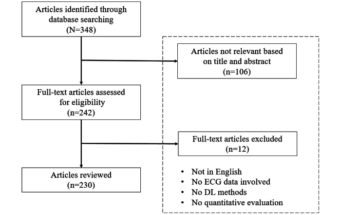
Flow diagram of the literature search. DL: deep learning; ECG: electrocardiogram.
Bibliometric Analysis
To obtain a clear picture of the literature search results, a co-occurrence analysis was conducted. For this purpose, the VOSviewer software tool (Nees Jan van Eck and Ludo Waltman) [53] was used to create and visualize 3 maps based on the bibliographic data of this study. Specifically, all keywords from the 230 relevant studies were grouped and linked to establish the impact of each keyword on the given scientific field and its interconnections with other keywords. In this way, 3 distinct clusters of keywords were formed, namely “clinical issues” (cluster 1), “methods and tools” (cluster 2), and “study characteristics” (cluster 3), as shown in Textbox 1, and an individual map was generated for each of the 3 categories. Figure 2 displays the co-occurrence network that corresponds to the “clinical issues” cluster of keywords. Cardiac arrhythmias and atrial fibrillation (AF) were identified as the major clinical issues in this review. Figure 3 presents the co-occurrence network for the “methods and tools” cluster, where ECG and DL constitute the network’s core. Finally, Figure 4 shows the co-occurrence network for the “study characteristics” cluster, where, as expected, humans are the center of attention.
Keyword cluster summary.
Cluster and keywords
-
Cluster 1
“arrhythmias, cardiac,” “atrial fibrillation,” “biometric identification,” “blood pressure determination,” “cardiomyopathy,” “cardiovascular diseases,” “coronary artery disease,” “covid-19,” “early diagnosis,” “fetal monitoring,” “heart diseases,” “heart failure,” “heartbeat classification,” “hypertension,” “monitoring, physiologic,” “myocardial infarction,” “sleep apnea,” “sudden cardiac death,” “ventricular fibrillation,” “ventricular function, left,” “ventricular premature complexes”
-
Cluster 2
“12-lead ecg,” “algorithms,” “artificial intelligence,” “attention mechanism,” “blood pressure,” “cardiology,” “continuous wavelet transform,” “convolutional neural networks, computer,” “data compression,” “deep learning,” “deep neural networks, computer,” “diagnosis, computer-assisted,” “echocardiography,” “electrocardiography,” “electroencephalography,” “feature extraction,” “feature fusion,” “heart,” “heart rate,” “heart rate variability,” “long short-term memory,” “machine learning,” “neural networks, computer,” “photoplethysmography,” “polysomnography,” “recurrent neural networks, computer,” “signal processing, computer-assisted,” “supervised machine learning,” “support vector machine,” “wavelet analysis,” “wearable electronic devices”
-
Cluster 3
“adult,” “aged,” “aged, 80 and over,” “cohort studies,” “databases, factual,” “female,” “humans,” “male,” “middle aged,” “predictive value of tests,” “pregnancy,” “reproducibility of results,” “retrospective studies,” “roc curve,” “sensitivity and specificity,” “young adult”
Figure 2.
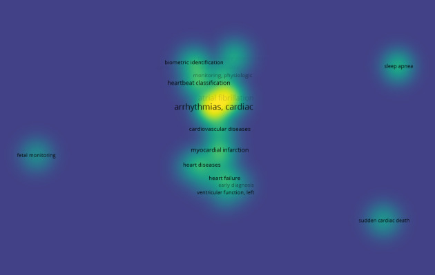
The co-occurrence network for the “clinical issues” cluster.
Figure 3.
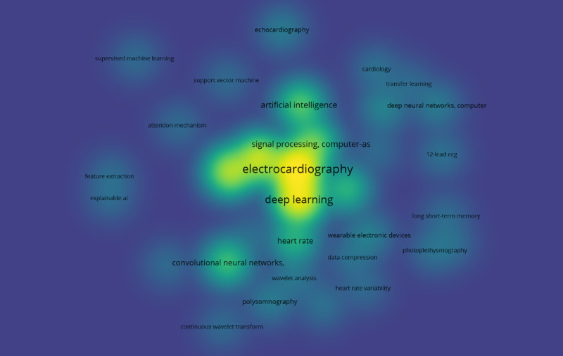
The co-occurrence network for the “methods and tools” cluster.
Figure 4.
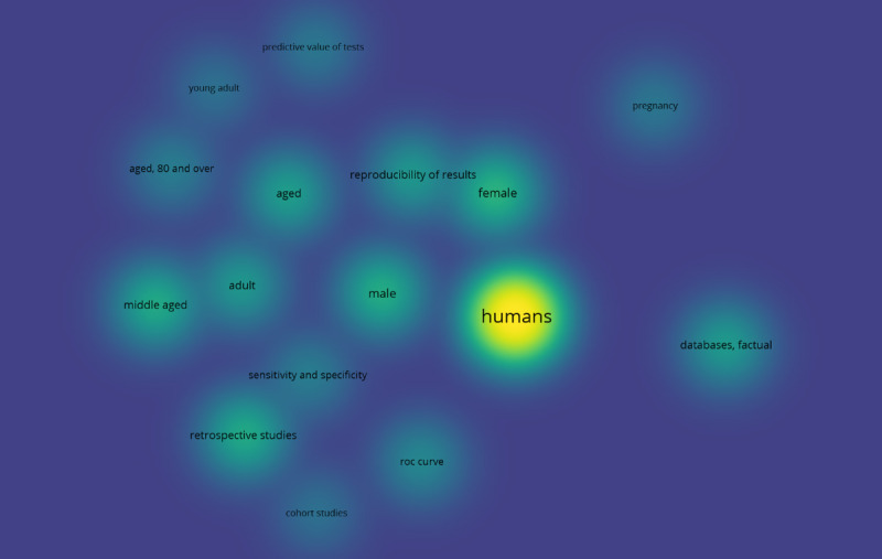
The co-occurrence network for the “study characteristics” cluster.
Results
ECG Data Sources
On the basis of the selected studies, multiple ECG data sources were identified, including several well-established publicly available databases. These data sources exhibit differences in the number of enrolled patients, number of recordings, ECG systems used to collect them, data duration, and sample rate. Their content is presented in Multimedia Appendix 1 [54-92], where the links to publicly available data are placed as hyperlinks on the name of each database.
The most commonly used databases were the Massachusetts Institute of Technology (MIT)–Beth Israel Hospital (BIH) Arrhythmia Database [80] (55/230, 23.9% studies), 2017 PhysioNet/CinC Challenge database [57] (31/230, 13.5% studies), the China Physiological Signal Challenge (CPSC) 2018 database [69] (26/230, 11.3% studies), the MIT-BIH Atrial Fibrillation Database [81] (17/230, 7.4% studies), and the Physikalisch Technische Bundesanstalt (PTB)–XL ECG data set [87] (17/230, 7.4% studies).
The MIT-BIH Arrhythmia Database contains 48 half-hour excerpts of 2-channel ambulatory ECG recordings obtained from 47 participants studied by the BIH Arrhythmia Laboratory between 1975 and 1979 with a sampling frequency of 360 Hz. Of these, 23 recordings were chosen at random from a set of 4000 recordings of 24-hour ambulatory ECG collected from a mixed population of inpatients (approximately 60%) and outpatients (approximately 40%) at Boston’s BIH, whereas the remaining 25 recordings were selected from the same set to include less common but clinically significant arrhythmias that would not be well represented in a small random sample. Finally, each recording was independently annotated by ≥2 cardiologists.
In contrast, the 2017 PhysioNet/CinC Challenge database contains 12,186 single-lead ECG recordings collected using a sampling frequency of 300 Hz. The training set contains 8528 single-lead ECG recordings lasting from 9 seconds to just >60 seconds, and the test set contains 3658 ECG recordings of similar lengths.
The CPSC 2018 database comprises ECG recordings collected from 11 hospitals by using a sampling frequency of 500 Hz. The training set contains 6877 (female: 3178; male: 3699) 12-lead ECG recordings lasting from 6 seconds to 60 seconds, and the test set, which is unavailable to the public for scoring purposes, contains 2954 ECG recordings of similar lengths.
Furthermore, the MIT-BIH Atrial Fibrillation Database includes 25 long-term ECG recordings of human patients with AF (mostly paroxysmal). The individual recordings are each 10 hours in duration and contain 2 ECG signals, each sampled at 250 Hz, whereas the rhythm annotation files were manually prepared and contain rhythm annotations of 4 types, namely, AFIB (AF), AFL (atrial flutter), J (AV junctional rhythm), and N (all other rhythms).
Finally, the PTB-XL ECG data set is a large data set of 21,837 clinical 12-lead ECGs from 18,885 patients with a duration of 10 seconds and a sampling frequency of 500 Hz. The raw waveform data were annotated by up to 2 cardiologists who assigned multiple ECG statements to each record.
Medical Applications
Overview
The 230 relevant articles identified during the literature search were grouped into several categories based on their study objectives. In particular, 6 distinct medical applications were identified: blood pressure (BP) estimation, CVD diagnosis, ECG analysis, biometric recognition, sleep analysis, and other clinical analyses.
Most of the studies use ECG signals for CVD diagnosis, mainly via signal or beat classification. Moreover, a significant portion of them uses DL algorithms to perform ECG analysis, as well as diagnosis of other clinical conditions.
In this study, the identified DL approaches are grouped per field of application, and the most notable approaches are discussed in detail. Moreover, Multimedia Appendix 2 [93-322] provides details regarding the author and the year of publication of each article, the medical task that each article refers to, data, data preprocessing, splitting strategy, DL algorithm applied in each study, and performance of each approach.
BP Estimation
Only 2.6% (6/230) of studies that applied DL methods to ECG data to perform BP estimation were identified in the literature search. A combined architecture of ResNets and LSTM was proposed twice (33.3%), once by Miao et al [94], who achieved a mean error of −0.22 (SD 5.82) mm Hg for systolic BP (SBP) prediction and of −0.75 (SD 5.62) mm Hg for diastolic BP (DBP) prediction using data that originated from a private database, and once by Paviglianiti et al [96], who achieved a mean average error of 4.118 mm Hg for SBP and 2.228 mm Hg for DBP prediction using the Medical Information Mart for Intensive Care database. By contrast, Jeong and Lim [98] exercised a CNN-LSTM network on the Medical Information Mart for Intensive Care database and managed to predict SBP and DBP with a mean error of 0.0 (SD 1.6) mm Hg and 0.2 (SD 1.3) mm Hg, respectively.
CVD Diagnosis
More than half (152/230, 66.1%) of the studies that were identified during the literature search applied DL methods to ECG data for CVD diagnosis. The most common data sources for CVD diagnosis are private (37%) and mixed public (25%) databases. However, a notable proportion (15%) of the aforementioned studies exclusively used the MIT-BIH Arrhythmia Database. Almost the half of them (10/23, 43.5%) applied a CNN structure.
Regarding the MIT-BIH Arrhythmia Database, the best accuracy (99.94%) was achieved by Wang et al [185], who introduced a fused autoencoder-CNN network to classify 6 different ECG rhythms. However, a high percentage of the studies that managed to classify data originating from the same database implemented a CNN structure. Lu et al [180] used a 1D-CNN for arrhythmia classification, achieving an accuracy of 99.31%, whereas Yu et al [219] used a 1D-CNN to detect premature ventricular contraction, achieving a classification accuracy of 99.70%.
On the contrary, a ResNet architecture was tested only 3 times on the MIT-BIH Arrhythmia Database; nonetheless, 0.9% (2/230) of these studies showed a high model performance. In particular, Li et al [146] proposed a ResNet model for heartbeat classification, achieving a classification accuracy of 99.38%, whereas Zhang et al [211] used a ResNet-101 structure to classify ECG beats with transfer learning and achieved an accuracy of 99.75%.
Regarding the rest of the databases, several noteworthy studies were identified in the literature. Specifically, Cai et al [101] implemented a densely connected DNN on a private ECG database for AF detection, achieving an accuracy between 97.74% and 99.35% for 3 different classification tasks, whereas Ghosh et al [103] applied a hierarchical extreme learning machine to ECG data from multiple public databases, achieving an accuracy of 99.40% in detecting AF.
Furthermore, Butun et al [125] proposed a 1D-capsule NN for the detection of coronary artery disease, achieving classification accuracies of 99.44% and 98.62% on 2-second and 5-second ECG segments, respectively, originating from a private ECG database. Another study by Thiagarajan et al [129] used multiple convolutional and pooling layers within a structure named DDxNet on ECG data from 2 public databases, achieving an accuracy of 98.50% for arrhythmia classification and 99.90% for myocardial infarction detection.
A study by Radhakrishnan et al [163] evaluated the performance (sensitivity 99.17%, specificity 99.18%, and accuracy 99.18%) of a 2D bidirectional LSTM network to detect AF in ECG signals from 4 public databases, whereas Petmezas et al [170] tested (sensitivity 97.87% and specificity 99.29%) a CNN-LSTM model on ECG signals originating from the MIT-BIH Atrial Fibrillation Database for the same medical task.
Moreover, Jahmunah et al [192] applied a CNN architecture to ECG data from several public ECG databases to detect coronary artery disease, myocardial infarction, and congestive heart failure, achieving an accuracy of 99.55%. Another study by Dai et al [195] proposed a CNN for CVD diagnosis using different intervals of ECG signals from the PTB Diagnostic ECG Database and achieved accuracies of 99.59%, 99.80%, and 99.84% for 1-, 2-, and 3-second ECG segments, respectively.
Finally, Ma et al [208] introduced an improved dilated causal CNN to classify ECG signals from the MIT-BIH Atrial Fibrillation Database, achieving a high model performance (sensitivity 98.79%, specificity 99.04%, and accuracy 98.65%), whereas Zhang et al [238] tested (sensitivity 99.65%, specificity 99.98%, and accuracy 99.84%) a CNN for AF detection on ECG signals from 2 major public databases.
ECG Analysis
In total, 12.6% (29/230) of studies that applied DL methods to ECG data to perform ECG analysis were identified during the literature search. Once again, CNN was the most commonly used DL method (11/29, 38%); nonetheless, the best model accuracy was achieved by studies using other DL methods. In particular, Teplitzky et al [251] tested (sensitivity 99.84% and positive predictive value 99.78%) a hybrid approach that combines 2 DL approaches, namely BeatNet and RhythmNet, to annotate ECG signals that originated from both public and private ECG databases, whereas Murat et al [258] used a CNN-LSTM approach on ECG data from the MIT-BIH Arrhythmia Database and achieved an accuracy of 99.26% in detecting 5 types of ECG beats.
By contrast, Vijayarangan et al [261] used a fused CNN-ResNet structure to perform R peak detection in ECG signals from several public ECG databases and achieved F1-scores between 96.32% and 99.65% for 3 testing data sets. Another study by Jimenez Perez et al [265] implemented a U-Net model to delineate 2-lead ECG signals originating from the QT Database and achieved sensitivities of 98.73%, 99.94%, and 99.88% for P wave, QRS complex, and T wave detection, respectively. Finally, a study by Strodthoff et al [274] used a ResNet for patient sex identification by using 12-lead ECG recordings lasting between 6 and 60 seconds from several public databases and achieved an area under the curve of 0.925 for the PTB-XL ECG data set and 0.974 for the CPSC 2018 database.
Biometric Recognition
Only 3% (7/230) of studies that applied DL methods to ECG data to perform biometric recognition were identified in the literature search. Although 57% (4/7) of the studies used a CNN architecture, only 29% (2/7) of them achieved high model performance. Specifically, Wu et al [284] achieved an identification rate of >99% by using ECG signals from 2 public databases, whereas Chiu et al [285] achieved an identification rate of 99.10% by using single-lead ECG recordings that originated from the PTB Diagnostic ECG Database.
On the contrary, Song et al [281] implemented a ResNet-50 architecture for person identification using multiple ECG, face, and fingerprint data from several public and private databases and achieved an accuracy of 98.97% for ID classification and 96.55% for gender classification. Finally, AlDuwaile and Islam [283] tested several pretrained models, including GoogleNet, ResNet, MobileNet, and EfficientNet, and a CNN model to perform human recognition using ECG signals that originated from 2 public databases and achieved an accuracy between 94.18% and 98.20% for ECG-ID mixed-session and multisession data sets.
Sleep Analysis
Approximately 5.2% (12/230) of studies that applied DL methods to ECG data to perform sleep analysis were identified during the literature search. Half (6/12, 50%) of the studies proposed a CNN model, some of which achieved high performance in several sleep analysis–related tasks. In particular, Chang et al [289] used 1-minute ECG segments from the Apnea-ECG Database and designed a CNN to detect sleep apnea, achieving an accuracy of 87.90% and 97.10% for per-minute and per-recording classification, respectively.
In addition, a study by Urtnasan et al [291] proposed a CNN for the identification of sleep apnea severity by using ECG segments from a private database and achieved an F1-score of 98.00%, whereas another study by Urtnasan et al [297] implemented a CNN to classify sleep disorders by using polysomnography recordings from the Cyclic Alternating Pattern Sleep Database and achieved F1-scores between 95% and 99% for 5 different sleep disorder categories. By contrast, Nasifoglu and Erogul [295] tested a fused CNN-ResNet approach for obstructive sleep apnea detection (accuracy 85.20%) and prediction (accuracy 82.30%) using data from a private database. Mukherjee et al [296] used a multilayer perceptron to detect sleep apnea from ECG recordings that originated from the Apnea-ECG Database, achieving an accuracy of 85.58%.
Other Clinical Analyses
Approximately 10.4% (24/230) of studies that applied DL methods to ECG data to perform other clinical analyses were identified during the literature search. Almost half (10/24, 42%) of the studies proposed a CNN approach, including Isasi et al [300], who used data from a private database to detect shockable and nonshockable rhythms during cardiopulmonary resuscitation with an accuracy of 96.10%, and Ozdemir et al [309], who used a private database to diagnose COVID-19 through ECG classification (accuracy 93.00%).
Other notable works include a study by Chang et al [311], which tested (sensitivity 84.60% and specificity 96.60%) an ECG12Net to detect digoxin toxicity by using private ECG signals from patients with digoxin toxicity and patients in the emergency room, and another study by Baghersalimi et al [313], which evaluated the performance (sensitivity 90.24% and specificity 91.58%) of a fused CNN-ResNet network to detect epileptic seizure events from single-lead ECG signals originating from a private database. Finally, Mazumder et al [318] implemented a CNN-LSTM structure for the detection of shockable rhythms in ECG signals from 2 public databases, achieving sensitivity scores between 94.68% and 99.21% and specificity scores between 92.77% and 99.68% for 2- and 8-second time windows, respectively.
Discussion
Principal Findings
DL has led to the creation of robust models that could potentially perform fast and reliable clinical diagnoses based on physiological signals. Remarkably, during the past 2 years, at least 230 studies that used DL on ECG data for various clinical applications were identified in the literature, which is a large number for such a short period, regardless of the application domain. This is mainly justified by the fact that DL methods can automatically capture distinctive features from ECG signals based on the trained models that achieve promising diagnostic performance, as shown in Multimedia Appendix 2 [93-322]. This constitutes a significant advantage compared with classical ML methods that perform manual feature selection and feature extraction—2 processes that conventionally require considerable effort and time [323]. Overall, CNN represents the most popular DL architecture and has been identified in most of the reviewed studies (142/230, 60.9% articles). On the contrary, 18.3% (42/230) of studies used LSTM architecture, whereas a ResNet architecture was used in 17.8% (41/230) of cases.
However, training a DL model is not always straightforward. Both architectural design choices and parameter tuning influence model performance; thus, multiple combinations must be considered. Furthermore, the training phase of DL algorithms typically involves complex computations that can be translated into long training times. This requires expensive state-of-the-art computer hardware, including graphics processing units that can dramatically accelerate the total execution time [324].
Another common problem with DL algorithms is overfitting; this occurs when the algorithm fits the noise and therefore performs well on the training set but fails to generalize its predictions to unseen data (ie, the testing set). For this reason, it is necessary to adopt an early stopping strategy during the training phase to prevent further training when the model’s performance on unknown data starts to deteriorate. This is usually done by implementing a separate data set, called the validation set, which most of the time is a small percentage of the training set that is held back from training to provide an unbiased evaluation of the model during training. Moreover, random data splitting can introduce bias; thus, k-fold cross-validation or leave-one-out cross-validation strategies are preferred when training DL models. In addition, it is important that different sets (ie, training, validation, and testing) contain different patients, also known as interpatient data splitting, so that the study’s results are more reliable. As concluded by this review and presented in Multimedia Appendix 2 [93-322], many researchers do not take this into consideration; hence, their results are questionable.
Another critical issue related to overfitting is the distribution of labels or predicted variables in the data set used for model development and validation. For instance, in the BP prediction problem, large stretches of constant BP from the same individual would bias the network toward a constant predictor with minimal error, with the network preferring to memorize patient-identifying features to predict the average BP for a patient rather than those which represent physiological metrics useful in predicting variable BP for the same patient. The resulting errors would be deceptively low if a patient’s nominal BP does not change but, critically, would not be clinically useful in the setting of hypertensive or hypotensive crisis or to guide patient care. None of the assessed papers described the results, indicating that the predicted BP follows meaningful trends.
Recent attention in the medical field to the concept of BP variability [325] rather than clinical spot checks highlights the need for ambulatory BP monitors that are both ergonomic for the patient to increase compliance and comfort, as well as reliable and well validated. A common pitfall in the use of calibrated techniques is that subsequent test data points do not differ significantly from the calibration value and thus yield small errors in prediction, whereas the data are presented as an aggregate pooled correlation plot or Bland-Altman plot with a correlation value that simply reflects the range of BPs across the population rather than patient-specific BP variation [326,327]. In our review of articles using DL for BP prediction, we did not encounter significant attempts to address the issue of BP variability in training data; in fact, many publications explicitly removed data points with hypertensive values or large pulse pressures from their data sets as “artifacts” [93-96,98].
In a calibration-less approach, a narrow range of variation would lead to a low prediction error even when predicting the population mean for each patient. If an ambulatory BP monitoring device plans to use AI-based techniques to measure variability, this variability must be represented in the training set for a model to learn to predict such changes adequately. A way of accomplishing this is to incorporate a variety of BP-modulating activities in the training data, which represent different sources of BP change and corresponding modulations in the feature space. For example, ice pressor tests may increase BP via peripheral vasoconstriction [328], whereas the valsalva maneuver increases chest pressure extrinsically [329] and may modulate input features such as heart rate in opposite ways, reducing the chance that bias-prone DL architectures learn misleading relationships.
In addition to the training and evaluation data, evaluation metrics and cost functions are areas with significant room for improvement. Mean squared error alone can be minimized with a constant predictor if the BP range does not vary significantly. Alternative cost functions such as cosine similarity, which is maximized with constant inputs, contrastive losses, or combinations thereof, have been successful in classification problems in imbalanced, rare event prediction problems such as critical events in patients with COVID-19 [330]. For other promising solutions, it would be prudent to examine similar trend prediction problems in other fields such as stock price movement, where progress has been made using intuitive data preparation and creative representation of the prediction targets, in this case, price changes, to generate trend deterministic predictions [331].
Furthermore, a vast majority of available ECG data sources experience data imbalance. This creates a major problem when trying to predict smaller classes that usually represent rare conditions or diseases that are as important as larger classes when designing health care decision support systems. To solve this problem, several oversampling techniques have been proposed, including random oversampling and undersampling, the synthetic minority oversampling technique [332], the adaptive synthetic sampling technique [333], the generative oversampling method [334], distribution-based balancing [335], and new loss functions such as focal loss [336], which can achieve both prediction error reduction and data imbalance handling. Papers addressing classification frequently use techniques to address class imbalance; however, evidence for such corrections in regression models does not appear as frequently or rigorously.
In addition, DL models are often characterized by black box behavior (lack of interpretability); that is, it is difficult for a human to understand why a particular result is generated by such complex architectures. This is crucial when training models for medical applications, as diagnoses based on unexplained model predictions are not usually accepted by medical experts. A possible solution to this problem is to take advantage of algorithms that are more easily interpretable, such as decision trees [337], additive models [338], attention-based networks [339], and sparse linear models [340], when designing a DL architecture. By contrast, several DL model interpretation approaches have been proposed in this direction, including permutation feature importance [341], partial dependence plots [342], and local interpretable model-agnostic explanations [343]. However, these techniques are rarely used in practice as they require additional time and effort. A useful technique that is used more often when dealing with medical images (and CNNs) is gradient-weighted class activation mapping [344], which makes CNN-based models more transparent by presenting visual explanations for their decisions.
Uncertainty quantification is another common problem associated with DL methods, which has recently drawn the attention of researchers. There are 2 main types of uncertainty: aleatoric (data uncertainty) and epistemic (knowledge uncertainty). It is important to evaluate the reliability and validity of DL methods before they can be tested in real-world applications; thus, uncertainty estimation should be provided. In the past few years, several uncertainty quantification techniques have been proposed, including deep Bayesian active learning [345], Monte Carlo dropout [346], Markov chain Monte Carlo [347], and Bayes by backprop [348].
Moreover, as presented in Multimedia Appendix 1 [54-92], there is no gold standard for data collection. As shown in Multimedia Appendix 2 [93-322], different studies used ECG data with distinct characteristics, namely, the number of leads, signal duration, and sample rate. In addition, many studies used multimodal data, such as photoplethysmograms, arterial BP, polysomnography, and electroencephalograms. Some studies used raw waveforms as input to their models, whereas others precomputed a set of features. This heterogeneity makes it difficult to compare study results; thus, finding the best algorithm is challenging, if not impossible.
Recent advancements [349] in materials and techniques to produce flexible, skin-integrated technology [350] have enabled the development of unique sensors and devices that can simultaneously measure both conventional and novel types of signals from the human body. Small wireless devices [351] such as these can extract continuous ECG; acceleration-based body orientation; physical activity [352]; vibrations such as heart sounds, breath sounds [353]; vocal processes [354]; and photoplethysmogram signals at multiple wavelengths and body locations. This wealth of physiological information that can be measured noninvasively and continuously throughout day-to-day life is potentially a treasure trove of useful insights into health status outside the rigidity of a clinical system. Tools such as DL have emerged as a tantalizing approach to take advantage of such multivariate data in the context of the increased complexity and unpredictability of ambulatory environments. With careful data curation and training approaches, as well as the use of intuitive, well-justified algorithms and network structures, explainable AI can also provide justifications for the use of novel features of underlying physiological relevance. Currently, the use of highly complex and computationally expensive DL models in wearable applications is limited. Generally, raw data are processed in a post hoc fashion after data have been uploaded to cloud servers, limiting real-time feedback. However, recently, there have been developments by chip manufacturers to enable “edge inferencing” by bringing AI-enabling computational acceleration to the low–power-integrated circuit level, opening up the possibilities for low-latency applications of DL algorithms. We strongly believe that the creation of robust DL models that can assist medical experts in clinical decision-making is an important direction for future investigations.
In general, we believe that with this study, we (1) provided a complete and systematic account of the current state-of-the-art DL methods applied to ECG data; (2) identified several ECG data sources used in clinical diagnosis, even some not so widely cited databases; and (3) identified important open research problems and provided suggestions for future research directions in the field of DL and ECG data. Several important relevant review studies have already presented novel DL methods that are used on ECG data [355-357]. Nonetheless, none of them combine all the aforementioned characteristics, which makes this study innovative.
By contrast, the limitations of this study could be summarized as the fact that owing to the enormous number of studies focusing on DL and ECG data, we performed a review based only on articles that have been published in various journals between January 2020 and December 2021.
Although the rationale behind this study was to identify all state-of-the-art DL methods that are applied to ECG data for various clinical applications, in the future, we intend to concentrate our efforts on providing a more complete account of DL methods that make good use of ECG data to address a specific clinical task (ie, congestive heart failure diagnosis).
Conclusions
In this study, we systematically reviewed 230 recently published articles on DL methods applied to ECG data for various clinical applications. We attempted to group the proposed DL approaches per field of application and summarize the most notable approaches among them. To the best of our knowledge, this is the first study that provides a complete account of the detailed strategy for designing each one of the proposed DL systems by recording the ECG data sources, data preprocessing techniques, model training, evaluation processes, and data splitting strategies that are implemented in each approach. Finally, open research problems and potential gaps were discussed to assess the future of the field and provide guidance to new researchers to design and implement reliable DL algorithms that can provide accurate diagnoses based on ECG data to support medical experts’ efforts for clinical decision-making.
Abbreviations
- AF
atrial fibrillation
- AI
artificial intelligence
- BIH
Beth Israel Hospital
- BP
blood pressure
- CNN
convolutional neural network
- CPSC
China Physiological Signal Challenge
- CVD
cardiovascular disease
- DBP
diastolic blood pressure
- DL
deep learning
- DNN
deep neural network
- ECG
electrocardiogram
- GRU
gated recurrent unit
- LSTM
long short-term memory
- MIT
Massachusetts Institute of Technology
- ML
machine learning
- NN
neural network
- PTB
Physikalisch Technische Bundesanstalt
- ResNet
residual neural network
- RNN
recurrent neural network
- SBP
systolic blood pressure
Summary of the major electrocardiogram databases.
Summary of works carried out using deep-learning algorithms and electrocardiogram signals.
Footnotes
Conflicts of Interest: None declared.
References
- 1.Schlant RC, Adolph RJ, DiMarco JP, Dreifus LS, Dunn MI, Fisch C, Garson A, Haywood LJ, Levine HJ, Murray JA. Guidelines for electrocardiography. A report of the American college of cardiology/American heart association task force on assessment of diagnostic and therapeutic cardiovascular procedures (committee on electrocardiography) Circulation. 1992 Mar;85(3):1221–8. doi: 10.1161/0cir.85.3.1221. doi: 10.1161/0cir.85.3.1221. [DOI] [PubMed] [Google Scholar]
- 2.Salerno SM, Alguire PC, Waxman HS, American College of Physicians Training and competency evaluation for interpretation of 12-lead electrocardiograms: recommendations from the American College of Physicians. Ann Intern Med. 2003 May 06;138(9):747–50. doi: 10.7326/0003-4819-138-9-200305060-00012. https://www.acpjournals.org/doi/abs/10.7326/0003-4819-138-9-200305060-00012?url_ver=Z39.88-2003&rfr_id=ori:rid:crossref.org&rfr_dat=cr_pub%3dpubmed .200305060-00012 [DOI] [PubMed] [Google Scholar]
- 3.Cook DA, Oh SY, Pusic MV. Accuracy of physicians' electrocardiogram interpretations: a systematic review and meta-analysis. JAMA Intern Med. 2020 Nov 01;180(11):1461–71. doi: 10.1001/jamainternmed.2020.3989. http://europepmc.org/abstract/MED/32986084 .2771093 [DOI] [PMC free article] [PubMed] [Google Scholar]
- 4.Ramesh AN, Kambhampati C, Monson JR, Drew PJ. Artificial intelligence in medicine. Ann R Coll Surg Engl. 2004 Sep;86(5):334–8. doi: 10.1308/147870804290. http://europepmc.org/abstract/MED/15333167 . [DOI] [PMC free article] [PubMed] [Google Scholar]
- 5.Johnson KW, Torres Soto J, Glicksberg BS, Shameer K, Miotto R, Ali M, Ashley E, Dudley JT. Artificial intelligence in cardiology. J Am Coll Cardiol. 2018 Jun 12;71(23):2668–79. doi: 10.1016/j.jacc.2018.03.521. https://linkinghub.elsevier.com/retrieve/pii/S0735-1097(18)34408-5 .S0735-1097(18)34408-5 [DOI] [PubMed] [Google Scholar]
- 6.Awan SE, Sohel F, Sanfilippo FM, Bennamoun M, Dwivedi G. Machine learning in heart failure: ready for prime time. Curr Opin Cardiol. 2018 Mar;33(2):190–5. doi: 10.1097/hco.0000000000000491. doi: 10.1097/hco.0000000000000491. [DOI] [PubMed] [Google Scholar]
- 7.Lai Y. A comparison of traditional machine learning and deep learning in image recognition. J Phys Conf Ser. 2019 Oct 01;1314(1):012148. doi: 10.1088/1742-6596/1314/1/012148. doi: 10.1088/1742-6596/1314/1/012148. [DOI] [Google Scholar]
- 8.LeCun Y, Bengio Y, Hinton G. Deep learning. Nature. 2015 May 28;521(7553):436–44. doi: 10.1038/nature14539. doi: 10.1038/nature14539.nature14539 [DOI] [PubMed] [Google Scholar]
- 9.Advanced Machine Learning Technologies and Applications. Singapore: Springer; 2021. Deep learning techniques: an overview. https://link.springer.com/chapter/10.1007/978-981-15-3383-9_54 . [Google Scholar]
- 10.LeCun Y, Boser B, Denker JS, Henderson D, Howard RE, Hubbard W, Jackel LD. Backpropagation applied to handwritten zip code recognition. Neural Comput. 1989 Dec;1(4):541–51. doi: 10.1162/neco.1989.1.4.541. [DOI] [Google Scholar]
- 11.Shape, Contour and Grouping in Computer Vision. Berlin, Heidelberg: Springer; 1999. Object recognition with gradient-based learning. [Google Scholar]
- 12.Anwar SM, Majid M, Qayyum A, Awais M, Alnowami M, Khan MK. Medical image analysis using convolutional neural networks: a review. J Med Syst. 2018 Oct 08;42(11):226. doi: 10.1007/s10916-018-1088-1. doi: 10.1007/s10916-018-1088-1.10.1007/s10916-018-1088-1 [DOI] [PubMed] [Google Scholar]
- 13.Kamaleswaran R, Mahajan R, Akbilgic O. A robust deep convolutional neural network for the classification of abnormal cardiac rhythm using single lead electrocardiograms of variable length. Physiol Meas. 2018 Mar 27;39(3):035006. doi: 10.1088/1361-6579/aaaa9d. doi: 10.1088/1361-6579/aaaa9d. [DOI] [PubMed] [Google Scholar]
- 14.Craik A, He Y, Contreras-Vidal JL. Deep learning for electroencephalogram (EEG) classification tasks: a review. J Neural Eng. 2019 Jun;16(3):031001. doi: 10.1088/1741-2552/ab0ab5. doi: 10.1088/1741-2552/ab0ab5. [DOI] [PubMed] [Google Scholar]
- 15.Bardou D, Zhang K, Ahmad SM. Lung sounds classification using convolutional neural networks. Artif Intell Med. 2018 Jun;88:58–69. doi: 10.1016/j.artmed.2018.04.008. doi: 10.1016/j.artmed.2018.04.008.S0933-3657(17)30205-1 [DOI] [PubMed] [Google Scholar]
- 16.Zhang Q, Fu L, Gu L. A cascaded convolutional neural network for assessing signal quality of dynamic ECG. Comput Math Methods Med. 2019;2019:7095137. doi: 10.1155/2019/7095137. doi: 10.1155/2019/7095137. [DOI] [PMC free article] [PubMed] [Google Scholar]
- 17.Wu H, Soraghan J, Lowit A, Di Caterina G. Convolutional neural networks for pathological voice detection. Annu Int Conf IEEE Eng Med Biol Soc. 2018 Jul;2018:1–4. doi: 10.1109/embc.2018.8513222. doi: 10.1109/embc.2018.8513222. [DOI] [PubMed] [Google Scholar]
- 18.Chriskos P, Frantzidis CA, Gkivogkli PT, Bamidis PD, Kourtidou-Papadeli C. Automatic sleep staging employing convolutional neural networks and cortical connectivity images. IEEE Trans Neural Netw Learning Syst. 2020 Jan;31(1):113–23. doi: 10.1109/tnnls.2019.2899781. doi: 10.1109/tnnls.2019.2899781. [DOI] [PubMed] [Google Scholar]
- 19.He K, Zhang X, Ren S, Sun J. Deep residual learning for image recognition. Proceedings of the 2016 IEEE Conference on Computer Vision and Pattern Recognition (CVPR); 2016 IEEE Conference on Computer Vision and Pattern Recognition (CVPR); Jun 27-30, 2016; Las Vegas, NV, USA. 2016. [DOI] [Google Scholar]
- 20.Xu H, Baxter JS, Akin O, Cantor-Rivera D. Prostate cancer detection using residual networks. Int J Comput Assist Radiol Surg. 2019 Oct;14(10):1647–50. doi: 10.1007/s11548-019-01967-5. http://europepmc.org/abstract/MED/30972686 .10.1007/s11548-019-01967-5 [DOI] [PMC free article] [PubMed] [Google Scholar]
- 21.Wang EK, Zhang X, Pan L, Cheng C, Dimitrakopoulou-Strauss A, Li Y, Zhe N. Multi-path dilated residual network for nuclei segmentation and detection. Cells. 2019 May 23;8(5):499. doi: 10.3390/cells8050499. https://www.mdpi.com/resolver?pii=cells8050499 .cells8050499 [DOI] [PMC free article] [PubMed] [Google Scholar]
- 22.Datong C, Minghui L, Cheng J, Yue S, Dongbin X, Yueming L. Coronary calcium detection based on improved deep residual network in mimics. J Med Syst. 2019 Mar 25;43(5):119. doi: 10.1007/s10916-019-1218-4. doi: 10.1007/s10916-019-1218-4.10.1007/s10916-019-1218-4 [DOI] [PubMed] [Google Scholar]
- 23.Nibali A, He Z, Wollersheim D. Pulmonary nodule classification with deep residual networks. Int J Comput Assist Radiol Surg. 2017 Oct;12(10):1799–808. doi: 10.1007/s11548-017-1605-6. doi: 10.1007/s11548-017-1605-6.10.1007/s11548-017-1605-6 [DOI] [PubMed] [Google Scholar]
- 24.Usama M, Ahmad B, Xiao W, Hossain MS, Muhammad G. Self-attention based recurrent convolutional neural network for disease prediction using healthcare data. Comput Methods Programs Biomed. 2020 Jul;190:105191. doi: 10.1016/j.cmpb.2019.105191. doi: 10.1016/j.cmpb.2019.105191.S0169-2607(19)31170-8 [DOI] [PubMed] [Google Scholar]
- 25.Chakravarty A, Sivaswamy J. RACE-Net: a recurrent neural network for biomedical image segmentation. IEEE J Biomed Health Inform. 2019 May;23(3):1151–62. doi: 10.1109/jbhi.2018.2852635. doi: 10.1109/jbhi.2018.2852635. [DOI] [PubMed] [Google Scholar]
- 26.Arsenali B, van Dijk J, Ouweltjes O, den Brinker B, Pevernagie D, Krijn R, van Gilst M, Overeem S. Recurrent neural network for classification of snoring and non-snoring sound events. Annu Int Conf IEEE Eng Med Biol Soc. 2018 Jul;2018:328–31. doi: 10.1109/embc.2018.8512251. doi: 10.1109/embc.2018.8512251. [DOI] [PubMed] [Google Scholar]
- 27.Rajeev R, Samath JA, Karthikeyan NK. An intelligent recurrent neural network with long short-term memory (LSTM) BASED batch normalization for medical image denoising. J Med Syst. 2019 Jun 15;43(8):234. doi: 10.1007/s10916-019-1371-9. doi: 10.1007/s10916-019-1371-9.10.1007/s10916-019-1371-9 [DOI] [PubMed] [Google Scholar]
- 28.Liu M, Cheng D, Yan W, Alzheimer’s Disease Neuroimaging Initiative Classification of alzheimer's disease by combination of convolutional and recurrent neural networks using FDG-PET images. Front Neuroinform. 2018;12:35. doi: 10.3389/fninf.2018.00035. doi: 10.3389/fninf.2018.00035. [DOI] [PMC free article] [PubMed] [Google Scholar]
- 29.Beeksma M, Verberne S, van den Bosch A, Das E, Hendrickx I, Groenewoud S. Predicting life expectancy with a long short-term memory recurrent neural network using electronic medical records. BMC Med Inform Decis Mak. 2019 Feb 28;19(1):36. doi: 10.1186/s12911-019-0775-2. https://bmcmedinformdecismak.biomedcentral.com/articles/10.1186/s12911-019-0775-2 .10.1186/s12911-019-0775-2 [DOI] [PMC free article] [PubMed] [Google Scholar]
- 30.Gao J, Zhang H, Lu P, Wang Z. An effective LSTM recurrent network to detect arrhythmia on imbalanced ECG dataset. J Healthc Eng. 2019;2019:6320651. doi: 10.1155/2019/6320651. doi: 10.1155/2019/6320651. [DOI] [PMC free article] [PubMed] [Google Scholar]
- 31.Ahmedt-Aristizabal D, Fookes C, Nguyen K, Sridharan S. Deep classification of epileptic signals. Annu Int Conf IEEE Eng Med Biol Soc. 2018 Jul;2018:332–5. doi: 10.1109/embc.2018.8512249. doi: 10.1109/embc.2018.8512249. [DOI] [PubMed] [Google Scholar]
- 32.Wollmann T, Gunkel M, Chung I, Erfle H, Rippe K, Rohr K. GRUU-Net: integrated convolutional and gated recurrent neural network for cell segmentation. Med Image Anal. 2019 Aug;56:68–79. doi: 10.1016/j.media.2019.04.011. doi: 10.1016/j.media.2019.04.011.S1361-8415(18)30675-3 [DOI] [PubMed] [Google Scholar]
- 33.Taheri Dezaki F, Liao Z, Luong C, Girgis H, Dhungel N, Abdi AH, Behnami D, Gin K, Rohling R, Abolmaesumi P, Tsang T. Cardiac phase detection in echocardiograms with densely gated recurrent neural networks and global extrema loss. IEEE Trans Med Imaging. 2019 Aug;38(8):1821–32. doi: 10.1109/tmi.2018.2888807. doi: 10.1109/tmi.2018.2888807. [DOI] [PubMed] [Google Scholar]
- 34.Vaswani A, Shazeer N, Parmar N, Uszkoreit J, Jones L, Gomez AN, Kaiser L, Polosukhin I. Attention is all you need. ArXiv. 2017 [Google Scholar]
- 35.Natarajan A, Chang Y, Mariani S, Rahman A, Boverman G, Vij S, Rubin J. A wide and deep transformer neural network for 12-lead ECG classification. Computing Cardiol. 2020;47 doi: 10.22489/cinc.2020.107. doi: 10.22489/cinc.2020.107. [DOI] [Google Scholar]
- 36.Wang L, Niu D, Zhao X, Wang X, Hao M, Che H. A comparative analysis of novel deep learning and ensemble learning models to predict the allergenicity of food proteins. Foods. 2021 Apr 09;10(4):809. doi: 10.3390/foods10040809. https://www.mdpi.com/resolver?pii=foods10040809 .foods10040809 [DOI] [PMC free article] [PubMed] [Google Scholar]
- 37.Yang F, Wang X, Ma H, Li J. Transformers-sklearn: a toolkit for medical language understanding with transformer-based models. BMC Med Inform Decis Mak. 2021 Jul 30;21(Suppl 2):90. doi: 10.1186/s12911-021-01459-0. https://bmcmedinformdecismak.biomedcentral.com/articles/10.1186/s12911-021-01459-0 .10.1186/s12911-021-01459-0 [DOI] [PMC free article] [PubMed] [Google Scholar]
- 38.Rajan K, Zielesny A, Steinbeck C. DECIMER 1.0: deep learning for chemical image recognition using transformers. J Cheminform. 2021 Aug 17;13(1):61. doi: 10.1186/s13321-021-00538-8. doi: 10.1186/s13321-021-00538-8.10.1186/s13321-021-00538-8 [DOI] [PMC free article] [PubMed] [Google Scholar]
- 39.Zhu Y, Wang MD, Tong L, Deshpande SR. Improved prediction on heart transplant rejection using convolutional autoencoder and multiple instance learning on whole-slide imaging. IEEE EMBS Int Conf Biomed Health Inform. 2019 May;2019 doi: 10.1109/bhi.2019.8834632. http://europepmc.org/abstract/MED/32577622 . [DOI] [PMC free article] [PubMed] [Google Scholar]
- 40.Song T, Sanchez V, EI Daly H, Rajpoot NM. Simultaneous cell detection and classification in bone marrow histology images. IEEE J Biomed Health Inform. 2019 Jul;23(4):1469–76. doi: 10.1109/jbhi.2018.2878945. doi: 10.1109/jbhi.2018.2878945. [DOI] [PubMed] [Google Scholar]
- 41.Xu X, Gu H, Wang Y, Wang J, Qin P. Autoencoder based feature selection method for classification of anticancer drug response. Front Genet. 2019;10:233. doi: 10.3389/fgene.2019.00233. doi: 10.3389/fgene.2019.00233. [DOI] [PMC free article] [PubMed] [Google Scholar]
- 42.Gordon M, Williams C. PVC detection using a convolutional autoencoder and random forest classifier. Biocomputing. 2019:42–53. doi: 10.1142/9789813279827_0005. doi: 10.1142/9789813279827_0005. [DOI] [PubMed] [Google Scholar]
- 43.Tong L, Wu H, Wang MD. CAESNet: convolutional AutoEncoder based Semi-supervised Network for improving multiclass classification of endomicroscopic images. J Am Med Inform Assoc. 2019 Nov 01;26(11):1286–96. doi: 10.1093/jamia/ocz089. http://europepmc.org/abstract/MED/31260038 .5526176 [DOI] [PMC free article] [PubMed] [Google Scholar]
- 44.Vaillant R. Original approach for the localisation of objects in images. IEE Proc Vis Image Process. 1994;141(4):245. doi: 10.1049/ip-vis:19941301. doi: 10.1049/ip-vis:19941301. [DOI] [Google Scholar]
- 45.Kiranyaz S, Avci O, Abdeljaber O, Ince T, Gabbouj M, Inman DJ. 1D convolutional neural networks and applications: a survey. Mech Syst Signal Process. 2021 Apr;151:107398. doi: 10.1016/j.ymssp.2020.107398. doi: 10.1016/j.ymssp.2020.107398. [DOI] [Google Scholar]
- 46.Rumelhart DE, Hinton GE, Williams RJ. Learning representations by back-propagating errors. Nature. 1986 Oct;323(6088):533–6. doi: 10.1038/323533a0. doi: 10.1038/323533a0. [DOI] [Google Scholar]
- 47.Hochreiter S, Schmidhuber J. Long short-term memory. Neural Comput. 1997 Nov 15;9(8):1735–80. doi: 10.1162/neco.1997.9.8.1735. doi: 10.1162/neco.1997.9.8.1735. [DOI] [PubMed] [Google Scholar]
- 48.Cho K, Merrienboer B, Gulcehre C, Bahdanau D, Bougares F, Schwenk H, Bengio Y. Learning phrase representations using RNN encoder–decoder for statistical machine translation. Proceedings of the 2014 Conference on Empirical Methods in Natural Language Processing (EMNLP); 2014 Conference on Empirical Methods in Natural Language Processing (EMNLP); Oct, 2014; Doha, Qatar. 2014. [DOI] [Google Scholar]
- 49.Parallel Distributed Processing: Explorations in the Microstructure of Cognition: Foundations. Cambridge, Massachusetts, United States: MIT Press; 1986. [Google Scholar]
- 50.Vincent P, Larochelle H, Bengio Y, Manzagol PA. Extracting and composing robust features with denoising autoencoders. Proceedings of the 25th international conference on Machine learning; ICML '08: The 25th Annual International Conference on Machine Learning held in conjunction with the 2007 International Conference on Inductive Logic Programming; Jul 5 - 9, 2008; Helsinki Finland. 2008. [DOI] [Google Scholar]
- 51.Kingma DP, Welling M. Auto-encoding variational bayes. arXiv. 2014 http://arxiv.org/abs/1312.6114 . [Google Scholar]
- 52.Li F, Qiao H, Zhang B. Discriminatively boosted image clustering with fully convolutional auto-encoders. Pattern Recognition. 2018 Nov;83:161–73. doi: 10.1016/j.patcog.2018.05.019. doi: 10.1016/j.patcog.2018.05.019. [DOI] [Google Scholar]
- 53.Welcome to VOSviewer. VOSviewer. [2021-11-09]. https://www.vosviewer.com/
- 54.Goldberger AL, Amaral LA, Glass L, Hausdorff JM, Ivanov PC, Mark RG, Mietus JE, Moody GB, Peng CK, Stanley HE. PhysioBank, PhysioToolkit, and PhysioNet: components of a new research resource for complex physiologic signals. Circulation. 2000 Jun 13;101(23):E215–20. doi: 10.1161/01.cir.101.23.e215. doi: 10.1161/01.cir.101.23.e215. [DOI] [PubMed] [Google Scholar]
- 55.Silva I, Behar J, Sameni R, Zhu T, Oster J, Clifford GD, Moody GB. Noninvasive fetal ECG: the PhysioNet/Computing in cardiology challenge 2013. Comput Cardiol (2010) 2013 Mar;40:149–52. http://europepmc.org/abstract/MED/25401167 . [PMC free article] [PubMed] [Google Scholar]
- 56.Clifford GD, Silva I, Moody B, Li Q, Kella D, Shahin A, Kooistra T, Mark RG. The PhysioNet/Computing in Cardiology Challenge 2015: reducing false arrhythmia alarms in the ICU. Proceedings of the 2015 Computing in Cardiology Conference (CinC); 2015 Computing in Cardiology Conference (CinC); Sep 06-09, 2015; Nice, France. 2015. [DOI] [PMC free article] [PubMed] [Google Scholar]
- 57.Clifford GD, Liu C, Moody B, Lehman L, Silva I, Li Q, Johnson AE, Mark RG. AF classification from a short single lead ECG recording: the physionet/computing in cardiology challenge 2017. Comput Cardiol. 2017;44 doi: 10.22489/cinc.2017.065-469. doi: 10.22489/cinc.2017.065-469. [DOI] [PMC free article] [PubMed] [Google Scholar]
- 58.Ghassemi MM, Moody BE, Lehman LW, Song C, Li Q, Sun H, Mark RG, Westover MB, Clifford GD. You snooze, you win: the PhysioNet/computing in cardiology challenge 2018. Comput Cardiol (2010) 2018 Sep;45 doi: 10.22489/cinc.2018.049. http://europepmc.org/abstract/MED/34796237 . [DOI] [PMC free article] [PubMed] [Google Scholar]
- 59.Perez Alday EA, Gu A, J Shah A, Robichaux C, Ian Wong A, Liu C, Liu F, Bahrami Rad A, Elola A, Seyedi S, Li Q, Sharma A, Clifford GD, Reyna MA. Classification of 12-lead ECGs: the PhysioNet/Computing in Cardiology Challenge 2020. Physiol Meas. 2021 Jan 01;41(12):124003. doi: 10.1088/1361-6579/abc960. http://europepmc.org/abstract/MED/33176294 . [DOI] [PMC free article] [PubMed] [Google Scholar]
- 60.Jezewski J, Matonia A, Kupka T, Roj D, Czabanski R. Determination of fetal heart rate from abdominal signals: evaluation of beat-to-beat accuracy in relation to the direct fetal electrocardiogram. Biomed Tech (Berl) 2012 Oct;57(5):383–94. doi: 10.1515/bmt-2011-0130. doi: 10.1515/bmt-2011-0130./j/bmte.2012.57.issue-5/bmt-2011-0130/bmt-2011-0130.xml [DOI] [PubMed] [Google Scholar]
- 61.Moody GE. Spontaneous termination of atrial fibrillation: a challenge from physionet and computers in cardiology 2004. Proceedings of the Computers in Cardiology, 2004; Computers in Cardiology, 2004; Sep 19-22, 2004; Chicago, IL, USA. 2004. [DOI] [Google Scholar]
- 62.Penzel T, Moody G, Mark RG, Goldberger AL, Peter JH. The apnea-ECG database. Proceedings of the Computers in Cardiology 2000. Vol.27 (Cat. 00CH37163); Computers in Cardiology 2000. Vol.27 (Cat. 00CH37163); Sep 24-27, 2000; Cambridge, MA, USA. 2000. [DOI] [Google Scholar]
- 63.Baim DS, Colucci WS, Monrad ES, Smith HS, Wright RF, Lanoue A, Gauthier DF, Ransil BJ, Grossman W, Braunwald E. Survival of patients with severe congestive heart failure treated with oral milrinone. J Am Coll Cardiol. 1986 Mar;7(3):661–70. doi: 10.1016/s0735-1097(86)80478-8. doi: 10.1016/s0735-1097(86)80478-8. [DOI] [PubMed] [Google Scholar]
- 64.Terzano MG, Parrino L, Sherieri A, Chervin R, Chokroverty S, Guilleminault C, Hirshkowitz M, Mahowald M, Moldofsky H, Rosa A, Thomas R, Walters A. Atlas, rules, and recording techniques for the scoring of cyclic alternating pattern (CAP) in human sleep. Sleep Med. 2001 Nov;2(6):537–53. doi: 10.1016/s1389-9457(01)00149-6. doi: 10.1016/s1389-9457(01)00149-6. [DOI] [PubMed] [Google Scholar]
- 65.Zheng J, Fu G, Anderson K, Chu H, Rakovski C. A 12-Lead ECG database to identify origins of idiopathic ventricular arrhythmia containing 334 patients. Sci Data. 2020 Mar 23;7(1):98. doi: 10.1038/s41597-020-0440-8. doi: 10.1038/s41597-020-0440-8.10.1038/s41597-020-0440-8 [DOI] [PMC free article] [PubMed] [Google Scholar]
- 66.Zheng J, Chu H, Struppa D, Zhang J, Yacoub SM, El-Askary H, Chang A, Ehwerhemuepha L, Abudayyeh I, Barrett A, Fu G, Yao H, Li D, Guo H, Rakovski C. Optimal multi-stage arrhythmia classification approach. Sci Rep. 2020 Feb 19;10(1):2898. doi: 10.1038/s41598-020-59821-7. doi: 10.1038/s41598-020-59821-7.10.1038/s41598-020-59821-7 [DOI] [PMC free article] [PubMed] [Google Scholar]
- 67.Zheng J, Zhang J, Danioko S, Yao H, Guo H, Rakovski C. A 12-lead electrocardiogram database for arrhythmia research covering more than 10,000 patients. Sci Data. 2020 Feb 12;7(1):48. doi: 10.1038/s41597-020-0386-x. doi: 10.1038/s41597-020-0386-x.10.1038/s41597-020-0386-x [DOI] [PMC free article] [PubMed] [Google Scholar]
- 68.Da Silva HP, Lourenço A, Fred A, Raposo N, Aires-de-Sousa M. Check your biosignals here: a new dataset for off-the-person ECG biometrics. Comput Methods Programs Biomed. 2014 Feb;113(2):503–14. doi: 10.1016/j.cmpb.2013.11.017. doi: 10.1016/j.cmpb.2013.11.017.S0169-2607(13)00389-1 [DOI] [PubMed] [Google Scholar]
- 69.Liu F, Liu C, Zhao L, Zhang X, Wu X, Xu X, Liu Y, Ma C, Wei S, He Z, Li J, Yin Kwee EN. An open access database for evaluating the algorithms of electrocardiogram rhythm and morphology abnormality detection. J Med Imaging Health Inform. 2018 Sep 01;8(7):1368–73. doi: 10.1166/jmihi.2018.2442. doi: 10.1166/jmihi.2018.2442. [DOI] [Google Scholar]
- 70.Gao H, Liu C, Wang X, Zhao L, Shen Q, Ng EY, Li J. An open-access ECG database for algorithm evaluation of QRS detection and heart rate estimation. J Med Imaging Health Inform. 2019 Dec 01;9(9):1853–8. doi: 10.1166/jmihi.2019.2800. doi: 10.1166/jmihi.2019.2800. [DOI] [Google Scholar]
- 71.Cai Z, Liu C, Gao H, Wang X, Zhao L, Shen Q, Ng EY, Li J. An open-access long-term wearable ECG database for premature ventricular contractions and supraventricular premature beat detection. J Med Imaging Health Inform. 2020 Nov 01;10(11):2663–7. doi: 10.1166/jmihi.2020.32892663. doi: 10.1166/jmihi.2020.32892663. [DOI] [Google Scholar]
- 72.Nolle FM. CREI-GARD: a new concept in computerized arrhythmia monitoring systems. Comput Cardiol. 1986;13:515–8. [Google Scholar]
- 73.Biometric human identification based on ECG. Saint-Petersburg, Russian Federation. [2022-07-26]. https://archive.physionet.org/physiobank/database/ecgiddb/images/
- 74.Iyengar N, Peng CK, Morin R, Goldberger AL, Lipsitz LA. Age-related alterations in the fractal scaling of cardiac interbeat interval dynamics. Am J Physiol Regul Integr Comp Physiol. 1996 Oct 01;271(4):R1078–84. doi: 10.1152/ajpregu.1996.271.4.r1078. [DOI] [PubMed] [Google Scholar]
- 75.Petrutiu S, Sahakian A, Swiryn S. Abrupt changes in fibrillatory wave characteristics at the termination of paroxysmal atrial fibrillation in humans. Europace. 2007 Jul;9(7):466–70. doi: 10.1093/europace/eum096.eum096 [DOI] [PubMed] [Google Scholar]
- 76.Moody GB, Mark RG. A database to support development and evaluation of intelligent intensive care monitoring. Proceedings of the Computers in Cardiology 1996; Computers in Cardiology 1996; Sep 8-11, 1996; Indianapolis, IN, USA. 1996. [DOI] [Google Scholar]
- 77.Saeed M, Villarroel M, Reisner AT, Clifford G, Lehman L, Moody G, Heldt T, Kyaw TH, Moody B, Mark RG. Multiparameter Intelligent Monitoring in Intensive Care II: a public-access intensive care unit database*. Crit Care Med. 2011;39(5):952–60. doi: 10.1097/ccm.0b013e31820a92c6. doi: 10.1097/ccm.0b013e31820a92c6. [DOI] [PMC free article] [PubMed] [Google Scholar]
- 78.MIMIC-III waveform database (version 1.0) PhysioNet. [2022-07-26]. [DOI]
- 79.Johnson AE, Pollard TJ, Shen L, Lehman LH, Feng M, Ghassemi M, Moody B, Szolovits P, Celi LA, Mark RG. MIMIC-III, a freely accessible critical care database. Sci Data. 2016 May 24;3(1):160035. doi: 10.1038/sdata.2016.35. doi: 10.1038/sdata.2016.35.sdata201635 [DOI] [PMC free article] [PubMed] [Google Scholar]
- 80.Moody G, Mark R. The impact of the MIT-BIH arrhythmia database. IEEE Eng Med Biol Mag. 2001;20(3):45–50. doi: 10.1109/51.932724. [DOI] [PubMed] [Google Scholar]
- 81.Moody GB, Mark RG. A new method for detecting atrial fibrillation using R-R intervals. Comput Cardiol. 1983:227. [Google Scholar]
- 82.The development and analysis of a ventricular fibrillation detector. Massachusetts Institute of Technology. 1986. [2022-07-26]. https://dspace.mit.edu/handle/1721.1/92988 .
- 83.Moody GB, Muldrow WE, Mark RG. A noise stress test for arrhythmia detectors. Comput Cardiol. 1984:381–4. [Google Scholar]
- 84.ST Segment Characterization for Long Term Automated ECG Analysis. Cambridge: Massachusetts Institute of Technology, Department of Electrical Engineering and Computer Science; 1983. [Google Scholar]
- 85.Moody G, Goldberger AL, McClennen S, Swiryn SP. Predicting the onset of paroxysmal atrial fibrillation: the Computers in Cardiology Challenge 2001. Proceedings of the Computers in Cardiology 2001. Vol.28 (Cat. No.01CH37287); Computers in Cardiology 2001. Vol.28 (Cat. No.01CH37287); Sep 23-26, 2001; Rotterdam, Netherlands. 2001. [DOI] [Google Scholar]
- 86.Bousseljot R, Kreiseler D, Schnabel A. Nutzung der EKG-Signaldatenbank CARDIODAT der PTB über das internet. De Gruyter. 1995;40(s1):317–8. doi: 10.1515/bmte.1995.40.s1.317. doi: 10.1515/bmte.1995.40.s1.317. [DOI] [Google Scholar]
- 87.Wagner P, Strodthoff N, Bousseljot RD, Kreiseler D, Lunze FI, Samek W, Schaeffter T. PTB-XL, a large publicly available electrocardiography dataset. Sci Data. 2020 May 25;7(1):154. doi: 10.1038/s41597-020-0495-6. doi: 10.1038/s41597-020-0495-6.10.1038/s41597-020-0495-6 [DOI] [PMC free article] [PubMed] [Google Scholar]
- 88.Laguna P, Mark RG, Goldberg A, Moody GB. A database for evaluation of algorithms for measurement of QT and other waveform intervals in the ECG. Proceedings of the Computers in Cardiology 1997; Computers in Cardiology 1997; Sep 07-10, 1997; Lund, Sweden. 1997. [DOI] [Google Scholar]
- 89.Melillo P, Izzo R, Orrico A, Scala P, Attanasio M, Mirra M, De Luca N, Pecchia L. Automatic prediction of cardiovascular and cerebrovascular events using heart rate variability analysis. PLoS One. 2015;10(3):e0118504. doi: 10.1371/journal.pone.0118504. https://dx.plos.org/10.1371/journal.pone.0118504 .PONE-D-14-34158 [DOI] [PMC free article] [PubMed] [Google Scholar]
- 90.St. Vincent's University Hospital / University College Dublin Sleep Apnea Database. PhysioNet. [2022-07-26]. https://physionet.org/content/ucddb/1.0.0/
- 91.Khamis H, Weiss R, Xie Y, Chang C, Lovell NH, Redmond SJ. QRS detection algorithm for telehealth electrocardiogram recordings. IEEE Trans Biomed Eng. 2016 Jul;63(7):1377–88. doi: 10.1109/tbme.2016.2549060. doi: 10.1109/tbme.2016.2549060. [DOI] [PubMed] [Google Scholar]
- 92.Bizzego A, Gabrieli G, Furlanello C, Esposito G. Comparison of wearable and clinical devices for acquisition of peripheral nervous system signals. Sensors (Basel) 2020 Nov 27;20(23):6778. doi: 10.3390/s20236778. https://www.mdpi.com/resolver?pii=s20236778 .s20236778 [DOI] [PMC free article] [PubMed] [Google Scholar]
- 93.Li YH, Harfiya LN, Purwandari K, Lin YD. Real-time cuffless continuous blood pressure estimation using deep learning model. Sensors (Basel) 2020 Sep 30;20(19):5606. doi: 10.3390/s20195606. https://www.mdpi.com/resolver?pii=s20195606 .s20195606 [DOI] [PMC free article] [PubMed] [Google Scholar]
- 94.Miao F, Wen B, Hu Z, Fortino G, Wang XP, Liu ZD, Tang M, Li Y. Continuous blood pressure measurement from one-channel electrocardiogram signal using deep-learning techniques. Artif Intell Med. 2020 Aug;108:101919. doi: 10.1016/j.artmed.2020.101919. doi: 10.1016/j.artmed.2020.101919.S0933-3657(19)30967-4 [DOI] [PubMed] [Google Scholar]
- 95.Hill BL, Rakocz N, Rudas Á, Chiang JN, Wang S, Hofer I, Cannesson M, Halperin E. Imputation of the continuous arterial line blood pressure waveform from non-invasive measurements using deep learning. Sci Rep. 2021 Aug 03;11(1):15755. doi: 10.1038/s41598-021-94913-y. doi: 10.1038/s41598-021-94913-y.10.1038/s41598-021-94913-y [DOI] [PMC free article] [PubMed] [Google Scholar]
- 96.Paviglianiti A, Randazzo V, Villata S, Cirrincione G, Pasero E. A comparison of deep learning techniques for arterial blood pressure prediction. Cognit Comput. 2021 Aug 27;:1–22. doi: 10.1007/s12559-021-09910-0. http://europepmc.org/abstract/MED/34466163 .9910 [DOI] [PMC free article] [PubMed] [Google Scholar]
- 97.Fan X, Wang H, Zhao Y, Li Y, Tsui KL. An adaptive weight learning-based multitask deep network for continuous blood pressure estimation using electrocardiogram signals. Sensors (Basel) 2021 Feb 25;21(5):1595. doi: 10.3390/s21051595. https://www.mdpi.com/resolver?pii=s21051595 .s21051595 [DOI] [PMC free article] [PubMed] [Google Scholar]
- 98.Jeong DU, Lim KM. Combined deep CNN-LSTM network-based multitasking learning architecture for noninvasive continuous blood pressure estimation using difference in ECG-PPG features. Sci Rep. 2021 Jun 29;11(1):13539. doi: 10.1038/s41598-021-92997-0. doi: 10.1038/s41598-021-92997-0.10.1038/s41598-021-92997-0 [DOI] [PMC free article] [PubMed] [Google Scholar]
- 99.Baalman SW, Schroevers FE, Oakley AJ, Brouwer TF, van der Stuijt W, Bleijendaal H, Ramos LA, Lopes RR, Marquering HA, Knops RE, de Groot JR. A morphology based deep learning model for atrial fibrillation detection using single cycle electrocardiographic samples. Int J Cardiol. 2020 Oct 01;316:130–6. doi: 10.1016/j.ijcard.2020.04.046. https://linkinghub.elsevier.com/retrieve/pii/S0167-5273(20)31153-0 .S0167-5273(20)31153-0 [DOI] [PubMed] [Google Scholar]
- 100.Gao Y, Wang H, Liu Z. An end-to-end atrial fibrillation detection by a novel residual-based temporal attention convolutional neural network with exponential nonlinearity loss. Knowledge Based Systems. 2021 Jan;212:106589. doi: 10.1016/j.knosys.2020.106589. doi: 10.1016/j.knosys.2020.106589. [DOI] [Google Scholar]
- 101.Cai W, Chen Y, Guo J, Han B, Shi Y, Ji L, Wang J, Zhang G, Luo J. Accurate detection of atrial fibrillation from 12-lead ECG using deep neural network. Comput Biol Med. 2020 Jan;116:103378. doi: 10.1016/j.compbiomed.2019.103378. doi: 10.1016/j.compbiomed.2019.103378.S0010-4825(19)30255-0 [DOI] [PubMed] [Google Scholar]
- 102.Shi H, Wang H, Qin C, Zhao L, Liu C. An incremental learning system for atrial fibrillation detection based on transfer learning and active learning. Comput Methods Programs Biomed. 2020 Apr;187:105219. doi: 10.1016/j.cmpb.2019.105219. doi: 10.1016/j.cmpb.2019.105219.S0169-2607(19)31256-8 [DOI] [PubMed] [Google Scholar]
- 103.Ghosh SK, Tripathy RK, Paternina MR, Arrieta JJ, Zamora-Mendez A, Naik GR. Detection of atrial fibrillation from single lead ECG signal using multirate cosine filter bank and deep neural network. J Med Syst. 2020 May 10;44(6):114. doi: 10.1007/s10916-020-01565-y. doi: 10.1007/s10916-020-01565-y.10.1007/s10916-020-01565-y [DOI] [PubMed] [Google Scholar]
- 104.Hsieh CH, Li YS, Hwang BJ, Hsiao CH. Detection of atrial fibrillation using 1D convolutional neural network. Sensors (Basel) 2020 Apr 10;20(7):2136. doi: 10.3390/s20072136. https://www.mdpi.com/resolver?pii=s20072136 .s20072136 [DOI] [PMC free article] [PubMed] [Google Scholar]
- 105.Mousavi S, Afghah F, Acharya UR. HAN-ECG: an interpretable atrial fibrillation detection model using hierarchical attention networks. Comput Biol Med. 2020 Dec;127:104057. doi: 10.1016/j.compbiomed.2020.104057. http://europepmc.org/abstract/MED/33126126 .S0010-4825(20)30388-7 [DOI] [PMC free article] [PubMed] [Google Scholar]
- 106.Tran L, Li Y, Nocera L, Shahabi C, Xiong L. MultiFusionNet: atrial fibrillation detection with deep neural networks. AMIA Jt Summits Transl Sci Proc. 2020;2020:654–63. http://europepmc.org/abstract/MED/32477688 . [PMC free article] [PubMed] [Google Scholar]
- 107.Abdelazez M, Rajan S, Chan AD. Transfer learning for detection of atrial fibrillation in deterministic compressive sensed ECG. Annu Int Conf IEEE Eng Med Biol Soc. 2020 Jul;2020:5398–401. doi: 10.1109/embc44109.2020.9175813. doi: 10.1109/embc44109.2020.9175813. [DOI] [PubMed] [Google Scholar]
- 108.Buscema PM, Grossi E, Massini G, Breda M, Della Torre F. Computer Aided Diagnosis for atrial fibrillation based on new artificial adaptive systems. Comput Methods Programs Biomed. 2020 Jul;191:105401. doi: 10.1016/j.cmpb.2020.105401. doi: 10.1016/j.cmpb.2020.105401.S0169-2607(19)31283-0 [DOI] [PubMed] [Google Scholar]
- 109.Oster J, Hopewell JC, Ziberna K, Wijesurendra R, Camm CF, Casadei B, Tarassenko L. Identification of patients with atrial fibrillation: a big data exploratory analysis of the UK Biobank. Physiol Meas. 2020 Mar 06;41(2):025001. doi: 10.1088/1361-6579/ab6f9a. doi: 10.1088/1361-6579/ab6f9a. [DOI] [PubMed] [Google Scholar]
- 110.Lai D, Bu Y, Su Y, Zhang X, Ma C. Non-standardized patch-based ECG lead together with deep learning based algorithm for automatic screening of atrial fibrillation. IEEE J Biomed Health Inform. 2020 Jun;24(6):1569–78. doi: 10.1109/jbhi.2020.2980454. doi: 10.1109/jbhi.2020.2980454. [DOI] [PubMed] [Google Scholar]
- 111.Kwon J, Cho Y, Jeon K, Cho S, Kim K, Baek SD, Jeung S, Park J, Oh B. A deep learning algorithm to detect anaemia with ECGs: a retrospective, multicentre study. Lancet Digital Health. 2020 Jul;2(7):e358–67. doi: 10.1016/s2589-7500(20)30108-4. doi: 10.1016/s2589-7500(20)30108-4. [DOI] [PubMed] [Google Scholar]
- 112.Kwon J, Lee SY, Jeon K, Lee Y, Kim K, Park J, Oh B, Lee M. Deep learning–based algorithm for detecting aortic stenosis using electrocardiography. J Am Heart Assoc. 2020 Apr 09;9(7):e014717. doi: 10.1161/jaha.119.014717. doi: 10.1161/jaha.119.014717. [DOI] [PMC free article] [PubMed] [Google Scholar]
- 113.Hsu PY, Cheng CK. Arrhythmia classification using deep learning and machine learning with features extracted from waveform-based signal processing. Annu Int Conf IEEE Eng Med Biol Soc. 2020 Jul;2020:292–5. doi: 10.1109/embc44109.2020.9176679. doi: 10.1109/embc44109.2020.9176679. [DOI] [PubMed] [Google Scholar]
- 114.Chen TM, Huang CH, Shih ES, Hu YF, Hwang MJ. Detection and classification of cardiac arrhythmias by a challenge-best deep learning neural network model. iScience. 2020 Mar 27;23(3):100886. doi: 10.1016/j.isci.2020.100886. https://linkinghub.elsevier.com/retrieve/pii/S2589-0042(20)30070-5 .S2589-0042(20)30070-5 [DOI] [PMC free article] [PubMed] [Google Scholar]
- 115.Cheng Y, Ye Y, Hou M, He W, Pan T. Multi-label arrhythmia classification from fixed-length compressed ECG segments in real-time wearable ECG monitoring. Annu Int Conf IEEE Eng Med Biol Soc. 2020 Jul;2020:580–3. doi: 10.1109/embc44109.2020.9176188. doi: 10.1109/embc44109.2020.9176188. [DOI] [PubMed] [Google Scholar]
- 116.Lennox C, Mahmud MS. Robust classification of cardiac arrhythmia using a deep neural network. Annu Int Conf IEEE Eng Med Biol Soc. 2020 Jul;2020:288–91. doi: 10.1109/embc44109.2020.9175213. doi: 10.1109/embc44109.2020.9175213. [DOI] [PubMed] [Google Scholar]
- 117.Chen M, Wang G, Ding Z, Li J, Yang H. Unsupervised domain adaptation for ECG arrhythmia classification. Annu Int Conf IEEE Eng Med Biol Soc. 2020 Jul;2020:304–7. doi: 10.1109/embc44109.2020.9175928. doi: 10.1109/embc44109.2020.9175928. [DOI] [PubMed] [Google Scholar]
- 118.Wang D, Meng Q, Chen D, Zhang H, Xu L. Automatic detection of arrhythmia based on multi-resolution representation of ECG signal. Sensors (Basel) 2020 Mar 12;20(6):1579. doi: 10.3390/s20061579. https://www.mdpi.com/resolver?pii=s20061579 .s20061579 [DOI] [PMC free article] [PubMed] [Google Scholar]
- 119.Liang Y, Yin S, Tang Q, Zheng Z, Elgendi M, Chen Z. Deep learning algorithm classifies heartbeat events based on electrocardiogram signals. Front Physiol. 2020;11:569050. doi: 10.3389/fphys.2020.569050. doi: 10.3389/fphys.2020.569050. [DOI] [PMC free article] [PubMed] [Google Scholar]
- 120.Wang R, Fan J, Li Y. Deep multi-scale fusion neural network for multi-class arrhythmia detection. IEEE J Biomed Health Inform. 2020 Sep;24(9):2461–72. doi: 10.1109/jbhi.2020.2981526. doi: 10.1109/jbhi.2020.2981526. [DOI] [PubMed] [Google Scholar]
- 121.Zhang J, Liu A, Gao M, Chen X, Zhang X, Chen X. ECG-based multi-class arrhythmia detection using spatio-temporal attention-based convolutional recurrent neural network. Artif Intell Med. 2020 Jun;106:101856. doi: 10.1016/j.artmed.2020.101856. doi: 10.1016/j.artmed.2020.101856.S0933-3657(19)31260-6 [DOI] [PubMed] [Google Scholar]
- 122.Sanjana K, Sowmya V, Gopalakrishnan EA, Soman KP. Explainable artificial intelligence for heart rate variability in ECG signal. Healthc Technol Lett. 2020 Dec;7(6):146–54. doi: 10.1049/htl.2020.0033. doi: 10.1049/htl.2020.0033.HTL.2020.0033 [DOI] [PMC free article] [PubMed] [Google Scholar]
- 123.Hata E, Seo C, Nakayama M, Iwasaki K, Ohkawauchi T, Ohya J. Classification of aortic stenosis using ECG by deep learning and its analysis using grad-CAM. Annu Int Conf IEEE Eng Med Biol Soc. 2020 Jul;2020:1548–51. doi: 10.1109/embc44109.2020.9175151. doi: 10.1109/embc44109.2020.9175151. [DOI] [PubMed] [Google Scholar]
- 124.Hu J, Zhao W, Jia D, Yan C, Wang H, Li Z, Fang J, Yang M. Deep multi-instance networks for bundle branch block detection from multi-lead ECG. Annu Int Conf IEEE Eng Med Biol Soc. 2020 Jul;2020:353–6. doi: 10.1109/embc44109.2020.9175909. doi: 10.1109/embc44109.2020.9175909. [DOI] [PubMed] [Google Scholar]
- 125.Butun E, Yildirim O, Talo M, Tan R, Rajendra Acharya U. 1D-CADCapsNet: one dimensional deep capsule networks for coronary artery disease detection using ECG signals. Phys Med. 2020 Feb;70:39–48. doi: 10.1016/j.ejmp.2020.01.007. doi: 10.1016/j.ejmp.2020.01.007.S1120-1797(20)30007-7 [DOI] [PubMed] [Google Scholar]
- 126.Kwon JM, Kim KH, Jeon KH, Lee SY, Park J, Oh BH. Artificial intelligence algorithm for predicting cardiac arrest using electrocardiography. Scand J Trauma Resusc Emerg Med. 2020 Oct 06;28(1):98. doi: 10.1186/s13049-020-00791-0. https://sjtrem.biomedcentral.com/articles/10.1186/s13049-020-00791-0 .10.1186/s13049-020-00791-0 [DOI] [PMC free article] [PubMed] [Google Scholar]
- 127.Yildirim O, Talo M, Ciaccio EJ, Tan RS, Acharya UR. Accurate deep neural network model to detect cardiac arrhythmia on more than 10,000 individual subject ECG records. Comput Methods Programs Biomed. 2020 Dec;197:105740. doi: 10.1016/j.cmpb.2020.105740. http://europepmc.org/abstract/MED/32932129 .S0169-2607(20)31573-X [DOI] [PMC free article] [PubMed] [Google Scholar]
- 128.Zhang X, Gu K, Miao S, Zhang X, Yin Y, Wan C, Yu Y, Hu J, Wang Z, Shan T, Jing S, Wang W, Ge Y, Chen Y, Guo J, Liu Y. Automated detection of cardiovascular disease by electrocardiogram signal analysis: a deep learning system. Cardiovasc Diagn Ther. 2020 Apr;10(2):227–35. doi: 10.21037/cdt.2019.12.10. doi: 10.21037/cdt.2019.12.10.cdt-10-02-227 [DOI] [PMC free article] [PubMed] [Google Scholar]
- 129.Thiagarajan JJ, Rajan D, Katoch S, Spanias A. DDxNet: a deep learning model for automatic interpretation of electronic health records, electrocardiograms and electroencephalograms. Sci Rep. 2020 Oct 02;10(1):16428. doi: 10.1038/s41598-020-73126-9. doi: 10.1038/s41598-020-73126-9.10.1038/s41598-020-73126-9 [DOI] [PMC free article] [PubMed] [Google Scholar]
- 130.Lin C, Lin C, Fang W, Hsu C, Chen S, Huang K, Lin W, Tsai C, Kuo C, Chau T, Yang SJ, Lin S. A deep-learning algorithm (ECG12Net) for detecting hypokalemia and hyperkalemia by electrocardiography: algorithm development. JMIR Med Inform. 2020 Mar 05;8(3):e15931. doi: 10.2196/15931. https://medinform.jmir.org/2020/3/e15931/ v8i3e15931 [DOI] [PMC free article] [PubMed] [Google Scholar]
- 131.Jeon E, Oh K, Kwon S, Son H, Yun Y, Jung E, Kim MS. A lightweight deep learning model for fast electrocardiographic beats classification with a wearable cardiac monitor: development and validation study. JMIR Med Inform. 2020 Mar 12;8(3):e17037. doi: 10.2196/17037. https://medinform.jmir.org/2020/3/e17037/ v8i3e17037 [DOI] [PMC free article] [PubMed] [Google Scholar]
- 132.Niu L, Chen C, Liu H, Zhou S, Shu M. A deep-learning approach to ECG classification based on adversarial domain adaptation. Healthcare (Basel) 2020 Oct 27;8(4):437. doi: 10.3390/healthcare8040437. https://www.mdpi.com/resolver?pii=healthcare8040437 .healthcare8040437 [DOI] [PMC free article] [PubMed] [Google Scholar]
- 133.Rincon JA, Guerra-Ojeda S, Carrascosa C, Julian V. An iot and fog computing-based monitoring system for cardiovascular patients with automatic ECG classification using deep neural networks. Sensors (Basel) 2020 Dec 21;20(24):7353. doi: 10.3390/s20247353. https://www.mdpi.com/resolver?pii=s20247353 .s20247353 [DOI] [PMC free article] [PubMed] [Google Scholar]
- 134.van de Leur R, Blom L, Gavves E, Hof I, van der Heijden J, Clappers N, Doevendans P, Hassink R, van Es R. Automatic triage of 12‐lead ECGs using deep convolutional neural networks. J Am Heart Assoc. 2020 May 18;9(10):e015138. doi: 10.1161/jaha.119.015138. doi: 10.1161/jaha.119.015138. [DOI] [PMC free article] [PubMed] [Google Scholar]
- 135.Niu J, Tang Y, Sun Z, Zhang W. Inter-patient ECG classification with symbolic representations and multi-perspective convolutional neural networks. IEEE J Biomed Health Inform. 2020 May;24(5):1321–32. doi: 10.1109/jbhi.2019.2942938. doi: 10.1109/jbhi.2019.2942938. [DOI] [PubMed] [Google Scholar]
- 136.Saadatnejad S, Oveisi M, Hashemi M. LSTM-based ECG classification for continuous monitoring on personal wearable devices. IEEE J Biomed Health Inform. 2020 Feb;24(2):515–23. doi: 10.1109/jbhi.2019.2911367. doi: 10.1109/jbhi.2019.2911367. [DOI] [PubMed] [Google Scholar]
- 137.Van Steenkiste G, van Loon G, Crevecoeur G. Transfer learning in ECG classification from human to horse using a novel parallel neural network architecture. Sci Rep. 2020 Jan 13;10(1):186. doi: 10.1038/s41598-019-57025-2. doi: 10.1038/s41598-019-57025-2.10.1038/s41598-019-57025-2 [DOI] [PMC free article] [PubMed] [Google Scholar]
- 138.Liu H, Zhao Z, Chen X, Yu R, She Q. Using the VQ-VAE to improve the recognition of abnormalities in short-duration 12-lead electrocardiogram records. Comput Methods Programs Biomed. 2020 Nov;196:105639. doi: 10.1016/j.cmpb.2020.105639. doi: 10.1016/j.cmpb.2020.105639.S0169-2607(20)31472-3 [DOI] [PubMed] [Google Scholar]
- 139.Vijayarangan S, Murugesan B, Vignesh R, Preejith SP, Joseph J, Sivaprakasam M. Interpreting deep neural networks for single-lead ECG arrhythmia classification. Proceedings of the 2020 42nd Annual International Conference of the IEEE Engineering in Medicine & Biology Society (EMBC); 2020 42nd Annual International Conference of the IEEE Engineering in Medicine & Biology Society (EMBC); Jul 20-24, 2020; Montreal, QC, Canada. 2020. [DOI] [PubMed] [Google Scholar]
- 140.Ribeiro AH, Ribeiro MH, Paixão GM, Oliveira DM, Gomes PR, Canazart JA, Ferreira MP, Andersson CR, Macfarlane PW, Meira W, Schön TB, Ribeiro AL. Author Correction: automatic diagnosis of the 12-lead ECG using a deep neural network. Nat Commun. 2020 May 01;11(1):2227. doi: 10.1038/s41467-020-16172-1. doi: 10.1038/s41467-020-16172-1.10.1038/s41467-020-16172-1 [DOI] [PMC free article] [PubMed] [Google Scholar]
- 141.Zhu H, Cheng C, Yin H, Li X, Zuo P, Ding J, Lin F, Wang J, Zhou B, Li Y, Hu S, Xiong Y, Wang B, Wan G, Yang X, Yuan Y. Automatic multilabel electrocardiogram diagnosis of heart rhythm or conduction abnormalities with deep learning: a cohort study. Lancet Digital Health. 2020 Jul;2(7):e348–57. doi: 10.1016/s2589-7500(20)30107-2. doi: 10.1016/s2589-7500(20)30107-2. [DOI] [PubMed] [Google Scholar]
- 142.Lih OS, Jahmunah V, San TR, Ciaccio EJ, Yamakawa T, Tanabe M, Kobayashi M, Faust O, Acharya UR. Comprehensive electrocardiographic diagnosis based on deep learning. Artif Intell Med. 2020 Mar;103:101789. doi: 10.1016/j.artmed.2019.101789. doi: 10.1016/j.artmed.2019.101789.S0933-3657(19)30903-0 [DOI] [PubMed] [Google Scholar]
- 143.Mousavi S, Fotoohinasab A, Afghah F. Single-modal and multi-modal false arrhythmia alarm reduction using attention-based convolutional and recurrent neural networks. PLoS One. 2020;15(1):e0226990. doi: 10.1371/journal.pone.0226990. https://dx.plos.org/10.1371/journal.pone.0226990 .PONE-D-19-25387 [DOI] [PMC free article] [PubMed] [Google Scholar]
- 144.Shahin M, Oo E, Ahmed B. Adversarial multi-task learning for robust end-to-end ECG-based heartbeat classification. Proceedings of the 2020 42nd Annual International Conference of the IEEE Engineering in Medicine & Biology Society (EMBC); 42nd Annual International Conference of the IEEE Engineering in Medicine & Biology Society (EMBC); Jul 20-24, 2020; Montreal, QC, Canada. 2020. [DOI] [PubMed] [Google Scholar]
- 145.Romdhane TF, Alhichri H, Ouni R, Atri M. Electrocardiogram heartbeat classification based on a deep convolutional neural network and focal loss. Comput Biol Med. 2020 Aug;123:103866. doi: 10.1016/j.compbiomed.2020.103866. doi: 10.1016/j.compbiomed.2020.103866.S0010-4825(20)30223-7 [DOI] [PubMed] [Google Scholar]
- 146.Li Z, Zhou D, Wan L, Li J, Mou W. Heartbeat classification using deep residual convolutional neural network from 2-lead electrocardiogram. J Electrocardiol. 2020;58:105–12. doi: 10.1016/j.jelectrocard.2019.11.046. doi: 10.1016/j.jelectrocard.2019.11.046.S0022-0736(19)30417-0 [DOI] [PubMed] [Google Scholar]
- 147.Soh DC, Ng EY, Jahmunah V, Oh SL, Tan RS, Acharya UR. Automated diagnostic tool for hypertension using convolutional neural network. Comput Biol Med. 2020 Nov;126:103999. doi: 10.1016/j.compbiomed.2020.103999. doi: 10.1016/j.compbiomed.2020.103999.S0010-4825(20)30330-9 [DOI] [PubMed] [Google Scholar]
- 148.Porumb M, Stranges S, Pescapè A, Pecchia L. Precision medicine and artificial intelligence: a pilot study on deep learning for hypoglycemic events detection based on ECG. Sci Rep. 2020 Jan 13;10(1):170. doi: 10.1038/s41598-019-56927-5. doi: 10.1038/s41598-019-56927-5.10.1038/s41598-019-56927-5 [DOI] [PMC free article] [PubMed] [Google Scholar]
- 149.Kwon J-M, Jeon K-H, Kim HM, Kim MJ, Lim SM, Kim K-H, Song PS, Park J, Choi RK, Oh B-H. Comparing the performance of artificial intelligence and conventional diagnosis criteria for detecting left ventricular hypertrophy using electrocardiography. Europace. 2020 Mar 01;22(3):412–9. doi: 10.1093/europace/euz324. doi: 10.1093/europace/euz324.5652054 [DOI] [PubMed] [Google Scholar]
- 150.Cho Y, Kwon J-M, Kim K-H, Medina-Inojosa JR, Jeon K-H, Cho S, Lee SY, Park J, Oh B-H. Artificial intelligence algorithm for detecting myocardial infarction using six-lead electrocardiography. Sci Rep. 2020 Nov 24;10(1):20495. doi: 10.1038/s41598-020-77599-6. doi: 10.1038/s41598-020-77599-6.10.1038/s41598-020-77599-6 [DOI] [PMC free article] [PubMed] [Google Scholar]
- 151.Makimoto H, Höckmann M, Lin T, Glöckner D, Gerguri S, Clasen L, Schmidt J, Assadi-Schmidt A, Bejinariu A, Müller P, Angendohr S, Babady M, Brinkmeyer C, Makimoto A, Kelm M. Performance of a convolutional neural network derived from an ECG database in recognizing myocardial infarction. Sci Rep. 2020 May 21;10(1):8445. doi: 10.1038/s41598-020-65105-x. doi: 10.1038/s41598-020-65105-x.10.1038/s41598-020-65105-x [DOI] [PMC free article] [PubMed] [Google Scholar]
- 152.Fu L, Lu B, Nie B, Peng Z, Liu H, Pi X. Hybrid network with attention mechanism for detection and location of myocardial infarction based on 12-lead electrocardiogram signals. Sensors (Basel) 2020 Feb 14;20(4):1020. doi: 10.3390/s20041020. https://www.mdpi.com/resolver?pii=s20041020 .s20041020 [DOI] [PMC free article] [PubMed] [Google Scholar]
- 153.Raghunath S, Ulloa Cerna AE, Jing L, vanMaanen DP, Stough J, Hartzel DN, Leader JB, Kirchner HL, Stumpe MC, Hafez A, Nemani A, Carbonati T, Johnson KW, Young K, Good CW, Pfeifer JM, Patel AA, Delisle BP, Alsaid A, Beer D, Haggerty CM, Fornwalt BK. Prediction of mortality from 12-lead electrocardiogram voltage data using a deep neural network. Nat Med. 2020 Jun;26(6):886–91. doi: 10.1038/s41591-020-0870-z. doi: 10.1038/s41591-020-0870-z.10.1038/s41591-020-0870-z [DOI] [PubMed] [Google Scholar]
- 154.Kwon J-M, Kim K-H, Akkus Z, Jeon K-H, Park J, Oh B-H. Artificial intelligence for detecting mitral regurgitation using electrocardiography. J Electrocardiol. 2020;59:151–7. doi: 10.1016/j.jelectrocard.2020.02.008. doi: 10.1016/j.jelectrocard.2020.02.008.S0022-0736(19)30704-6 [DOI] [PubMed] [Google Scholar]
- 155.Missel R, Gyawali PK, Murkute JV, Li Z, Zhou S, AbdelWahab A, Davis J, Warren J, Sapp JL, Wang L. A hybrid machine learning approach to localizing the origin of ventricular tachycardia using 12-lead electrocardiograms. Comput Biol Med. 2020 Nov;126:104013. doi: 10.1016/j.compbiomed.2020.104013. http://europepmc.org/abstract/MED/33002841 .S0010-4825(20)30344-9 [DOI] [PMC free article] [PubMed] [Google Scholar]
- 156.Çınar A, Tuncer SA. Classification of normal sinus rhythm, abnormal arrhythmia and congestive heart failure ECG signals using LSTM and hybrid CNN-SVM deep neural networks. Comput Methods Biomech Biomed Engin. 2021 Feb;24(2):203–14. doi: 10.1080/10255842.2020.1821192. doi: 10.1080/10255842.2020.1821192. [DOI] [PubMed] [Google Scholar]
- 157.Cho J, Lee B, Kwon J-M, Lee Y, Park H, Oh B-H, Jeon K-H, Park J, Kim K-H. Artificial intelligence algorithm for screening heart failure with reduced ejection fraction using electrocardiography. ASAIO J. 2021 Mar 01;67(3):314–21. doi: 10.1097/mat.0000000000001218. doi: 10.1097/mat.0000000000001218.00002480-202103000-00015 [DOI] [PubMed] [Google Scholar]
- 158.Gumpfer N, Grün D, Hannig J, Keller T, Guckert M. Detecting myocardial scar using electrocardiogram data and deep neural networks. Biol Chem. 2021 Jul 27;402(8):911–23. doi: 10.1515/hsz-2020-0169. https://www.degruyter.com/document/doi/10.1515/hsz-2020-0169 .hsz-2020-0169 [DOI] [PubMed] [Google Scholar]
- 159.Noseworthy PA, Attia ZI, Brewer LC, Hayes SN, Yao X, Kapa S, Friedman PA, Lopez-Jimenez F. Assessing and mitigating bias in medical artificial intelligence. Circ Arrhythmia Electrophysiology. 2020 Mar;13(3):e007988. doi: 10.1161/circep.119.007988. doi: 10.1161/circep.119.007988. [DOI] [PMC free article] [PubMed] [Google Scholar]
- 160.Han C, Song Y, Lim H, Tae Y, Jang J, Lee BT, Lee Y, Bae W, Yoon D. Automated detection of acute myocardial infarction using asynchronous electrocardiogram signals-preview of implementing artificial intelligence with multichannel electrocardiographs obtained from smartwatches: retrospective study. J Med Internet Res. 2021 Sep 10;23(9):e31129. doi: 10.2196/31129. https://www.jmir.org/2021/9/e31129/ v23i9e31129 [DOI] [PMC free article] [PubMed] [Google Scholar]
- 161.Ivaturi P, Gadaleta M, Pandey A, Pazzani M, Steinhubl Sr, Quer G. A comprehensive explanation framework for biomedical time series classification. IEEE J Biomed Health Inform. 2021 Jul;25(7):2398–408. doi: 10.1109/jbhi.2021.3060997. doi: 10.1109/jbhi.2021.3060997. [DOI] [PMC free article] [PubMed] [Google Scholar]
- 162.Baek Y-S, Lee S-C, Choi W, Kim D-H. A new deep learning algorithm of 12-lead electrocardiogram for identifying atrial fibrillation during sinus rhythm. Sci Rep. 2021 Jun 17;11(1):12818. doi: 10.1038/s41598-021-92172-5. doi: 10.1038/s41598-021-92172-5.10.1038/s41598-021-92172-5 [DOI] [PMC free article] [PubMed] [Google Scholar]
- 163.Radhakrishnan T, Karhade J, Ghosh S, Muduli P, Tripathy R, Acharya UR. AFCNNet: automated detection of AF using chirplet transform and deep convolutional bidirectional long short term memory network with ECG signals. Comput Biol Med. 2021 Oct;137:104783. doi: 10.1016/j.compbiomed.2021.104783. doi: 10.1016/j.compbiomed.2021.104783.S0010-4825(21)00577-1 [DOI] [PubMed] [Google Scholar]
- 164.Tutuko B, Nurmaini S, Tondas AE, Rachmatullah MN, Darmawahyuni A, Esafri R, Firdaus F, Sapitri AI. AFibNet: an implementation of atrial fibrillation detection with convolutional neural network. BMC Med Inform Decis Mak. 2021 Jul 14;21(1):216. doi: 10.1186/s12911-021-01571-1. https://bmcmedinformdecismak.biomedcentral.com/articles/10.1186/s12911-021-01571-1 .10.1186/s12911-021-01571-1 [DOI] [PMC free article] [PubMed] [Google Scholar]
- 165.Salinas-Martínez R, de Bie J, Marzocchi N, Sandberg F. Detection of brief episodes of atrial fibrillation based on electrocardiomatrix and convolutional neural network. Front Physiol. 2021;12:673819. doi: 10.3389/fphys.2021.673819. doi: 10.3389/fphys.2021.673819. [DOI] [PMC free article] [PubMed] [Google Scholar]
- 166.Seo H-C, Oh S, Kim H, Joo S. ECG data dependency for atrial fibrillation detection based on residual networks. Sci Rep. 2021 Sep 14;11(1):18256. doi: 10.1038/s41598-021-97308-1. doi: 10.1038/s41598-021-97308-1.10.1038/s41598-021-97308-1 [DOI] [PMC free article] [PubMed] [Google Scholar]
- 167.Jo Y-Y, Cho Y, Lee SY, Kwon J-M, Kim K-H, Jeon K-H, Cho S, Park J, Oh B-H. Explainable artificial intelligence to detect atrial fibrillation using electrocardiogram. Int J Cardiol. 2021 Apr 01;328:104–10. doi: 10.1016/j.ijcard.2020.11.053. doi: 10.1016/j.ijcard.2020.11.053.S0167-5273(20)34222-4 [DOI] [PubMed] [Google Scholar]
- 168.Zhang X, Li J, Cai Z, Zhang L, Chen Z, Liu C. Over-fitting suppression training strategies for deep learning-based atrial fibrillation detection. Med Biol Eng Comput. 2021 Jan;59(1):165–73. doi: 10.1007/s11517-020-02292-9. doi: 10.1007/s11517-020-02292-9.10.1007/s11517-020-02292-9 [DOI] [PubMed] [Google Scholar]
- 169.Zhang H, Dong Z, Sun M, Gu H, Wang Z. TP-CNN: a detection method for atrial fibrillation based on transposed projection signals with compressed sensed ECG. Comput Methods Programs Biomed. 2021 Oct;210:106358. doi: 10.1016/j.cmpb.2021.106358. doi: 10.1016/j.cmpb.2021.106358.S0169-2607(21)00432-6 [DOI] [PubMed] [Google Scholar]
- 170.Petmezas G, Haris K, Stefanopoulos L, Kilintzis V, Tzavelis A, Rogers J, Katsaggelos A, Maglaveras N. Automated atrial fibrillation detection using a hybrid CNN-LSTM network on imbalanced ECG datasets. Biomedical Signal Process Control. 2021 Jan;63:102194. doi: 10.1016/j.bspc.2020.102194. doi: 10.1016/j.bspc.2020.102194. [DOI] [Google Scholar]
- 171.Nishimori M, Kiuchi K, Nishimura K, Kusano K, Yoshida A, Adachi K, Hirayama Y, Miyazaki Y, Fujiwara R, Sommer P, El Hamriti M, Imada H, Takemoto M, Takami M, Shinohara M, Toh R, Fukuzawa K, Hirata K-I. Accessory pathway analysis using a multimodal deep learning model. Sci Rep. 2021 Apr 13;11(1):8045. doi: 10.1038/s41598-021-87631-y. doi: 10.1038/s41598-021-87631-y.10.1038/s41598-021-87631-y [DOI] [PMC free article] [PubMed] [Google Scholar]
- 172.Sawano S, Kodera S, Katsushika S, Nakamoto M, Ninomiya K, Shinohara H, Higashikuni Y, Nakanishi K, Nakao T, Seki T, Takeda N, Fujiu K, Daimon M, Akazawa H, Morita H, Komuro I. Deep learning model to detect significant aortic regurgitation using electrocardiography. J Cardiol. 2022 Mar;79(3):334–41. doi: 10.1016/j.jjcc.2021.08.029. https://linkinghub.elsevier.com/retrieve/pii/S0914-5087(21)00232-X .S0914-5087(21)00232-X [DOI] [PubMed] [Google Scholar]
- 173.Yang X, Zhang X, Yang M, Zhang L. 12-Lead ECG arrhythmia classification using cascaded convolutional neural network and expert feature. J Electrocardiol. 2021;67:56–62. doi: 10.1016/j.jelectrocard.2021.04.016. doi: 10.1016/j.jelectrocard.2021.04.016.S0022-0736(21)00088-1 [DOI] [PubMed] [Google Scholar]
- 174.Kiyasseh D, Zhu T, Clifton D. A clinical deep learning framework for continually learning from cardiac signals across diseases, time, modalities, and institutions. Nat Commun. 2021 Jul 09;12(1):4221. doi: 10.1038/s41467-021-24483-0. doi: 10.1038/s41467-021-24483-0.10.1038/s41467-021-24483-0 [DOI] [PMC free article] [PubMed] [Google Scholar]
- 175.Che C, Zhang P, Zhu M, Qu Y, Jin B. Constrained transformer network for ECG signal processing and arrhythmia classification. BMC Med Inform Decis Mak. 2021 Jun 09;21(1):184. doi: 10.1186/s12911-021-01546-2. https://bmcmedinformdecismak.biomedcentral.com/articles/10.1186/s12911-021-01546-2 .10.1186/s12911-021-01546-2 [DOI] [PMC free article] [PubMed] [Google Scholar]
- 176.Jo Y-Y, Kwon J-M, Jeon K-H, Cho Y-H, Shin J-H, Lee Y-J, Jung M-S, Ban J-H, Kim K-H, Lee SY, Park J, Oh B-H. Detection and classification of arrhythmia using an explainable deep learning model. J Electrocardiol. 2021;67:124–32. doi: 10.1016/j.jelectrocard.2021.06.006. doi: 10.1016/j.jelectrocard.2021.06.006.S0022-0736(21)00124-2 [DOI] [PubMed] [Google Scholar]
- 177.Mousavi S, Afghah F, Khadem F, Acharya UR. ECG Language processing (ELP): a new technique to analyze ECG signals. Comput Methods Programs Biomed. 2021 Apr;202:105959. doi: 10.1016/j.cmpb.2021.105959. https://linkinghub.elsevier.com/retrieve/pii/S0169-2607(21)00034-1 .S0169-2607(21)00034-1 [DOI] [PMC free article] [PubMed] [Google Scholar]
- 178.Jang J-H, Kim TY, Yoon D. Effectiveness of transfer learning for deep learning-based electrocardiogram analysis. Healthc Inform Res. 2021 Jan;27(1):19–28. doi: 10.4258/hir.2021.27.1.19. https://www.e-hir.org/DOIx.php?id=10.4258/hir.2021.27.1.19 .hir.2021.27.1.19 [DOI] [PMC free article] [PubMed] [Google Scholar]
- 179.Jiang M, Gu J, Li Y, Wei B, Zhang J, Wang Z, Xia L. HADLN: hybrid attention-based deep learning network for automated arrhythmia classification. Front Physiol. 2021 Jul 5;12:683025. doi: 10.3389/fphys.2021.683025. doi: 10.3389/fphys.2021.683025. [DOI] [PMC free article] [PubMed] [Google Scholar]
- 180.Lu P, Gao Y, Xi H, Zhang Y, Gao C, Zhou B, Zhang H, Chen L, Mao X. KecNet: a light neural network for arrhythmia classification based on knowledge reinforcement. J Healthc Eng. 2021 Apr 24;2021:6684954–10. doi: 10.1155/2021/6684954. doi: 10.1155/2021/6684954. [DOI] [PMC free article] [PubMed] [Google Scholar]
- 181.Lee H, Shin M. Learning explainable time-morphology patterns for automatic arrhythmia classification from short single-lead ECGs. Sensors (Basel) 2021 Jun 24;21(13):4331. doi: 10.3390/s21134331. https://www.mdpi.com/resolver?pii=s21134331 .s21134331 [DOI] [PMC free article] [PubMed] [Google Scholar]
- 182.Zhang J, Liang D, Liu A, Gao M, Chen X, Zhang X, Chen X. MLBF-Net: a multi-lead-branch fusion network for multi-class arrhythmia classification using 12-lead ECG. IEEE J Transl Eng Health Med. 2021;9:1–11. doi: 10.1109/jtehm.2021.3064675. doi: 10.1109/jtehm.2021.3064675. [DOI] [PMC free article] [PubMed] [Google Scholar]
- 183.Luo X, Yang L, Cai H, Tang R, Chen Y, Li W. Multi-classification of arrhythmias using a HCRNet on imbalanced ECG datasets. Comput Methods Programs Biomed. 2021 Sep;208:106258. doi: 10.1016/j.cmpb.2021.106258. doi: 10.1016/j.cmpb.2021.106258.S0169-2607(21)00332-1 [DOI] [PubMed] [Google Scholar]
- 184.Zhang H, Liu C, Zhang Z, Xing Y, Liu X, Dong R, He Y, Xia L, Liu F. Recurrence plot-based approach for cardiac arrhythmia classification using inception-ResNet-v2. Front Physiol. 2021;12:648950. doi: 10.3389/fphys.2021.648950. doi: 10.3389/fphys.2021.648950. [DOI] [PMC free article] [PubMed] [Google Scholar]
- 185.Wang J, Li R, Li R, Fu B, Xiao C, Chen Dz. Towards interpretable arrhythmia classification with human-machine collaborative knowledge representation. IEEE Trans Biomed Eng. 2021 Jul;68(7):2098–109. doi: 10.1109/tbme.2020.3024970. doi: 10.1109/tbme.2020.3024970. [DOI] [PubMed] [Google Scholar]
- 186.Chang KC, Hsieh PH, Wu MY, Wang YC, Chen JY, Tsai FJ, Shih ES, Hwang MJ, Huang TC. Usefulness of machine learning-based detection and classification of cardiac arrhythmias with 12-lead electrocardiograms. Can J Cardiol. 2021 Jan;37(1):94–104. doi: 10.1016/j.cjca.2020.02.096. doi: 10.1016/j.cjca.2020.02.096.S0828-282X(20)30216-6 [DOI] [PubMed] [Google Scholar]
- 187.Elul Y, Rosenberg AA, Schuster A, Bronstein AM, Yaniv Y. Meeting the unmet needs of clinicians from AI systems showcased for cardiology with deep-learning-based ECG analysis. Proc Natl Acad Sci U S A. 2021 Jun 15;118(24):e2020620118. doi: 10.1073/pnas.2020620118. http://europepmc.org/abstract/MED/34099565 .2020620118 [DOI] [PMC free article] [PubMed] [Google Scholar]
- 188.Nannavecchia A, Girardi F, Fina PR, Scalera M, Dimauro G. Personal heart health monitoring based on 1D convolutional neural network. J Imaging. 2021 Feb 05;7(2):26. doi: 10.3390/jimaging7020026. https://www.mdpi.com/resolver?pii=jimaging7020026 .jimaging7020026 [DOI] [PMC free article] [PubMed] [Google Scholar]
- 189.Yoo J, Jun TJ, Kim Y. xECGNet: fine-tuning attention map within convolutional neural network to improve detection and explainability of concurrent cardiac arrhythmias. Comput Methods Programs Biomed. 2021 Sep;208:106281. doi: 10.1016/j.cmpb.2021.106281.S0169-2607(21)00355-2 [DOI] [PubMed] [Google Scholar]
- 190.Mori H, Inai K, Sugiyama H, Muragaki Y. Diagnosing atrial septal defect from electrocardiogram with deep learning. Pediatr Cardiol. 2021 Aug;42(6):1379–87. doi: 10.1007/s00246-021-02622-0. doi: 10.1007/s00246-021-02622-0.10.1007/s00246-021-02622-0 [DOI] [PubMed] [Google Scholar]
- 191.Liu C, Liu C, Hu K, Tseng VS, Chang S, Lin Y, Lo L, Chung F, Chao T, Tuan T, Liao J, Lin C, Chang T, Shen-Jang Fann C, Higa S, Yagi N, Hu Y, Chen S. A deep learning-enabled electrocardiogram model for the identification of a rare inherited arrhythmia: Brugada syndrome. Can J Cardiol. 2022 Feb;38(2):152–9. doi: 10.1016/j.cjca.2021.08.014. doi: 10.1016/j.cjca.2021.08.014.S0828-282X(21)00659-0 [DOI] [PubMed] [Google Scholar]
- 192.Jahmunah V, Ng EY, San TR, Acharya UR. Automated detection of coronary artery disease, myocardial infarction and congestive heart failure using GaborCNN model with ECG signals. Comput Biol Med. 2021 Jul;134:104457. doi: 10.1016/j.compbiomed.2021.104457.S0010-4825(21)00251-1 [DOI] [PubMed] [Google Scholar]
- 193.Bender T, Seidler T, Bengel P, Sax U, Krefting D. Application of pre-trained deep learning models for clinical ECGs. Stud Health Technol Inform. 2021 Sep 21;283:39–45. doi: 10.3233/shti210539. doi: 10.3233/shti210539.SHTI210539 [DOI] [PubMed] [Google Scholar]
- 194.Fu Z, Hong S, Zhang R, Du S. Artificial-intelligence-enhanced mobile system for cardiovascular health management. Sensors (Basel) 2021 Jan 24;21(3):773. doi: 10.3390/s21030773. https://www.mdpi.com/resolver?pii=s21030773 .s21030773 [DOI] [PMC free article] [PubMed] [Google Scholar]
- 195.Dai H, Hwang HG, Tseng VS. Convolutional neural network based automatic screening tool for cardiovascular diseases using different intervals of ECG signals. Comput Methods Programs Biomed. 2021 May;203:106035. doi: 10.1016/j.cmpb.2021.106035. doi: 10.1016/j.cmpb.2021.106035.S0169-2607(21)00110-3 [DOI] [PubMed] [Google Scholar]
- 196.Deevi SA, Kaniraja CP, Mani VD, Mishra D, Ummar S, Satheesh C. HeartNetEC: a deep representation learning approach for ECG beat classification. Biomed Eng Lett. 2021 Feb 08;11(1):69–84. doi: 10.1007/s13534-021-00184-x. http://europepmc.org/abstract/MED/33747604 .184 [DOI] [PMC free article] [PubMed] [Google Scholar]
- 197.Chen CY, Lin YT, Lee SJ, Tsai WC, Huang TC, Liu YH, Cheng MC, Dai CY. Automated ECG classification based on 1D deep learning network. Methods. 2022 Jun;202:127–35. doi: 10.1016/j.ymeth.2021.04.021. doi: 10.1016/j.ymeth.2021.04.021.S1046-2023(21)00113-4 [DOI] [PubMed] [Google Scholar]
- 198.Wang J, Qiao X, Liu C, Wang X, Liu Y, Yao L, Zhang H. Automated ECG classification using a non-local convolutional block attention module. Comput Methods Programs Biomed. 2021 May;203:106006. doi: 10.1016/j.cmpb.2021.106006.S0169-2607(21)00081-X [DOI] [PubMed] [Google Scholar]
- 199.Wang T, Lu C, Sun Y, Yang M, Liu C, Ou C. Automatic ECG classification using continuous wavelet transform and convolutional neural network. Entropy (Basel) 2021 Jan 18;23(1):119. doi: 10.3390/e23010119. https://www.mdpi.com/resolver?pii=e23010119 .e23010119 [DOI] [PMC free article] [PubMed] [Google Scholar]
- 200.Pokaprakarn T, Kitzmiller RR, Moorman JR, Lake DE, Krishnamurthy AK, Kosorok MR. Sequence to sequence ECG cardiac rhythm classification using convolutional recurrent neural networks. IEEE J Biomed Health Inform. 2022 Feb;26(2):572–80. doi: 10.1109/jbhi.2021.3098662. doi: 10.1109/jbhi.2021.3098662. [DOI] [PMC free article] [PubMed] [Google Scholar]
- 201.Weimann K, Conrad TO. Transfer learning for ECG classification. Sci Rep. 2021 Mar 04;11(1):5251. doi: 10.1038/s41598-021-84374-8. doi: 10.1038/s41598-021-84374-8.10.1038/s41598-021-84374-8 [DOI] [PMC free article] [PubMed] [Google Scholar]
- 202.Zhang D, Yang S, Yuan X, Zhang P. Interpretable deep learning for automatic diagnosis of 12-lead electrocardiogram. iScience. 2021 Apr 23;24(4):102373. doi: 10.1016/j.isci.2021.102373. https://linkinghub.elsevier.com/retrieve/pii/S2589-0042(21)00341-2 .S2589-0042(21)00341-2 [DOI] [PMC free article] [PubMed] [Google Scholar]
- 203.Mishra S, Khatwani G, Patil R, Sapariya D, Shah V, Parmar D, Dinesh S, Daphal P, Mehendale N. ECG paper record digitization and diagnosis using deep learning. J Med Biol Eng. 2021;41(4):422–32. doi: 10.1007/s40846-021-00632-0. http://europepmc.org/abstract/MED/34149335 .632 [DOI] [PMC free article] [PubMed] [Google Scholar]
- 204.van de Leur RR, Taha K, Bos MN, van der Heijden JF, Gupta D, Cramer MJ, Hassink RJ, van der Harst P, Doevendans PA, Asselbergs FW, van Es R. Discovering and visualizing disease-specific electrocardiogram features using deep learning. Circ Arrhythmia Electrophysiol. 2021 Feb;14(2):e009056. doi: 10.1161/circep.120.009056. doi: 10.1161/circep.120.009056. [DOI] [PMC free article] [PubMed] [Google Scholar]
- 205.Zhang J, Liu A, Liang D, Chen X, Gao M. Interpatient ECG heartbeat classification with an adversarial convolutional neural network. J Healthc Eng. 2021;2021:9946596. doi: 10.1155/2021/9946596. doi: 10.1155/2021/9946596. [DOI] [PMC free article] [PubMed] [Google Scholar]
- 206.Ammour N, Alhichri H, Bazi Y, Alajlan N. LwF-ECG: learning-without-forgetting approach for electrocardiogram heartbeat classification based on memory with task selector. Comput Biol Med. 2021 Oct;137:104807. doi: 10.1016/j.compbiomed.2021.104807. https://linkinghub.elsevier.com/retrieve/pii/S0010-4825(21)00601-6 .S0010-4825(21)00601-6 [DOI] [PubMed] [Google Scholar]
- 207.Wu M, Lu Y, Yang W, Wong SY. A study on arrhythmia via ECG signal classification using the convolutional neural network. Front Comput Neurosci. 2020;14:564015. doi: 10.3389/fncom.2020.564015. doi: 10.3389/fncom.2020.564015. [DOI] [PMC free article] [PubMed] [Google Scholar]
- 208.Ma H, Chen C, Zhu Q, Yuan H, Chen L, Shu M. An ECG signal classification method based on dilated causal convolution. Comput Math Methods Med. 2021;2021:6627939. doi: 10.1155/2021/6627939. doi: 10.1155/2021/6627939. [DOI] [PMC free article] [PubMed] [Google Scholar]
- 209.Siontis KC, Liu K, Bos JM, Attia ZI, Cohen-Shelly M, Arruda-Olson AM, Zanjirani Farahani N, Friedman PA, Noseworthy PA, Ackerman MJ. Detection of hypertrophic cardiomyopathy by an artificial intelligence electrocardiogram in children and adolescents. Int J Cardiol. 2021 Oct 01;340:42–7. doi: 10.1016/j.ijcard.2021.08.026. doi: 10.1016/j.ijcard.2021.08.026.S0167-5273(21)01238-9 [DOI] [PubMed] [Google Scholar]
- 210.Huang Y, Li H, Yu X. A multiview feature fusion model for heartbeat classification. Physiol Meas. 2021 Jun 29;42(6) doi: 10.1088/1361-6579/ac010f. [DOI] [PubMed] [Google Scholar]
- 211.Zhang Y, Li J, Wei S, Zhou F, Li D. Heartbeats classification using hybrid time-frequency analysis and transfer learning based on ResNet. IEEE J Biomed Health Inform. 2021 Nov;25(11):4175–84. doi: 10.1109/jbhi.2021.3085318. doi: 10.1109/jbhi.2021.3085318. [DOI] [PubMed] [Google Scholar]
- 212.Wang C-X, Zhang Y-C, Kong Q-L, Wu Z-L, Yang P-P, Zhu C-H, Chen S-L, Wu T, Wu Q-H, Chen Q. Development and validation of a deep learning model to screen hypokalemia from electrocardiogram in emergency patients. Chin Med J (Engl) 2021 Sep 02;134(19):2333–9. doi: 10.1097/CM9.0000000000001650. http://europepmc.org/abstract/MED/34483253 .00029330-202110050-00011 [DOI] [PMC free article] [PubMed] [Google Scholar]
- 213.Paragliola G, Coronato A. An hybrid ECG-based deep network for the early identification of high-risk to major cardiovascular events for hypertension patients. J Biomed Inform. 2021 Jan;113:103648. doi: 10.1016/j.jbi.2020.103648. https://linkinghub.elsevier.com/retrieve/pii/S1532-0464(20)30276-8 .S1532-0464(20)30276-8 [DOI] [PubMed] [Google Scholar]
- 214.Sun J-Y, Qiu Y, Guo H-C, Hua Y, Shao B, Qiao Y-C, Guo J, Ding H-L, Zhang Z-Y, Miao L-F, Wang N, Zhang Y-M, Chen Y, Lu J, Dai M, Zhang C-Y, Wang R-X. A method to screen left ventricular dysfunction through ECG based on convolutional neural network. J Cardiovasc Electrophysiol. 2021 Apr;32(4):1095–102. doi: 10.1111/jce.14936. doi: 10.1111/jce.14936. [DOI] [PubMed] [Google Scholar]
- 215.Attia IZ, Tseng AS, Benavente ED, Medina-Inojosa JR, Clark TG, Malyutina S, Kapa S, Schirmer H, Kudryavtsev AV, Noseworthy PA, Carter RE, Ryabikov A, Perel P, Friedman PA, Leon DA, Lopez-Jimenez F. External validation of a deep learning electrocardiogram algorithm to detect ventricular dysfunction. Int J Cardiol. 2021 Apr 15;329:130–5. doi: 10.1016/j.ijcard.2020.12.065. https://linkinghub.elsevier.com/retrieve/pii/S0167-5273(20)34313-8 .S0167-5273(20)34313-8 [DOI] [PMC free article] [PubMed] [Google Scholar]
- 216.Bigler MR, Seiler C. Detection of myocardial ischemia by intracoronary ECG using convolutional neural networks. Eur Heart J. 2021;42(Supplement_1) doi: 10.1093/eurheartj/ehab724.3049. doi: 10.1093/eurheartj/ehab724.3049. [DOI] [PMC free article] [PubMed] [Google Scholar]
- 217.Raghunath S, Pfeifer JM, Ulloa-Cerna AE, Nemani A, Carbonati T, Jing L, vanMaanen DP, Hartzel DN, Ruhl JA, Lagerman BF, Rocha DB, Stoudt NJ, Schneider G, Johnson KW, Zimmerman N, Leader JB, Kirchner HI, Griessenauer CJ, Hafez A, Good CW, Fornwalt BK, Haggerty CM. Deep neural networks can predict new-onset atrial fibrillation from the 12-lead ECG and help identify those at risk of atrial fibrillation–related stroke. Circulation. 2021 Mar 30;143(13):1287–98. doi: 10.1161/circulationaha.120.047829. doi: 10.1161/circulationaha.120.047829. [DOI] [PMC free article] [PubMed] [Google Scholar]
- 218.Yang J, Cai W, Wang M. Premature beats detection based on a novel convolutional neural network. Physiol Meas. 2021 Jul 28;42(7) doi: 10.1088/1361-6579/ac0e82. doi: 10.1088/1361-6579/ac0e82. [DOI] [PubMed] [Google Scholar]
- 219.Yu J, Wang X, Chen X, Guo J. Automatic premature ventricular contraction detection using deep metric learning and KNN. Biosensors (Basel) 2021 Mar 04;11(3):69. doi: 10.3390/bios11030069. https://www.mdpi.com/resolver?pii=bios11030069 .bios11030069 [DOI] [PMC free article] [PubMed] [Google Scholar]
- 220.Naz M, Shah JH, Khan MA, Sharif M, Raza M, Damaševičius R. From ECG signals to images: a transformation based approach for deep learning. PeerJ Comput Sci. 2021;7:e386. doi: 10.7717/peerj-cs.386. http://europepmc.org/abstract/MED/33817032 .cs-386 [DOI] [PMC free article] [PubMed] [Google Scholar]
- 221.Petryshak B, Kachko I, Maksymenko M, Dobosevych O. Robust deep learning pipeline for PVC beats localization. Technol Health Care. 2021 Mar 25;29:475–86. doi: 10.3233/thc-218045. doi: 10.3233/thc-218045. [DOI] [PMC free article] [PubMed] [Google Scholar]
- 222.Sabut S, Pandey O, Mishra BS, Mohanty M. Detection of ventricular arrhythmia using hybrid time-frequency-based features and deep neural network. Phys Eng Sci Med. 2021 Mar;44(1):135–45. doi: 10.1007/s13246-020-00964-2. doi: 10.1007/s13246-020-00964-2.10.1007/s13246-020-00964-2 [DOI] [PubMed] [Google Scholar]
- 223.Liu W-T, Lin C-S, Tsao T-P, Lee C-C, Cheng C-C, Chen J-T, Tsai C-S, Lin W-S, Lin C. A deep-learning algorithm-enhanced system integrating electrocardiograms and chest X-rays for diagnosing aortic dissection. Can J Cardiol. 2022 Feb;38(2):160–8. doi: 10.1016/j.cjca.2021.09.028. doi: 10.1016/j.cjca.2021.09.028.S0828-282X(21)00749-2 [DOI] [PubMed] [Google Scholar]
- 224.Li Z, Wang H, Liu X. A one-dimensional Siamese few-shot learning approach for ECG classification under limited data. Proceedings of the 2021 43rd Annual International Conference of the IEEE Engineering in Medicine & Biology Society (EMBC); 2021 43rd Annual International Conference of the IEEE Engineering in Medicine & Biology Society (EMBC); Nov 1-5, 2021; Mexico. 2021. [DOI] [PubMed] [Google Scholar]
- 225.Liu X, Wang H, Li Z. An approach for deep learning in ECG classification tasks in the presence of noisy labels. Proceedngs of the 2021 43rd Annual International Conference of the IEEE Engineering in Medicine & Biology Society (EMBC); 2021 43rd Annual International Conference of the IEEE Engineering in Medicine & Biology Society (EMBC); Nov 1-5, 2021; Mexico. 2021. [DOI] [PubMed] [Google Scholar]
- 226.Liu W-C, Lin C, Lin C-S, Tsai M-C, Chen S-J, Tsai S-H, Lin W-S, Lee C-C, Tsao T-P, Cheng C-C. An artificial intelligence-based alarm strategy facilitates management of acute myocardial infarction. J Pers Med. 2021 Nov 04;11(11):1149. doi: 10.3390/jpm11111149. https://www.mdpi.com/resolver?pii=jpm11111149 .jpm11111149 [DOI] [PMC free article] [PubMed] [Google Scholar]
- 227.Krasteva V, Christov I, Naydenov S, Stoyanov T, Jekova I. Application of dense neural networks for detection of atrial fibrillation and ranking of augmented ECG feature set. Sensors (Basel) 2021 Oct 15;21(20):6848. doi: 10.3390/s21206848. https://www.mdpi.com/resolver?pii=s21206848 .s21206848 [DOI] [PMC free article] [PubMed] [Google Scholar]
- 228.Ramesh J, Solatidehkordi Z, Aburukba R, Sagahyroon A. Atrial fibrillation classification with smart wearables using short-term heart rate variability and deep convolutional neural networks. Sensors (Basel) 2021 Oct 30;21(21):7233. doi: 10.3390/s21217233. https://www.mdpi.com/resolver?pii=s21217233 .s21217233 [DOI] [PMC free article] [PubMed] [Google Scholar]
- 229.Xie Y, Qin L, Tan H, Li X, Liu B, Wang H. Automatic 12-leading electrocardiogram classification network with deformable convolution. Proceedings of the 2021 43rd Annual International Conference of the IEEE Engineering in Medicine & Biology Society (EMBC); 43rd Annual International Conference of the IEEE Engineering in Medicine & Biology Society (EMBC); Nov 1-5, 2021; Mexico. 2021. [DOI] [PubMed] [Google Scholar]
- 230.Liu Y, Li Q, Wang K, Liu J, He R, Yuan Y, Zhang H. Automatic multi-label ECG classification with category imbalance and cost-sensitive thresholding. Biosensors (Basel) 2021 Nov 14;11(11):453. doi: 10.3390/bios11110453. https://www.mdpi.com/resolver?pii=bios11110453 .bios11110453 [DOI] [PMC free article] [PubMed] [Google Scholar]
- 231.Ullah W, Siddique I, Zulqarnain RM, Alam MM, Ahmad I, Raza UA. Classification of arrhythmia in heartbeat detection using deep learning. Comput Intell Neurosci. 2021;2021:2195922. doi: 10.1155/2021/2195922. doi: 10.1155/2021/2195922. [DOI] [PMC free article] [PubMed] [Google Scholar]
- 232.Tadesse GA, Javed H, Weldemariam K, Liu Y, Liu J, Chen J, Zhu T. DeepMI: deep multi-lead ECG fusion for identifying myocardial infarction and its occurrence-time. Artif Intell Med. 2021 Nov;121:102192. doi: 10.1016/j.artmed.2021.102192. doi: 10.1016/j.artmed.2021.102192.S0933-3657(21)00185-8 [DOI] [PubMed] [Google Scholar]
- 233.Adedinsewo DA, Johnson PW, Douglass EJ, Attia IZ, Phillips SD, Goswami RM, Yamani MH, Connolly HM, Rose CH, Sharpe EE, Blauwet L, Lopez-Jimenez F, Friedman PA, Carter RE, Noseworthy PA. Detecting cardiomyopathies in pregnancy and the postpartum period with an electrocardiogram-based deep learning model. Eur Heart J Digit Health. 2021 Dec;2(4):586–96. doi: 10.1093/ehjdh/ztab078. http://europepmc.org/abstract/MED/34993486 .ztab078 [DOI] [PMC free article] [PubMed] [Google Scholar]
- 234.Chen L, Yu H, Huang Y, Jin H. ECG signal-enabled automatic diagnosis technology of heart failure. J Healthc Eng. 2021;2021:5802722. doi: 10.1155/2021/5802722. doi: 10.1155/2021/5802722. [DOI] [PMC free article] [PubMed] [Google Scholar] [Retracted]
- 235.Akbilgic O, Butler L, Karabayir I, Chang PP, Kitzman DW, Alonso A, Chen LY, Soliman EZ. ECG-AI: electrocardiographic artificial intelligence model for prediction of heart failure. Eur Heart J Digit Health. 2021;2(4):626–34. doi: 10.1093/ehjdh/ztab080. doi: 10.1093/ehjdh/ztab080. [DOI] [PMC free article] [PubMed] [Google Scholar]
- 236.Khurshid S, Friedman S, Reeder C, Di Achille P, Diamant N, Singh P, Harrington LX, Wang X, Al-Alusi MA, Sarma G, Foulkes AS, Ellinor PT, Anderson CD, Ho JE, Philippakis A, Batra P, Lubitz SA. ECG-based deep learning and clinical risk factors to predict atrial fibrillation. Circulation. 2022 Jan 11;145(2):122–33. doi: 10.1161/circulationaha.121.057480. doi: 10.1161/circulationaha.121.057480. [DOI] [PMC free article] [PubMed] [Google Scholar]
- 237.Gibson CM, Mehta S, Ceschim MR, Frauenfelder A, Vieira D, Botelho R, Fernandez F, Villagran C, Niklitschek S, Matheus CI, Pinto G, Vallenilla I, Lopez C, Acosta MI, Munguia A, Fitzgerald C, Mazzini J, Pisana L, Quintero S. Evolution of single-lead ECG for STEMI detection using a deep learning approach. Int J Cardiol. 2022 Jan 01;346:47–52. doi: 10.1016/j.ijcard.2021.11.039. doi: 10.1016/j.ijcard.2021.11.039.S0167-5273(21)01854-4 [DOI] [PubMed] [Google Scholar]
- 238.Zhang P, Ma C, Sun Y, Fan G, Song F, Feng Y, Zhang G. Global hybrid multi-scale convolutional network for accurate and robust detection of atrial fibrillation using single-lead ECG recordings. Comput Biol Med. 2021 Dec;139:104880. doi: 10.1016/j.compbiomed.2021.104880. doi: 10.1016/j.compbiomed.2021.104880.S0010-4825(21)00674-0 [DOI] [PubMed] [Google Scholar]
- 239.Bizzego A, Gabrieli G, Neoh MJ, Esposito G. Improving the efficacy of deep-learning models for heart beat detection on heterogeneous datasets. Bioengineering (Basel) 2021 Nov 28;8(12):193. doi: 10.3390/bioengineering8120193. https://www.mdpi.com/resolver?pii=bioengineering8120193 .bioengineering8120193 [DOI] [PMC free article] [PubMed] [Google Scholar]
- 240.Li H, Wang X, Liu C, Li P, Jiao Y. Integrating multi-domain deep features of electrocardiogram and phonocardiogram for coronary artery disease detection. Comput Biol Med. 2021 Nov;138:104914. doi: 10.1016/j.compbiomed.2021.104914. doi: 10.1016/j.compbiomed.2021.104914.S0010-4825(21)00708-3 [DOI] [PubMed] [Google Scholar]
- 241.Li Y, Qian R, Li K. Inter-patient arrhythmia classification with improved deep residual convolutional neural network. Comput Methods Programs Biomed. 2022 Feb;214:106582. doi: 10.1016/j.cmpb.2021.106582. https://linkinghub.elsevier.com/retrieve/pii/S0169-2607(21)00656-8 .S0169-2607(21)00656-8 [DOI] [PubMed] [Google Scholar]
- 242.Lai C, Zhou S, Trayanova NA. Optimal ECG-lead selection increases generalizability of deep learning on ECG abnormality classification. Philos Trans A Math Phys Eng Sci. 2021 Dec 13;379(2212):20200258. doi: 10.1098/rsta.2020.0258. doi: 10.1098/rsta.2020.0258. [DOI] [PMC free article] [PubMed] [Google Scholar]
- 243.Tzou H-A, Lin S-F, Chen P-S. Paroxysmal atrial fibrillation prediction based on morphological variant P-wave analysis with wideband ECG and deep learning. Comput Methods Programs Biomed. 2021 Nov;211:106396. doi: 10.1016/j.cmpb.2021.106396. doi: 10.1016/j.cmpb.2021.106396.S0169-2607(21)00470-3 [DOI] [PubMed] [Google Scholar]
- 244.Bollepalli SC, Sevakula RK, Au‐Yeung WM, Kassab MB, Merchant FM, Bazoukis G, Boyer R, Isselbacher EM, Armoundas AA. Real‐time arrhythmia detection using hybrid convolutional neural networks. J Am Heart Assoc. 2021 Dec 07;10(23):e023222. doi: 10.1161/jaha.121.023222. doi: 10.1161/jaha.121.023222. [DOI] [PMC free article] [PubMed] [Google Scholar]
- 245.Malik J, Devecioglu OC, Kiranyaz S, Ince T, Gabbouj M. Real-time patient-specific ECG classification by 1d self-operational neural networks. IEEE Trans Biomed Eng. 2022 May;69(5):1788–801. doi: 10.1109/tbme.2021.3135622. doi: 10.1109/tbme.2021.3135622. [DOI] [PubMed] [Google Scholar]
- 246.Luo C, Wang G, Ding Z, Chen H, Yang F. Segment origin prediction: a self-supervised learning method for electrocardiogram arrhythmia classification. Proceedings of the 2021 43rd Annual International Conference of the IEEE Engineering in Medicine & Biology Society (EMBC); 2021 43rd Annual International Conference of the IEEE Engineering in Medicine & Biology Society (EMBC); Nov 1-5, 2021; Mexico. 2021. [DOI] [PubMed] [Google Scholar]
- 247.Lee BT, Kong ST, Song Y, Lee Y. Self-supervised learning with electrocardiogram delineation for arrhythmia detection. Proceedings of the 2021 43rd Annual International Conference of the IEEE Engineering in Medicine & Biology Society (EMBC); 2021 43rd Annual International Conference of the IEEE Engineering in Medicine & Biology Society (EMBC); Nov 1-5, 2021; Mexico. 2021. [DOI] [PubMed] [Google Scholar]
- 248.Rasmussen SM, Jensen M, Meyhoff CS, Aasvang EK, Słrensen H. Semi-supervised analysis of the electrocardiogram using deep generative models. Proceedings of the 2021 43rd Annual International Conference of the IEEE Engineering in Medicine & Biology Society (EMBC); 2021 43rd Annual International Conference of the IEEE Engineering in Medicine & Biology Society (EMBC); Nov 1-5, 2021; Mexico. 2021. [DOI] [PubMed] [Google Scholar]
- 249.Park J, An J, Kim J, Jung S, Gil Y, Jang Y, Lee K, Oh I-Y. Study on the use of standard 12-lead ECG data for rhythm-type ECG classification problems. Comput Methods Programs Biomed. 2022 Feb;214:106521. doi: 10.1016/j.cmpb.2021.106521. https://linkinghub.elsevier.com/retrieve/pii/S0169-2607(21)00595-2 .S0169-2607(21)00595-2 [DOI] [PubMed] [Google Scholar]
- 250.Vaid A, Johnson KW, Badgeley MA, Somani SS, Bicak M, Landi I, Russak A, Zhao S, Levin MA, Freeman RS, Charney AW, Kukar A, Kim B, Danilov T, Lerakis S, Argulian E, Narula J, Nadkarni GN, Glicksberg BS. Using deep-learning algorithms to simultaneously identify right and left ventricular dysfunction from the electrocardiogram. JACC Cardiovasc Imaging. 2022 Mar;15(3):395–410. doi: 10.1016/j.jcmg.2021.08.004. https://linkinghub.elsevier.com/retrieve/pii/S1936-878X(21)00627-6 .S1936-878X(21)00627-6 [DOI] [PMC free article] [PubMed] [Google Scholar]
- 251.Teplitzky BA, McRoberts M, Ghanbari H. Deep learning for comprehensive ECG annotation. Heart Rhythm. 2020 May;17(5 Pt B):881–8. doi: 10.1016/j.hrthm.2020.02.015. https://linkinghub.elsevier.com/retrieve/pii/S1547-5271(20)30117-X .S1547-5271(20)30117-X [DOI] [PMC free article] [PubMed] [Google Scholar]
- 252.Li Y, Qu Q, Wang M, Yu L, Wang J, Shen L, He K. Deep learning for digitizing highly noisy paper-based ECG records. Comput Biol Med. 2020 Dec;127:104077. doi: 10.1016/j.compbiomed.2020.104077. https://linkinghub.elsevier.com/retrieve/pii/S0010-4825(20)30408-X .S0010-4825(20)30408-X [DOI] [PubMed] [Google Scholar]
- 253.Cao F, Budhota A, Chen H, Singh Rajput K. Feature matching based ECG generative network for arrhythmia event augmentation. Proceedings of the 2020 42nd Annual International Conference of the IEEE Engineering in Medicine & Biology Society (EMBC); 2020 42nd Annual International Conference of the IEEE Engineering in Medicine & Biology Society (EMBC); Jul 20-24, 2020; Montreal, QC, Canada. 2020. [DOI] [PubMed] [Google Scholar]
- 254.Herraiz AH, Martínez-Rodrigo A, Bertomeu-González V, Quesada A, Rieta JJ, Alcaraz R. A deep learning approach for featureless robust quality assessment of intermittent atrial fibrillation recordings from portable and wearable devices. Entropy (Basel) 2020 Jul 01;22(7):733. doi: 10.3390/e22070733. https://www.mdpi.com/resolver?pii=e22070733 .e22070733 [DOI] [PMC free article] [PubMed] [Google Scholar]
- 255.Fotiadou E, Konopczyński T, Hesser J, Vullings R. End-to-end trained encoder-decoder convolutional neural network for fetal electrocardiogram signal denoising. Physiol Meas. 2020 Feb 05;41(1):015005. doi: 10.1088/1361-6579/ab69b9. doi: 10.1088/1361-6579/ab69b9. [DOI] [PubMed] [Google Scholar]
- 256.Fotiadou E, Vullings R. Multi-channel fetal ECG denoising with deep convolutional neural networks. Front Pediatr. 2020;8:508. doi: 10.3389/fped.2020.00508. doi: 10.3389/fped.2020.00508. [DOI] [PMC free article] [PubMed] [Google Scholar]
- 257.Vo K, Le T, Rahmani AM, Dutt N, Cao H. An efficient and robust deep learning method with 1-D octave convolution to extract fetal electrocardiogram. Sensors (Basel) 2020 Jul 04;20(13):3757. doi: 10.3390/s20133757. https://www.mdpi.com/resolver?pii=s20133757 .s20133757 [DOI] [PMC free article] [PubMed] [Google Scholar]
- 258.Murat F, Yildirim O, Talo M, Baloglu UB, Demir Y, Acharya UR. Application of deep learning techniques for heartbeats detection using ECG signals-analysis and review. Comput Biol Med. 2020 May;120:103726. doi: 10.1016/j.compbiomed.2020.103726. doi: 10.1016/j.compbiomed.2020.103726.S0010-4825(20)30110-4 [DOI] [PubMed] [Google Scholar]
- 259.Silva P, Luz E, Silva G, Moreira G, Wanner E, Vidal F, Menotti D. Towards better heartbeat segmentation with deep learning classification. Sci Rep. 2020 Nov 26;10(1):20701. doi: 10.1038/s41598-020-77745-0. doi: 10.1038/s41598-020-77745-0.10.1038/s41598-020-77745-0 [DOI] [PMC free article] [PubMed] [Google Scholar]
- 260.Hao C, Wibowo S, Singh rajput K. Compressive sampling based multi-spectrum deep learning for sub-nyquist pacemaker ECG analysis. Proceedings of the 2020 42nd Annual International Conference of the IEEE Engineering in Medicine & Biology Society (EMBC); 2020 42nd Annual International Conference of the IEEE Engineering in Medicine & Biology Society (EMBC); Jul 20-24, 2020; Montreal, QC, Canada. 2020. [DOI] [PubMed] [Google Scholar]
- 261.Vijayarangan S, Vignesh R, Murugesan B, Preejith SP, Joseph J, Sivaprakasam M. RPnet: a deep learning approach for robust R peak detection in noisy ECG. Proceedings of the 2020 42nd Annual International Conference of the IEEE Engineering in Medicine & Biology Society (EMBC); 2020 42nd Annual International Conference of the IEEE Engineering in Medicine & Biology Society (EMBC); Jul 20-24, 2020; Montreal, QC, Canada. 2020. [DOI] [PubMed] [Google Scholar]
- 262.Zaman SD, Morshed BI. Estimating reliability of signal quality of physiological data from data statistics itself for real-time wearables. Proceedings of the 2020 42nd Annual International Conference of the IEEE Engineering in Medicine & Biology Society (EMBC); 2020 42nd Annual International Conference of the IEEE Engineering in Medicine & Biology Society (EMBC); Jul 20-24, 2020; Montreal, QC, Canada. 2020. [DOI] [PubMed] [Google Scholar]
- 263.Hicks SA, Isaksen JL, Thambawita V, Ghouse J, Ahlberg G, Linneberg A, Grarup N, Strumke I, Ellervik C, Olesen MS, Hansen T, Graff C, Holstein-Rathlou N-H, Halvorsen P, Maleckar MM, Riegler MA, Kanters JK. Explaining deep neural networks for knowledge discovery in electrocardiogram analysis. medRxiv. 2021 doi: 10.1038/s41598-021-90285-5. doi: 10.1038/s41598-021-90285-5. [DOI] [PMC free article] [PubMed] [Google Scholar]
- 264.Gyawali PK, Murkute JV, Toloubidokhti M, Jiang X, Horacek BM, Sapp JL, Wang L. Learning to disentangle inter-subject anatomical variations in electrocardiographic data. IEEE Trans Biomed Eng. 2022 Feb;69(2):860–70. doi: 10.1109/tbme.2021.3108164. doi: 10.1109/tbme.2021.3108164. [DOI] [PMC free article] [PubMed] [Google Scholar]
- 265.Jimenez-Perez G, Alcaine A, Camara O. Delineation of the electrocardiogram with a mixed-quality-annotations dataset using convolutional neural networks. Sci Rep. 2021 Jan 13;11(1):863. doi: 10.1038/s41598-020-79512-7. doi: 10.1038/s41598-020-79512-7.10.1038/s41598-020-79512-7 [DOI] [PMC free article] [PubMed] [Google Scholar]
- 266.Kuznetsov V V, Moskalenko V A, Gribanov D V, Zolotykh NY. Interpretable feature generation in ECG using a variational autoencoder. Front Genet. 2021;12:638191. doi: 10.3389/fgene.2021.638191. doi: 10.3389/fgene.2021.638191. [DOI] [PMC free article] [PubMed] [Google Scholar]
- 267.Liu G, Han X, Tian L, Zhou W, Liu H. ECG quality assessment based on hand-crafted statistics and deep-learned S-transform spectrogram features. Comput Methods Programs Biomed. 2021 Sep;208:106269. doi: 10.1016/j.cmpb.2021.106269. doi: 10.1016/j.cmpb.2021.106269.S0169-2607(21)00343-6 [DOI] [PubMed] [Google Scholar]
- 268.Seeuws N, De Vos M, Bertrand A. Electrocardiogram quality assessment using unsupervised deep learning. IEEE Trans Biomed Eng. 2022 Feb;69(2):882–93. doi: 10.1109/tbme.2021.3108621. doi: 10.1109/tbme.2021.3108621. [DOI] [PubMed] [Google Scholar]
- 269.Bacoyannis T, Ly B, Cedilnik N, Cochet H, Sermesant M. Deep learning formulation of electrocardiographic imaging integrating image and signal information with data-driven regularization. Europace. 2021 Mar 04;23(23 Suppl 1):i55–62. doi: 10.1093/europace/euaa391. doi: 10.1093/europace/euaa391.6158552 [DOI] [PubMed] [Google Scholar]
- 270.Rjoob K, Bond R, Finlay D, McGilligan V, J Leslie S, Rababah A, Iftikhar A, Guldenring D, Knoery C, McShane A, Peace A. Reliable deep learning-based detection of misplaced chest electrodes during electrocardiogram recording: algorithm development and validation. JMIR Med Inform. 2021 Apr 16;9(4):e25347. doi: 10.2196/25347. https://medinform.jmir.org/2021/4/e25347/ v9i4e25347 [DOI] [PMC free article] [PubMed] [Google Scholar]
- 271.Fotiadou E, van Sloun R J, van Laar JO, Vullings R. A dilated inception CNN-LSTM network for fetal heart rate estimation. Physiol Meas. 2021 May 13;42(4):045007. doi: 10.1088/1361-6579/abf7db. doi: 10.1088/1361-6579/abf7db. [DOI] [PubMed] [Google Scholar]
- 272.Giudicessi JR, Schram M, Bos JM, Galloway CD, Shreibati JB, Johnson PW, Carter RE, Disrud LW, Kleiman R, Attia ZI, Noseworthy PA, Friedman PA, Albert DE, Ackerman MJ. Artificial intelligence–enabled assessment of the heart rate corrected QT interval using a mobile electrocardiogram device. Circulation. 2021 Mar 30;143(13):1274–86. doi: 10.1161/circulationaha.120.050231. doi: 10.1161/circulationaha.120.050231. [DOI] [PubMed] [Google Scholar]
- 273.Ganapathy N, Swaminathan R, Deserno TM. Adaptive learning and cross training improves R-wave detection in ECG. Comput Methods Programs Biomed. 2021 Mar;200:105931. doi: 10.1016/j.cmpb.2021.105931. doi: 10.1016/j.cmpb.2021.105931.S0169-2607(21)00005-5 [DOI] [PubMed] [Google Scholar]
- 274.Strodthoff N, Wagner P, Schaeffter T, Samek W. Deep learning for ECG analysis: benchmarks and insights from PTB-XL. IEEE J Biomed Health Inform. 2021 May;25(5):1519–28. doi: 10.1109/jbhi.2020.3022989. doi: 10.1109/jbhi.2020.3022989. [DOI] [PubMed] [Google Scholar]
- 275.Śmigiel S, Pałczyński K, Ledziński D. Deep learning techniques in the classification of ECG signals using r-peak detection based on the PTB-XL dataset. Sensors (Basel) 2021 Dec 07;21(24):8174. doi: 10.3390/s21248174. https://www.mdpi.com/resolver?pii=s21248174 .s21248174 [DOI] [PMC free article] [PubMed] [Google Scholar]
- 276.Pool MD, de Vos BD, Winter MM, Isgum I. Deep learning-based data-point precise R-peak detection in single-lead electrocardiograms. Proceedings of the 2021 43rd Annual International Conference of the IEEE Engineering in Medicine & Biology Society (EMBC); 2021 43rd Annual International Conference of the IEEE Engineering in Medicine & Biology Society (EMBC); Nov 1-5, 2021; Mexico. 2021. [DOI] [PubMed] [Google Scholar]
- 277.Spicher N, Klingenberg A, Purrucker V, Deserno TM. Edge computing in 5G cellular networks for real-time analysis of electrocardiography recorded with wearable textile sensors. Proceedings of the 2021 43rd Annual International Conference of the IEEE Engineering in Medicine & Biology Society (EMBC); 2021 43rd Annual International Conference of the IEEE Engineering in Medicine & Biology Society (EMBC); Nov 1-5, 2021; Mexico. 2021. [DOI] [PubMed] [Google Scholar]
- 278.Venton J, Harris PM, Sundar A, Smith NA, Aston PJ. Robustness of convolutional neural networks to physiological electrocardiogram noise. Philos Trans A Math Phys Eng Sci. 2021 Dec 13;379(2212):20200262. doi: 10.1098/rsta.2020.0262. https://royalsocietypublishing.org/doi/abs/10.1098/rsta.2020.0262?url_ver=Z39.88-2003&rfr_id=ori:rid:crossref.org&rfr_dat=cr_pub%3dpubmed . [DOI] [PMC free article] [PubMed] [Google Scholar]
- 279.Mehari T, Strodthoff N. Self-supervised representation learning from 12-lead ECG data. Comput Biol Med. 2022 Feb;141:105114. doi: 10.1016/j.compbiomed.2021.105114. https://linkinghub.elsevier.com/retrieve/pii/S0010-4825(21)00908-2 .S0010-4825(21)00908-2 [DOI] [PubMed] [Google Scholar]
- 280.M Jomaa R, Mathkour H, Bazi Y, Islam MS. End-to-end deep learning fusion of fingerprint and electrocardiogram signals for presentation attack detection. Sensors (Basel) 2020 Apr 07;20(7):2085. doi: 10.3390/s20072085. https://www.mdpi.com/resolver?pii=s20072085 .s20072085 [DOI] [PMC free article] [PubMed] [Google Scholar]
- 281.Song H-K, AlAlkeem E, Yun J, Kim T-H, Yoo H, Heo D, Chae M, Yeob Yeun C. Deep user identification model with multiple biometric data. BMC Bioinformatics. 2020 Jul 16;21(1):315. doi: 10.1186/s12859-020-03613-3. https://bmcbioinformatics.biomedcentral.com/articles/10.1186/s12859-020-03613-3 .10.1186/s12859-020-03613-3 [DOI] [PMC free article] [PubMed] [Google Scholar]
- 282.Belo D, Bento N, Silva H, Fred A, Gamboa H. ECG biometrics using deep learning and relative score threshold classification. Sensors (Basel) 2020 Jul 22;20(15):4078. doi: 10.3390/s20154078. https://www.mdpi.com/resolver?pii=s20154078 .s20154078 [DOI] [PMC free article] [PubMed] [Google Scholar]
- 283.AlDuwaile DA, Islam MS. Using convolutional neural network and a single heartbeat for ECG biometric recognition. Entropy (Basel) 2021 Jun 09;23(6):733. doi: 10.3390/e23060733. https://www.mdpi.com/resolver?pii=e23060733 .e23060733 [DOI] [PMC free article] [PubMed] [Google Scholar]
- 284.Wu S, Wei S, Chang C, Swindlehurst AL, Chiu J. A scalable open-set ECG identification system based on compressed CNNs. IEEE Trans Neural Netw Learning Syst. 2021:1–15. doi: 10.1109/tnnls.2021.3127497. doi: 10.1109/tnnls.2021.3127497. [DOI] [PubMed] [Google Scholar]
- 285.Chiu JK, Chang CS, Wu SC. ECG-based biometric recognition without QRS segmentation: a deep learning-based approach. Proceedings of the 2021 43rd Annual International Conference of the IEEE Engineering in Medicine & Biology Society (EMBC); 2021 43rd Annual International Conference of the IEEE Engineering in Medicine & Biology Society (EMBC); Nov 1-5, 2021; Mexico. 2021. [DOI] [PubMed] [Google Scholar]
- 286.Ghazarian A, Zheng J, El-Askary H, Chu H, Fu G, Rakovski C. Increased risks of re-identification for patients posed by deep learning-based ECG identification algorithms. Proceedings of the 2021 43rd Annual International Conference of the IEEE Engineering in Medicine & Biology Society (EMBC); 2021 43rd Annual International Conference of the IEEE Engineering in Medicine & Biology Society (EMBC); Nov 1-5, 2021; Mexico. 2021. [DOI] [PubMed] [Google Scholar]
- 287.Fonseca P, van Gilst MM, Radha M, Ross M, Moreau A, Cerny A, Anderer P, Long X, van Dijk JP, Overeem S. Automatic sleep staging using heart rate variability, body movements, and recurrent neural networks in a sleep disordered population. Sleep. 2020 Sep 14;43(9):zsaa048. doi: 10.1093/sleep/zsaa048. doi: 10.1093/sleep/zsaa048.5811423 [DOI] [PubMed] [Google Scholar]
- 288.Sridhar N, Shoeb A, Stephens P, Kharbouch A, Shimol DB, Burkart J, Ghoreyshi A, Myers L. Deep learning for automated sleep staging using instantaneous heart rate. NPJ Digit Med. 2020 Aug 20;3(1):106. doi: 10.1038/s41746-020-0291-x. doi: 10.1038/s41746-020-0291-x. [DOI] [PMC free article] [PubMed] [Google Scholar]
- 289.Chang H-Y, Yeh C-Y, Lee C-T, Lin C-C. A sleep apnea detection system based on a one-dimensional deep convolution neural network model using single-lead electrocardiogram. Sensors (Basel) 2020 Jul 26;20(15):4157. doi: 10.3390/s20154157. https://www.mdpi.com/resolver?pii=s20154157 .s20154157 [DOI] [PMC free article] [PubMed] [Google Scholar]
- 290.Sharan RV, Berkovsky S, Xiong H, Coiera E. ECG-derived heart rate variability interpolation and 1-D convolutional neural networks for detecting sleep apnea. Proceedings of the 2020 42nd Annual International Conference of the IEEE Engineering in Medicine & Biology Society (EMBC); 2020 42nd Annual International Conference of the IEEE Engineering in Medicine & Biology Society (EMBC); Jul 20-24, 2020; Montreal, QC, Canada. 2020. [DOI] [PubMed] [Google Scholar]
- 291.Urtnasan E, Park JU, Joo EY, Lee KJ. Identification of sleep apnea severity based on deep learning from a short-term normal ECG. J Korean Med Sci. 2020 Dec 07;35(47):e399. doi: 10.3346/jkms.2020.35.e399. https://jkms.org/DOIx.php?id=10.3346/jkms.2020.35.e399 .35.e399 [DOI] [PMC free article] [PubMed] [Google Scholar]
- 292.Jarchi D, Andreu-Perez J, Kiani M, Vysata O, Kuchynka J, Prochazka A, Sanei S. Recognition of patient groups with sleep related disorders using bio-signal processing and deep learning. Sensors (Basel) 2020 May 02;20(9):2594. doi: 10.3390/s20092594. https://www.mdpi.com/resolver?pii=s20092594 .s20092594 [DOI] [PMC free article] [PubMed] [Google Scholar]
- 293.Li A, Chen S, Quan SF, Powers LS, Roveda JM. A deep learning-based algorithm for detection of cortical arousal during sleep. Sleep. 2020 Dec 14;43(12):zsaa120. doi: 10.1093/sleep/zsaa120. http://europepmc.org/abstract/MED/32556242 .5859167 [DOI] [PMC free article] [PubMed] [Google Scholar]
- 294.Mashrur FR, Islam MS, Saha DK, Islam SR, Moni MA. SCNN: scalogram-based convolutional neural network to detect obstructive sleep apnea using single-lead electrocardiogram signals. Comput Biol Med. 2021 Jul;134:104532. doi: 10.1016/j.compbiomed.2021.104532. doi: 10.1016/j.compbiomed.2021.104532.S0010-4825(21)00326-7 [DOI] [PubMed] [Google Scholar]
- 295.Nasifoglu H, Erogul O. Obstructive sleep apnea prediction from electrocardiogram scalograms and spectrograms using convolutional neural networks. Physiol Meas. 2021 Jun 29;42(6) doi: 10.1088/1361-6579/ac0a9c. doi: 10.1088/1361-6579/ac0a9c. [DOI] [PubMed] [Google Scholar]
- 296.Mukherjee D, Dhar K, Schwenker F, Sarkar R. Ensemble of deep learning models for sleep apnea detection: an experimental study. Sensors (Basel) 2021 Aug 11;21(16):5425. doi: 10.3390/s21165425. https://www.mdpi.com/resolver?pii=s21165425 .s21165425 [DOI] [PMC free article] [PubMed] [Google Scholar]
- 297.Urtnasan E, Joo EY, Lee KH. Ai-enabled algorithm for automatic classification of sleep disorders based on single-lead electrocardiogram. Diagnostics (Basel) 2021 Nov 05;11(11):2054. doi: 10.3390/diagnostics11112054. https://www.mdpi.com/resolver?pii=diagnostics11112054 .diagnostics11112054 [DOI] [PMC free article] [PubMed] [Google Scholar]
- 298.Yang Q, Zou L, Wei K, Liu G. Obstructive sleep apnea detection from single-lead electrocardiogram signals using one-dimensional squeeze-and-excitation residual group network. Comput Biol Med. 2021 Dec 06;140:105124. doi: 10.1016/j.compbiomed.2021.105124fully. doi: 10.1016/j.compbiomed.2021.105124fully.S0010-4825(21)00918-5 [DOI] [PubMed] [Google Scholar]
- 299.Krasteva V, Ménétré S, Didon J-P, Jekova I. Fully convolutional deep neural networks with optimized hyperparameters for detection of shockable and non-shockable rhythms. Sensors (Basel) 2020 May 19;20(10):2875. doi: 10.3390/s20102875. https://www.mdpi.com/resolver?pii=s20102875 .s20102875 [DOI] [PMC free article] [PubMed] [Google Scholar]
- 300.Isasi I, Irusta U, Aramendi E, Eftestøl T, Kramer-Johansen J, Wik L. Rhythm analysis during cardiopulmonary resuscitation using convolutional neural networks. Entropy (Basel) 2020 May 27;22(6):595. doi: 10.3390/e22060595. https://www.mdpi.com/resolver?pii=e22060595 .e22060595 [DOI] [PMC free article] [PubMed] [Google Scholar]
- 301.Miura K, Goto S, Katsumata Y, Ikura H, Shiraishi Y, Sato K, Fukuda K. Feasibility of the deep learning method for estimating the ventilatory threshold with electrocardiography data. NPJ Digit Med. 2020;3:141. doi: 10.1038/s41746-020-00348-6. doi: 10.1038/s41746-020-00348-6.348 [DOI] [PMC free article] [PubMed] [Google Scholar]
- 302.Kwon J-M, Kim K-H, Medina-Inojosa J, Jeon K-H, Park J, Oh B-H. Artificial intelligence for early prediction of pulmonary hypertension using electrocardiography. J Heart Lung Transplant. 2020 Aug;39(8):805–14. doi: 10.1016/j.healun.2020.04.009. doi: 10.1016/j.healun.2020.04.009.S1053-2498(20)31516-3 [DOI] [PubMed] [Google Scholar]
- 303.Wang L, Mu Y, Zhao J, Wang X, Che H. IGRNet: a deep learning model for non-invasive, real-time diagnosis of prediabetes through electrocardiograms. Sensors (Basel) 2020 Apr 30;20(9):2556. doi: 10.3390/s20092556. https://www.mdpi.com/resolver?pii=s20092556 .s20092556 [DOI] [PMC free article] [PubMed] [Google Scholar]
- 304.Ahmad Z, Khan NM. Multi-level stress assessment using multi-domain fusion of ECG signal. Proceedings of the 2020 42nd Annual International Conference of the IEEE Engineering in Medicine & Biology Society (EMBC); 2020 42nd Annual International Conference of the IEEE Engineering in Medicine & Biology Society (EMBC); Jul 20-24, 2020; Montreal, QC, Canada. 2020. [DOI] [PubMed] [Google Scholar]
- 305.Hajeb‐M S, Cascella A, Valentine M, Chon K. Deep neural network approach for continuous ECG‐based automated external defibrillator shock advisory system during cardiopulmonary resuscitation. J Am Heart Assoc. 2021 Mar 16;10(6):e019065. doi: 10.1161/jaha.120.019065. doi: 10.1161/jaha.120.019065. [DOI] [PMC free article] [PubMed] [Google Scholar]
- 306.Jekova I, Krasteva V. Optimization of end-to-end convolutional neural networks for analysis of out-of-hospital cardiac arrest rhythms during cardiopulmonary resuscitation. Sensors (Basel) 2021 Jun 15;21(12):4150. doi: 10.3390/s21124105. https://www.mdpi.com/resolver?pii=s21124105 .s21124105 [DOI] [PMC free article] [PubMed] [Google Scholar]
- 307.Dunn AJ, ElRefai MH, Roberts PR, Coniglio S, Wiles BM, Zemkoho AB. Deep learning methods for screening patients' S-ICD implantation eligibility. Artif Intell Med. 2021 Sep;119:102139. doi: 10.1016/j.artmed.2021.102139. https://linkinghub.elsevier.com/retrieve/pii/S0933-3657(21)00132-9 .S0933-3657(21)00132-9 [DOI] [PubMed] [Google Scholar]
- 308.Kwon J-M, Jung M-S, Kim K-H, Jo Y-Y, Shin J-H, Cho Y-H, Lee Y-J, Ban J-H, Jeon K-H, Lee SY, Park J, Oh B-H. Artificial intelligence for detecting electrolyte imbalance using electrocardiography. Ann Noninvasive Electrocardiol. 2021 May;26(3):e12839. doi: 10.1111/anec.12839. http://europepmc.org/abstract/MED/33719135 . [DOI] [PMC free article] [PubMed] [Google Scholar]
- 309.Ozdemir MA, Ozdemir GD, Guren O. Classification of COVID-19 electrocardiograms by using hexaxial feature mapping and deep learning. BMC Med Inform Decis Mak. 2021 May 25;21(1):170. doi: 10.1186/s12911-021-01521-x. https://bmcmedinformdecismak.biomedcentral.com/articles/10.1186/s12911-021-01521-x .10.1186/s12911-021-01521-x [DOI] [PMC free article] [PubMed] [Google Scholar]
- 310.Noor ST, Asad ST, Khan MM, Gaba GS, Al-Amri JF, Masud M. Predicting the risk of depression based on ECG using RNN. Comput Intell Neurosci. 2021;2021:1299870. doi: 10.1155/2021/1299870. doi: 10.1155/2021/1299870. [DOI] [PMC free article] [PubMed] [Google Scholar] [Retracted]
- 311.Chang D-W, Lin C-S, Tsao T-P, Lee C-C, Chen J-T, Tsai C-S, Lin W-S, Lin C. Detecting digoxin toxicity by artificial intelligence-assisted electrocardiography. Int J Environ Res Public Health. 2021 Apr 06;18(7):3839. doi: 10.3390/ijerph18073839. https://www.mdpi.com/resolver?pii=ijerph18073839 .ijerph18073839 [DOI] [PMC free article] [PubMed] [Google Scholar]
- 312.Lin C, Lee Y, Fang W, Lou Y, Kuo F, Lee C, Lin C. Deep learning algorithm for management of diabetes mellitus via electrocardiogram-based glycated hemoglobin (ECG-HbA1c): a retrospective cohort study. J Pers Med. 2021 Jul 27;11(8):725. doi: 10.3390/jpm11080725. https://www.mdpi.com/resolver?pii=jpm11080725 .jpm11080725 [DOI] [PMC free article] [PubMed] [Google Scholar]
- 313.Baghersalimi S, Teijeiro T, Atienza D, Aminifar A. Personalized real-time federated learning for epileptic seizure detection. IEEE J Biomed Health Inform. 2022 Feb;26(2):898–909. doi: 10.1109/jbhi.2021.3096127. doi: 10.1109/jbhi.2021.3096127. [DOI] [PubMed] [Google Scholar]
- 314.Russell B, McDaid A, Toscano W, Hume P. Predicting fatigue in long duration mountain events with a single sensor and deep learning model. Sensors (Basel) 2021 Aug 12;21(16):5442. doi: 10.3390/s21165442. doi: 10.3390/s21165442.s21165442 [DOI] [PMC free article] [PubMed] [Google Scholar]
- 315.Bleijendaal H, Ramos LA, Lopes RR, Verstraelen TE, Baalman SW, Oudkerk Pool MD, Tjong FV, Melgarejo-Meseguer FM, Gimeno-Blanes FJ, Gimeno-Blanes JR, Amin AS, Winter MM, Marquering HA, Kok WE, Zwinderman AH, Wilde AA, Pinto YM. Computer versus cardiologist: is a machine learning algorithm able to outperform an expert in diagnosing a phospholamban p.Arg14del mutation on the electrocardiogram? Heart Rhythm. 2021 Jan;18(1):79–87. doi: 10.1016/j.hrthm.2020.08.021. https://linkinghub.elsevier.com/retrieve/pii/S1547-5271(20)30861-4 .S1547-5271(20)30861-4 [DOI] [PubMed] [Google Scholar]
- 316.Lopes RR, Bleijendaal H, Ramos LA, Verstraelen TE, Amin AS, Wilde AA, Pinto YM, de Mol BA, Marquering HA. Improving electrocardiogram-based detection of rare genetic heart disease using transfer learning: an application to phospholamban p.Arg14del mutation carriers. Comput Biol Med. 2021 Apr;131:104262. doi: 10.1016/j.compbiomed.2021.104262. https://linkinghub.elsevier.com/retrieve/pii/S0010-4825(21)00056-1 .S0010-4825(21)00056-1 [DOI] [PubMed] [Google Scholar]
- 317.Lin C, Lin C-S, Lee D-J, Lee C-C, Chen S-J, Tsai S-H, Kuo F-C, Chau T, Lin S-H. Artificial intelligence-assisted electrocardiography for early diagnosis of thyrotoxic periodic paralysis. J Endocr Soc. 2021 Sep 01;5(9):bvab120. doi: 10.1210/jendso/bvab120. http://europepmc.org/abstract/MED/34308091 .bvab120 [DOI] [PMC free article] [PubMed] [Google Scholar]
- 318.Mazumder O, Banerjee R, Roy D, Mukherjee A, Ghose A, Khandelwal S, Sinha A. Computational model for therapy optimization of wearable cardioverter defibrillator: shockable rhythm detection and optimal electrotherapy. Front Physiol. 2021 Dec 10;12:787180. doi: 10.3389/fphys.2021.787180. doi: 10.3389/fphys.2021.787180. [DOI] [PMC free article] [PubMed] [Google Scholar]
- 319.He Z, Liu X, He H, Wang H. Dual attention convolutional neural network based on adaptive parametric ReLU for denoising ECG signals with strong noise. Proceedings of the 2021 43rd Annual International Conference of the IEEE Engineering in Medicine & Biology Society (EMBC); 2021 43rd Annual International Conference of the IEEE Engineering in Medicine & Biology Society (EMBC); Nov 1-5, 2021; Mexico. 2021. [DOI] [PubMed] [Google Scholar]
- 320.Li W-C, Yang C-J, Liu B-T, Fang W-C. A real-time affective computing platform integrated with AI system-on-chip design and multimodal signal processing system. Proceedings of the 2021 43rd Annual International Conference of the IEEE Engineering in Medicine & Biology Society (EMBC); 2021 43rd Annual International Conference of the IEEE Engineering in Medicine & Biology Society (EMBC); Nov 1-5, 2021; Mexico. 2021. [DOI] [PubMed] [Google Scholar]
- 321.Kwon J-M, Lee YR, Jung M-S, Lee Y-J, Jo Y-Y, Kang D-Y, Lee SY, Cho Y-H, Shin J-H, Ban J-H, Kim K-H. Deep-learning model for screening sepsis using electrocardiography. Scand J Trauma Resusc Emerg Med. 2021 Oct 03;29(1):145. doi: 10.1186/s13049-021-00953-8. https://sjtrem.biomedcentral.com/articles/10.1186/s13049-021-00953-8 .10.1186/s13049-021-00953-8 [DOI] [PMC free article] [PubMed] [Google Scholar]
- 322.Sarkar P, Lobmaier S, Fabre B, González D, Mueller A, Frasch MG, Antonelli MC, Etemad A. Detection of maternal and fetal stress from the electrocardiogram with self-supervised representation learning. Sci Rep. 2021 Dec 17;11(1):24146. doi: 10.1038/s41598-021-03376-8. doi: 10.1038/s41598-021-03376-8.10.1038/s41598-021-03376-8 [DOI] [PMC free article] [PubMed] [Google Scholar]
- 323.Machine Learning Refined: Foundations, Algorithms, and Applications (2nd edition) Cambridge: Cambridge University Press; 2020. https://www.cambridge.org/gr/academic/subjects/engineering/communications-and-signal-processing/machine-learning-refined-foundations-algorithms-and-applications-2nd-edition?format=HB . [Google Scholar]
- 324.Jeon W, Ko G, Lee J, Lee H, Ha D, Ro HW. Advances in Computers. Amsterdam: Elsevier Science; 2021. Deep learning with GPUs. [DOI] [Google Scholar]
- 325.Schutte AE, Kollias A, Stergiou GS. Blood pressure and its variability: classic and novel measurement techniques. Nat Rev Cardiol. 2022 Apr 19;:1–12. doi: 10.1038/s41569-022-00690-0. (forthcoming) http://europepmc.org/abstract/MED/35440738.10.1038/s41569-022-00690-0 [DOI] [PMC free article] [PubMed] [Google Scholar]
- 326.Stergiou GS, Mukkamala R, Avolio A, Kyriakoulis KG, Mieke S, Murray A, Parati G, Schutte AE, Sharman JE, Asmar R, McManus RJ, Asayama K, De La Sierra A, Head G, Kario K, Kollias A, Myers M, Niiranen T, Ohkubo T, Wang J, Wuerzner G, O'Brien E, Kreutz R, Palatini P, European Society of Hypertension Working Group on Blood Pressure MonitoringCardiovascular Variability Cuffless blood pressure measuring devices: review and statement by the European Society of Hypertension Working Group on Blood Pressure Monitoring and Cardiovascular Variability. J Hypertens. 2022 Jun 17; doi: 10.1097/hjh.0000000000003224. doi: 10.1097/hjh.0000000000003224.00004872-990000000-00007 [DOI] [PubMed] [Google Scholar]
- 327.Mukkamala R, Yavarimanesh M, Natarajan K, Hahn J, Kyriakoulis KG, Avolio AP, Stergiou GS. Evaluation of the accuracy of cuffless blood pressure measurement devices: challenges and proposals. Hypertension. 2021 Nov;78(5):1161–7. doi: 10.1161/hypertensionaha.121.17747. doi: 10.1161/hypertensionaha.121.17747. [DOI] [PMC free article] [PubMed] [Google Scholar]
- 328.Silverthorn DU, Michael J. Cold stress and the cold pressor test. Adv Physiol Educ. 2013 Mar;37(1):93–6. doi: 10.1152/advan.00002.2013. https://journals.physiology.org/doi/10.1152/advan.00002.2013?url_ver=Z39.88-2003&rfr_id=ori:rid:crossref.org&rfr_dat=cr_pub%3dpubmed .37/1/93 [DOI] [PubMed] [Google Scholar]
- 329.Goldstein DS, Cheshire WP. Beat-to-beat blood pressure and heart rate responses to the Valsalva maneuver. Clin Auton Res. 2017 Dec;27(6):361–7. doi: 10.1007/s10286-017-0474-y. http://europepmc.org/abstract/MED/29052077 .10.1007/s10286-017-0474-y [DOI] [PMC free article] [PubMed] [Google Scholar]
- 330.Wanyan T, Honarvar H, Jaladanki SK, Zang C, Naik N, Somani S, De Freitas JK, Paranjpe I, Vaid A, Zhang J, Miotto R, Wang Z, Nadkarni GN, Zitnik M, Azad A, Wang F, Ding Y, Glicksberg BS. Contrastive learning improves critical event prediction in COVID-19 patients. Patterns (N Y) 2021 Dec 10;2(12):100389. doi: 10.1016/j.patter.2021.100389. https://linkinghub.elsevier.com/retrieve/pii/S2666-3899(21)00256-7 .S2666-3899(21)00256-7 [DOI] [PMC free article] [PubMed] [Google Scholar]
- 331.Patel J, Shah S, Thakkar P, Kotecha K. Predicting stock and stock price index movement using Trend Deterministic Data Preparation and machine learning techniques. Expert Syst Applications. 2015 Jan;42(1):259–68. doi: 10.1016/j.eswa.2014.07.040. doi: 10.1016/j.eswa.2014.07.040. [DOI] [Google Scholar]
- 332.Chawla NV, Bowyer KW, Hall LO, Kegelmeyer WP. SMOTE: synthetic minority over-sampling technique. J Artif Intell Res. 2002 Jun 01;16:321–57. doi: 10.1613/jair.953. doi: 10.1613/jair.953. [DOI] [Google Scholar]
- 333.He H, Bai Y, Garcia EA, Shutao L. ADASYN: adaptive synthetic sampling approach for imbalanced learning. Proceedings of the 2008 IEEE International Joint Conference on Neural Networks (IEEE World Congress on Computational Intelligence); 2008 IEEE International Joint Conference on Neural Networks (IEEE World Congress on Computational Intelligence); Jun 01-08, 2008; Hong Kong. 2008. [DOI] [Google Scholar]
- 334.Sanabila HR, Kusuma I, Jatmiko W. Generative oversampling method (GenOMe) for imbalanced data on apnea detection using ECG data. Proceedings of the 2016 International Conference on Advanced Computer Science and Information Systems (ICACSIS); 2016 International Conference on Advanced Computer Science and Information Systems (ICACSIS); Oct 15-16, 2016; Malang, Indonesia. 2016. [DOI] [Google Scholar]
- 335.Rajesh KN, Dhuli R. Classification of imbalanced ECG beats using re-sampling techniques and AdaBoost ensemble classifier. Biomedical Signal Process Control. 2018 Mar;41:242–54. doi: 10.1016/j.bspc.2017.12.004. doi: 10.1016/j.bspc.2017.12.004. [DOI] [Google Scholar]
- 336.Lin T, Goyal P, Girshick R, He K, Dollar P. Focal loss for dense object detection. IEEE Trans Pattern Anal Mach Intell. 2020 Feb 1;42(2):318–27. doi: 10.1109/tpami.2018.2858826. doi: 10.1109/tpami.2018.2858826. [DOI] [PubMed] [Google Scholar]
- 337.Classification And Regression Trees. Milton Park, Abingdon-on-Thames, Oxfordshire, England, UK: Routledge; 1984. Regression trees. [Google Scholar]
- 338.Friedman Jh, Stuetzle W. Projection Pursuit Regression. J Am Statistical Assoc. 1981 Dec;76(376):817–23. doi: 10.1080/01621459.1981.10477729. doi: 10.1080/01621459.1981.10477729. [DOI] [Google Scholar]
- 339.Bahdanau D, Cho K, Bengio Y. Neural machine translation by jointly learning to align and translate. ArXiv. 2014 [Google Scholar]
- 340.340 TR. Regression shrinkage and selection via the lasso. J Royal Statistical Soc Series B (Methodological) 1996;58(1):267–88. doi: 10.1111/j.2517-6161.1996.tb02080.x. doi: 10.1111/j.2517-6161.1996.tb02080.x. [DOI] [Google Scholar]
- 341.Altmann A, Toloşi L, Sander O, Lengauer T. Permutation importance: a corrected feature importance measure. Bioinformatics. 2010 May 15;26(10):1340–7. doi: 10.1093/bioinformatics/btq134. doi: 10.1093/bioinformatics/btq134.btq134 [DOI] [PubMed] [Google Scholar]
- 342.Friedman JH. Greedy function approximation: a gradient boosting machine. Ann Statist. 2001 Oct 1;29(5):1189–232. doi: 10.1214/aos/1013203451. doi: 10.1214/aos/1013203451. [DOI] [Google Scholar]
- 343.Ribeiro M, Singh S, Guestrin C. “Why should I trust you?”: explaining the predictions of any classifier. Proceedings of the 2016 Conference of the North American Chapter of the Association for Computational Linguistics: Demonstrations; 2016 Conference of the North American Chapter of the Association for Computational Linguistics: Demonstrations; Jun, 2016; San Diego, California. 2016. [DOI] [Google Scholar]
- 344.Selvaraju RR, Cogswell M, Das A, Vedantam R, Parikh D, Batra D. Grad-CAM: visual explanations from deep networks via gradient-based localization. Proceedings of the 2017 IEEE International Conference on Computer Vision (ICCV); 2017 IEEE International Conference on Computer Vision (ICCV); Oct 22-29, 2017; Venice, Italy. 2017. [DOI] [Google Scholar]
- 345.GAL Y, Islam R, Ghahramani Z. Deep Bayesian active learning with image data. Proceedings of the 34th International Conference on Machine Learning - Volume 70; ICML'17: Proceedings of the 34th International Conference on Machine Learning - Volume 70; Aug 6 - 11, 2017; Sydney NSW Australia. 2017. https://proceedings.mlr.press/v70/gal17a.html . [Google Scholar]
- 346.Srivastava N, Hinton G, Krizhevsky A, Sutskever I, Salakhutdinov R. Dropout: a simple way to prevent neural networks from overfitting. J Mach Learn Res. 2014;15:1929–58. [Google Scholar]
- 347.Kupinski MA, Hoppin JW, Clarkson E, Barrett HH. Ideal-observer computation in medical imaging with use of Markov-chain Monte Carlo techniques. J Opt Soc Am A Opt Image Sci Vis. 2003 Mar;20(3):430–8. doi: 10.1364/josaa.20.000430. http://europepmc.org/abstract/MED/12630829 . [DOI] [PMC free article] [PubMed] [Google Scholar]
- 348.Blundell C, Cornebise J, Kavukcuoglu K, Wierstra D. Weight uncertainty in neural networks. Proceedings of the 32nd International Conference on International Conference on Machine Learning - Volume 37; ICML'15: Proceedings of the 32nd International Conference on International Conference on Machine Learning - Volume 37; Jul 6 - 11, 2015; Lille France. 2015. [DOI] [Google Scholar]
- 349.Ray TR, Choi J, Bandodkar AJ, Krishnan S, Gutruf P, Tian L, Ghaffari R, Rogers JA. Bio-integrated wearable systems: a comprehensive review. Chem Rev. 2019 Apr 24;119(8):5461–533. doi: 10.1021/acs.chemrev.8b00573. doi: 10.1021/acs.chemrev.8b00573. [DOI] [PubMed] [Google Scholar]
- 350.Kwak SS, Yoo S, Avila R, Chung HU, Jeong H, Liu C, Vogl JL, Kim J, Yoon H-J, Park Y, Ryu H, Lee G, Kim J, Koo J, Oh YS, Kim S, Xu S, Zhao Z, Xie Z, Huang Y, Rogers JA. Skin-integrated devices with soft, holey architectures for wireless physiological monitoring, with applications in the neonatal intensive care unit. Adv Mater. 2021 Nov;33(44):e2103974. doi: 10.1002/adma.202103974. doi: 10.1002/adma.202103974. [DOI] [PubMed] [Google Scholar]
- 351.Chung HU, Rwei AY, Hourlier-Fargette A, Xu S, Lee K, Dunne EC, Xie Z, Liu C, Carlini A, Kim DH, Ryu D, Kulikova E, Cao J, Odland IC, Fields KB, Hopkins B, Banks A, Ogle C, Grande D, Park JB, Kim J, Irie M, Jang H, Lee J, Park Y, Kim J, Jo HH, Hahm H, Avila R, Xu Y, Namkoong M, Kwak JW, Suen E, Paulus MA, Kim RJ, Parsons BV, Human KA, Kim SS, Patel M, Reuther W, Kim HS, Lee SH, Leedle JD, Yun Y, Rigali S, Son T, Jung I, Arafa H, Soundararajan VR, Ollech A, Shukla A, Bradley A, Schau M, Rand CM, Marsillio LE, Harris ZL, Huang Y, Hamvas A, Paller AS, Weese-Mayer DE, Lee JY, Rogers JA. Skin-interfaced biosensors for advanced wireless physiological monitoring in neonatal and pediatric intensive-care units. Nat Med. 2020 Mar;26(3):418–29. doi: 10.1038/s41591-020-0792-9. http://europepmc.org/abstract/MED/32161411 .10.1038/s41591-020-0792-9 [DOI] [PMC free article] [PubMed] [Google Scholar]
- 352.Jeong H, Lee JY, Lee K, Kang YJ, Kim J-T, Avila R, Tzavelis A, Kim J, Ryu H, Kwak SS, Kim JU, Banks A, Jang H, Chang J-K, Li S, Mummidisetty CK, Park Y, Nappi S, Chun KS, Lee YJ, Kwon K, Ni X, Chung HU, Luan H, Kim J-H, Wu C, Xu S, Banks A, Jayaraman A, Huang Y, Rogers JA. Differential cardiopulmonary monitoring system for artifact-canceled physiological tracking of athletes, workers, and COVID-19 patients. Sci Adv. 2021 May;7(20):eabg3092. doi: 10.1126/sciadv.abg3092. https:///www.science.org/doi/10.1126/sciadv.abg3092?url_ver=Z39.88-2003&rfr_id=ori:rid:crossref.org&rfr_dat=cr_pub%3dpubmed .7/20/eabg3092 [DOI] [PMC free article] [PubMed] [Google Scholar]
- 353.Lee K, Ni X, Lee JY, Arafa H, Pe DJ, Xu S, Avila R, Irie M, Lee JH, Easterlin RL, Kim DH, Chung HU, Olabisi OO, Getaneh S, Chung E, Hill M, Bell J, Jang H, Liu C, Park JB, Kim J, Kim S, Mehta S, Pharr M, Tzavelis A, Reeder JT, Huang I, Deng Y, Xie Z, Davies CR, Huang Y, Rogers JA. Mechano-acoustic sensing of physiological processes and body motions via a soft wireless device placed at the suprasternal notch. Nat Biomed Eng. 2020 Feb;4(2):148–58. doi: 10.1038/s41551-019-0480-6. http://europepmc.org/abstract/MED/31768002 .10.1038/s41551-019-0480-6 [DOI] [PMC free article] [PubMed] [Google Scholar]
- 354.Ni X, Ouyang W, Jeong H, Kim J, Tzaveils A, Mirzazadeh A, Wu C, Lee JY, Keller M, Mummidisetty CK, Patel M, Shawen N, Huang J, Chen H, Ravi S, Chang J, Lee K, Wu Y, Lie F, Kang YJ, Kim JU, Chamorro LP, Banks AR, Bharat A, Jayaraman A, Xu S, Rogers JA. Automated, multiparametric monitoring of respiratory biomarkers and vital signs in clinical and home settings for COVID-19 patients. Proc Natl Acad Sci U S A. 2021 May 11;118(19):e2026610118. doi: 10.1073/pnas.2026610118. https://www.pnas.org/doi/abs/10.1073/pnas.2026610118?url_ver=Z39.88-2003&rfr_id=ori:rid:crossref.org&rfr_dat=cr_pub%3dpubmed .2026610118 [DOI] [PMC free article] [PubMed] [Google Scholar]
- 355.Chen SW, Wang SL, Qi XZ, Samuri SM, Yang C. Review of ECG detection and classification based on deep learning: coherent taxonomy, motivation, open challenges and recommendations. Biomedical Signal Process Control. 2022 Apr;74:103493. doi: 10.1016/j.bspc.2022.103493. doi: 10.1016/j.bspc.2022.103493. [DOI] [Google Scholar]
- 356.Hammad M, Kandala RN, Abdelatey A, Abdar M, Zomorodi‐Moghadam M, Tan RS, Acharya UR, Pławiak J, Tadeusiewicz R, Makarenkov V, Sarrafzadegan N, Khosravi A, Nahavandi S, EL-Latif AA, Pławiak P. Automated detection of shockable ECG signals: a review. Inf Sci. 2021 Sep;571:580–604. doi: 10.1016/j.ins.2021.05.035. doi: 10.1016/j.ins.2021.05.035. [DOI] [Google Scholar]
- 357.Liu X, Wang H, Li Z, Qin L. Deep learning in ECG diagnosis: a review. Knowl Based Syst. 2021 Sep;227:107187. doi: 10.1016/j.knosys.2021.107187. doi: 10.1016/j.knosys.2021.107187. [DOI] [Google Scholar]
Associated Data
This section collects any data citations, data availability statements, or supplementary materials included in this article.
Supplementary Materials
Summary of the major electrocardiogram databases.
Summary of works carried out using deep-learning algorithms and electrocardiogram signals.