ABSTRACT
Staphylococcus aureus is a major human and animal pathogen, colonizing diverse ecological niches within its hosts. Predicting whether an isolate will infect a specific host and its subsequent clinical fate remains unknown. In this study, we investigated the S. aureus pangenome using a curated set of 356 strains, spanning a wide range of hosts, origins, and clinical display and antibiotic resistance profiles. We used genome-wide association study (GWAS) and random forest (RF) algorithms to discriminate strains based on their origins and clinical sources. Here, we show that the presence of sak and scn can discriminate strains based on their host specificity, while other genes such as mecA are often associated with virulent outcomes. Both GWAS and RF indicated the importance of intergenic regions (IGRs) and coding DNA sequence (CDS) but not sRNAs in forecasting an outcome. Additional transcriptomic analyses performed on the most prevalent clonal complex 8 (CC8) clonal types, in media mimicking nasal colonization or bacteremia, indicated three RNAs as potential RNA markers to forecast infection, followed by 30 others that could serve as infection severity predictors. Our report shows that genetic association and transcriptomics are complementary approaches that will be combined in a single analytical framework to improve our understanding of bacterial pathogenesis and ultimately identify potential predictive molecular markers.
IMPORTANCE Predicting the outcome of bacterial colonization and infections, based on extensive genomic and transcriptomic data from a given pathogen, would be of substantial help for clinicians in treating and curing patients. In this report, genome-wide association studies and random forest algorithms have defined gene combinations that differentiate human from animal strains, colonization from diseases, and nonsevere from severe diseases, while it revealed the importance of IGRs and CDS, but not small RNAs (sRNAs), in anticipating an outcome. In addition, transcriptomic analyses performed on the most prevalent clonal types, in media mimicking either nasal colonization or bacteremia, revealed significant differences and therefore potent RNA markers. Overall, the use of both genomic and transcriptomic data in a single analytical framework can enhance our understanding of bacterial pathogenesis.
KEYWORDS: Staphylococcus aureus, genomics, GWAS, coding and noncoding regions, random forest, trancriptomics, genetic markers, bacteremia, nasal colonization
INTRODUCTION
Staphylococcus aureus is a widespread bacterium, colonizing around 30% of humans (1) along with domestic and wild animals (2). In humans, the spectrum of S. aureus infections ranges from superficial skin infections to life-threatening conditions such as bone and joint infections, endocarditis, and bacteremia (3). The severity of these infections is significant, with up to 50% mortality associated with S. aureus bacteremia (4, 5). A key factor in the outcomes of bacterial infections is the virulence of the pathogen, in addition to the host immune status and response. Virulence depends on the bacterial genome arsenal and the efficiency of gene expression regulation in response to the host defenses or antibiotic treatments. S. aureus nasal colonization usually plays a key role as a starting point of infections that may lead to considerable outbreaks (6). There is a continuum between colonization and infection (7), although among colonized people, only a minority will develop infections. Comparative genomic studies and genome-wide association studies (GWAS) have focused on predicting S. aureus virulence (8), on identifying clonal differences in bacteremia-associated mortality (9), on subtle genetic differences between infective endocarditis and bacteremia strains (10), on the identification of strain-specific metabolic capabilities (11), on phyleoepidemiology (12), or, recently, on defining antimicrobial resistance determinants associated with bacteremia (13, 14). Being able to predict in given colonized individuals if they will contract infection and, if so, anticipating how severe the outcome will be should provide a substantial advance notification to set up the appropriate treatment. When human pathogens switch from colonization to infection, they reprogram their gene expression pattern to respond and adapt to the host defense mechanisms and to the multiple stresses encountered (15). Whereas a large number of toxins, transcription factors, and small RNAs (sRNAs) participate in S. aureus virulence (16), a breach in host immunity is fertile ground for staphylococcal infections. On the other hand, the presence of hypervirulent (17) and highly adaptable clones (18) suggests that specific and unknown features contribute to the severity of disease (19). Unraveling the genetic factors and their expression that allow some strains to spread aggressively, whereas others remain asymptomatic, is of substantial interest. The genetic changes accompanying the transition from nasal colonization to bloodstream infection in the same individual have been investigated previously in S. aureus (20). Interestingly, very few mutations occur, with half producing premature termination codons among some protein-encoding genes and one involved in the truncation of a virulence gene transcription regulator from the AraC family (21). Recently, bacterial fitness studies in human serum showed an inverse correlation between toxicity and disease severity for isolates implicated in invasive staphylococcal diseases (22). Additionally, whole-genome sequencing (WGS) was used to predict the presence of long-term carriers as outbreak sources (23). Despite many studies on S. aureus genomics, very few examined whether S. aureus colonization could be differentiated from infection at a genome scale and even fewer at the transcriptome level. The advent of next-generation sequencing (NGS) offers the possibility of detecting nucleic acid determinants (DNAs, mRNAs, or sRNAs) that could discriminate the clinical onset of a strain. Taking advantage of hundreds of high-quality S. aureus genomes available from both animal and human isolates around the world, we searched for genetic determinants that could predict host preference and, for human strains, colonization, virulence, and disease phenotypes. Knowing the clinical outcome of each isolate included in our data set, we used genome-wide and transcriptome sequencing (RNA-seq) technologies to identify molecular determinants or approaches that could forecast the fate of a colonization or an infection by S. aureus. We performed genome-wide association studies followed by the use of the random forest (RF) algorithm to define gene combinations that could differentiate human from animal strains, colonization from diseases, and nonsevere from severe diseases. We found that intergenic regions (IGRs) and coding DNA sequences (CDSs) were critical for accurate strain classification and should serve as templates for future machine learning (ML) strategies. To test whether our approach could be improved by using clonal complex (CC) matrices, we created a second data set solely composed of the most prevalent clonal complex (CC8). However, the reduction in number of available isolates was detrimental for an accurate prediction of unknown strains. Finally, a transcriptomics approach employed under conditions that mimic transition from colonization to bacteremia enabled the identification of several transcripts, including mRNAs and sRNAs, that are specifically expressed in colonizing or infecting strains. Those transcripts could therefore serve as predictive biomarkers to evaluate disease acuteness.
RESULTS
Constitution of a multicountry Staphylococcus aureus genome library with clinical information.
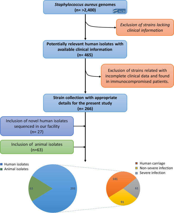
To generate an extensive and harmonized collection associated with the metadata, we first performed literature investigations. Fig. 1 illustrates the whole strategy. More than 2,400 S. aureus genomes were collected and analyzed. After two independent rounds of reviews devoted to the exclusion of irrelevant studies, 266 sequenced S. aureus genomes from humans with reliable clinical information were conserved. Then, we added 27 new S. aureus genomes sequenced in our laboratory and collected from different facilities, as well as 63 genomes sequenced from animal strains, to obtain a final data set of 356 strains (see Table S1 in the supplemental material). Of these, 293 were isolated from human hosts, among which 141 came from colonization and 152 from infections. This last category could be divided into 61 nonsevere and 91 severe infections (Fig. 1). All 356 sequenced strains included in our multicountry study were originally collected from 1943 to 2013 (Fig. S1 and Table S1), except those from Africa, the Middle East, Russia, and Greenland. Most strains (61%) were collected in Europe, with United Kingdom and France accounting for the majority. Eighteen percent of the isolates belong to the North American continent, followed by Latin America (11%), Asia (9%), and Oceania (1%).
FIG 1.
Overview of the incremental construction of a Staphylococcus aureus genome set composed of extensive clinical metadata.
Geographical location of the 356 S. aureus sequenced strains with reliable metadata. The color code is indicated in the right panel. Dot sizes reflects the number of strains per country. Download FIG S1, PDF file, 0.4 MB (427.5KB, pdf) .
Copyright © 2022 Sassi et al.
This content is distributed under the terms of the Creative Commons Attribution 4.0 International license.
Overview of the S. aureus strains used in this study. Abbreviations: CC, clonal complex; MRSA, methicillin-resistant Staphylococcus aureus. Download Table S1, XLSX file, 0.04 MB (40KB, xlsx) .
Copyright © 2022 Sassi et al.
This content is distributed under the terms of the Creative Commons Attribution 4.0 International license.
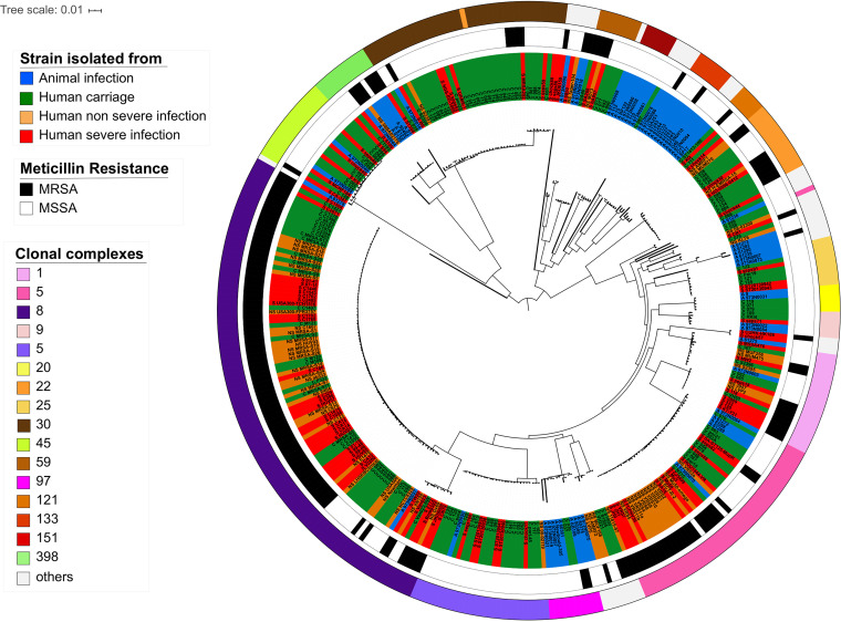
We performed a phylogenetic reconstruction based on genome sequences (Fig. 2) and subsequently conducted multilocus sequence typing (MLST) and methicillin-resistant S. aureus (MRSA) analyses. MLST analysis predicted 97 different sequence types belonging to 41 clonal complexes (Fig. 2; Fig. S2 and Table S1). In our data set, 27% of the strains belong to clonal complex 8 (CC8), which is one of the most prevalent clonal complexes worldwide both inside and outside health care settings in the United States and Europe (24). The second most prevalent CC was CC5 (Fig. 2; Fig. S2), in agreement with its worldwide distribution (see Fig. S3 at https://biochpharma.univ-rennes1.fr/supplemental-data) (25). Eleven CCs were exclusively limited to strains isolated from animals (n = 27), such as CC133 (n = 7), CC151 (n = 6), and CC130 (n = 3) (Table S1). Based on genome analysis, 43% of the strains were methicillin resistant. The distribution of MRSA and MSSA isolates was not random with respect to CCs. Eighty-three percent of the MRSA isolates were concentrated in only five CCs (CC1, -5, -8, -22, and -30). In those five CCs, approximately half of the strains were MRSA, except for CC8, where the proportion exceeded 80% (Fig. 2; Table S1).
FIG 2.
Phylogenetic reconstruction of multiclonal Staphylococcus aureus lineages. Maximum likelihood tree based on 356 genomes. Colors of strain names indicate the type of associated metadata: blue, animal infection; green, human colonization; orange, human nonsevere infection; red, human severe infection. The first outer circle represents the methicillin resistance profile (MRSA for resistance and MSSA for susceptibility). The second outer circle indicates the clonal complex of each strain.
Minimum spanning tree representing the repartition and diversity of our data set. The size of the circles is a function of the number of clonal isolates. Colors indicate the clonal complex diversity of the data set. Download FIG S2, PDF file, 0.1 MB (76KB, pdf) .
Copyright © 2022 Sassi et al.
This content is distributed under the terms of the Creative Commons Attribution 4.0 International license.
Pangenomic and genome-wide association study. (i) S. aureus genes, sRNAs, and IGR content.
The size of the pangenome, which includes coding sequences, sRNA genes, and intergenic regions (IGRs) of the S. aureus species, was derived from the 356 genomes listed in Table S1. First, the CDS analysis performed with Panaroo (26) revealed that from a total of 8,827 predicted groups of orthologs (pan), 1,489 groups of orthologs (54%) belong to the core genome. Within the accessory genome (n = 7,338), 2,813 unique genes were identified which corresponded to ~8 singletons per genome (see Fig. S4 at https://biochpharma.univ-rennes1.fr/supplemental-data and Table S2 in the supplemental material). Second, The pan-sRNome (the entire set of sRNAs) was predicted to contain 632 sRNA genes as reported in the SRD database (27), with around 50 of them confirmed experimentally (28). The core of this pan-sRNome contains 271 predicted srna genes (42%) (see Fig. S5 at https://biochpharma.univ-rennes1.fr/supplemental-data), indicating that more than half of the predicted sRNA genes are among a variable set of accessory genes, inherited laterally (29). Only one sRNA singleton was identified from multidrug-resistant clone JKD6008 (30). Third, the remaining parts of the genomes are IGRs, which contain regulatory elements. A total of 25,983 independent IGRs constituted our pangenome. Only 137 IGRs were common to all 356 strains (core IGR), suggesting that they contain essential regulatory elements for gene expression. Overall, a total of 8,558 IGRs fell within the accessory genome and 17,288 IGRs were strain specific (see Fig. S6 at https://biochpharma.univ-rennes1.fr/supplemental-data), indicating high variability in those regions.
Analysis of the pangenome using Panaroo. Download Table S2, XLSX file, 10.5 MB (10.8MB, xlsx) .
Copyright © 2022 Sassi et al.
This content is distributed under the terms of the Creative Commons Attribution 4.0 International license.
(ii) Global functional analysis to investigate differences according to the origin of the strains and their clinical display.
To address whether livestock-, colonization-, or infection-associated (severe and nonsevere) genomes encode determinants specific to their origins, an in-depth functional analysis was conducted on the pangenome. To that aim, each genome was annotated using the COG database (31), and each gene was assigned to functional COG classes and categories. The abundances of genes present in each COG were compared. Significant differences (P < 0.01) were found between animal and human isolates for genes encoding the mobilome (i.e., prophages encoding genes and transposons; COG category “X”) (see Fig. S7 at https://biochpharma.univ-rennes1.fr/supplemental-data). The genomes of human isolates significantly contain more genes (P < 0.01) assigned to the mobilome (mean number of genes, 45) than those of the animal isolates (mean number of genes = 33). Within human isolates, significant differences (P < 0.01) were also found within this category between colonization and nonsevere infection isolates. The genomes of nonsevere infection isolates contain more genes from the mobilome (mean number of genes = 56) than colonization isolates (mean number of genes = 43) (see Fig. S8 at https://biochpharma.univ-rennes1.fr/supplemental-data). Conversely, no significant differences were found between isolates from colonization versus global infection (severe and nonsevere), from colonization versus severe infections, and from nonsevere versus severe infections (see Fig. S8 and S9 at https://biochpharma.univ-rennes1.fr/supplemental-data). Together, these findings show that significant stochastic variation at the mobilome level occurs between livestock and human isolates but also between colonization and nonsevere isolates.
(iii) GWAS to discriminate strains.
To identify genomic specificities that underline host-associated or severity outcomes, two GWAS approaches were conducted, as was recently done for biofilm-associated genotypes in multidrug-resistant (MDR) Pseudomonas aeruginosa (32), but using more restrictive parameters to narrow to small outputs. First, we investigated the presence/absence of CDSs, IGRs, and sRNAs using statistical tools (R and Scoary) (Table S3). Second, to avoid identifying characteristics of the population structure rather than causative loci that would be truly predictive of bacterial epidemiology, pyseer was utilized (Table S4). Overall, significant differences were retrieved for the CDS and IGR analyses but not for sRNAs, as described below (Tables S3 and S4).
Identification of significant differences based on host preference or clinical status using Scoary. Download Table S3, XLSX file, 0.04 MB (46.1KB, xlsx) .
Copyright © 2022 Sassi et al.
This content is distributed under the terms of the Creative Commons Attribution 4.0 International license.
Identification of significant differences based on host preference or clinical status using pyseer. Download Table S4, XLSX file, 0.2 MB (251.5KB, xlsx) .
Copyright © 2022 Sassi et al.
This content is distributed under the terms of the Creative Commons Attribution 4.0 International license.
(iv) Discrimination based on host specificity.
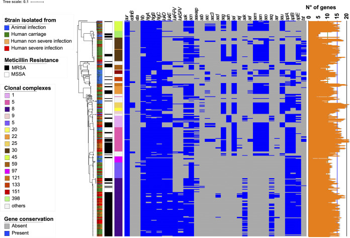
The implementation of a CDS presence/absence matrix with Scoary identified significant differences between animal and human isolates (n = 48, Bonferroni’s P < 10−5), with the virulence genes sak and scn being among the most significant (Table S3) (P = 2.85 × 10⁻³⁵ and 2.39 × 10⁻¹⁷, respectively). Other genes with strong P values (i.e., ≤10−12) included recT (a recombinase gene), group 3780, or group 3965. The second approach with pyseer identified 75 genes significantly associated with human isolates, with scn and sak being again among the most significant (likelihood ratio test, P = 3.53 × 10⁻⁸ and 4.67 × 10⁻⁵, respectively) and a total of seven genes in common with Scoary (genes coding for 2 transposases and 3 phage proteins besides scn and sak) (Table S3). Since sak and scn belong to the virulome, we generated a matrix of the presence/absence of these genes (Fig. 3). This analysis showed that although the presence of both sak and scn indicates human as a host, a few strains isolated from animals but known to belong to sequence types (STs) able to infect both animals and humans can harbor these two genes (e.g., strain A 61278 on the first line of Fig. 3). This clearly indicates that these two genes are absent in STs that were never reported to infect humans.
FIG 3.
Distribution of virulence genes within the multiclonal data set. The ML phylogenetic tree is annotated with three colored strips representing the type of isolation of each strain, their methicillin resistance status, and their clonal complex. The binary heat map represents the presence (blue) or absence (gray) of virulence genes identified with the pangenome study. The orange strip on the right indicates the sum of virulence genes present in each isolate.
Studies of IGRs revealed better consistency between the two GWAS approaches. Scoary identified 32 significant IGRs between animal and human isolates with P values up to 5.50 × 10⁻²⁹ for cluster 15862, located upstream from a putative nonheme iron containing ferritin that belongs to the ferric uptake regulator-like protein PerR and member of the FUR family (Tables S3 and S5). Other clusters with P values of <1.10−15 were clusters 15817, 24735, 16230, 11006, and 24115. Among IGRs identified as significant with pyseer, 18/48 were also present in Scoary when livestock strains and human isolates were compared, with clusters 16230, 11006, and 24115 being again recovered but not clusters 15817 and 24735. Cluster 16230 stands for the IGR that corresponds to the promoter region of sak, already identified as significant in the CDS analysis (Tables S3 and S5). Cluster 11006 is located between genes encoding a hypothetical protein and a phage major tail protein, whereas cluster 24115 corresponds to the region of integration of prophage Φ13 in NCTC_8325 that truncates a gene encoding a β-hemolysin (SAOUHSC_02240).
Intergenic region matrix of the data set to identify and locate differences. Download Table S5, XLSX file, 13.6 MB (13.9MB, xlsx) .
Copyright © 2022 Sassi et al.
This content is distributed under the terms of the Creative Commons Attribution 4.0 International license.
(v) Discrimination based on clinical outcome.
Strains isolated from humans were further classified based on clinical status. GWAS was also performed to investigate colonization versus infection, colonization versus nonsevere infection, colonization versus severe infection, and nonsevere versus severe genetic traits. For CDS, Scoary identified significant differences in clinical status (Table S3) between colonizations and infections (number of sequences = 15, Bonferroni’s P < 10−4), colonizations and nonsevere infections (number of sequences = 4, Bonferroni’s P < 10−4), and colonizations and severe infections (number of sequences = 5, Bonferroni’s P < 10−4) but not between nonsevere and severe infections. pyseer revealed 6 to 47 genes associated with a clinical category (P < 10−5) (Table S4). Twenty-seven genes were associated with strains isolated from an infection versus strains isolated from colonization, with seven being found in common with the Scoary output (Tables S3 and S4). Among genes identified with both GWAS approaches, mecA was associated with infection (P = 2.28 × 10⁻⁶ and P = 1.60 × 10⁻¹⁰ for pyseer and Scoary, respectively). Similarly, mecA was again identified (P = 7.25 × 10⁻⁶ and P = 5.75 × 10⁻⁵ for pyseer and Scoary, respectively) when colonizations were compared with severe infections.
For IGRs using Scoary, significant differences were found between (Table S3) colonization and infection (number of sequences = 25), colonization and nonsevere infection (number of sequences = 4), and colonization and severe infection (number of sequences = 8) but not between nonsevere and severe infections, as for CDSs. There was again a better consistency between Scoary and pyseer using IGRs, with 20/25 in common between colonization and infection, 1/4 in common between colonization and nonsevere infections, and finally, 8/9 in common between colonization and severe infections. Between colonization and infection, two of the most significant IGRs in common were within tRNA clusters (clusters 25463 and 21327), while cluster 21904 was situated next to the 3′ end of mecA (Tables S3 and S5). Together, these findings suggest that IGRs might be more critical for differentiating isolates relative to their isolation status and clinical manifestation of disease than other genetic markers/categories.
(vi) SNP-based discrimination.
We then investigated whether single nucleotide polymorphisms (SNP) were more appropriate for discriminating isolates from colonization and infection and ultimately to be correlated with clinical outputs. For this purpose, a total of 26,577 core genome SNPs, identified in our data set, were examined. Only four significant SNPs (Bonferroni’s P < 0.0001) were identified using Scoary between animal and human strains, while none were found for the other categories of strains (Table S3). In contrast, pyseer identified a total of 889 SNPs (likelihood ratio test [lrt] P < 0.0001) associated with one of the groups of strains (Table S4). One hundred fifty-nine are significantly different between animal- versus human-isolated strains, 66 between colonization versus global infection isolates, 102 between colonization versus nonsevere isolates, 538 between colonization and severe infection, and 24 between nonsevere and severe infections. However, none of the SNPs correlated with sak, scn, or mecA, which were the most discriminating genes using both Scoary and pyseer. Among SNPs identified between colonization and severe infection, the most significant (lrt P = 4.66 × 10⁻⁶) was located in sbnE (NCTC8325_85880_T_C) (Table S4), which is involved in siderophore biosynthesis, suggesting that the battle for iron during infection may be a discriminant feature.
(vii) Homoplastic SNPs, selection, and horizontal gene transfer.
In order to detect whether some SNPs or genes undergo positive selection, and more importantly are associated with a clinical outcome or animal host, we implemented an automated homoplasy analysis with the HomoplasyFinder software (33). In Table S6, we present a list of the 129 most homoplastic SNPs (Homoplasy Index > 0.9). Among them, nucleic acid changes are detected for csoR, a gene that regulates copper resistance mechanisms and is often found on plasmids (34), graR, which is involved in resistance against cationic antimicrobial peptides and vancomycin (35), and norB, which chromosomally encodes an efflux pump whose overexpression can confer MDR to quinolones or tetracyclines (36, 37). Yet, no strong phylogenetic or SNP associations were detected in relation with the goal of this study, underpinning the stochastic nature of local selection, recombination events, and horizontal gene transfers.
List of homoplastic single nucleotide polymorphisms of the data set. Download Table S6, XLSX file, 0.2 MB (140.4KB, xlsx) .
Copyright © 2022 Sassi et al.
This content is distributed under the terms of the Creative Commons Attribution 4.0 International license.
RF as a powerful method to separate strains according to their origins.
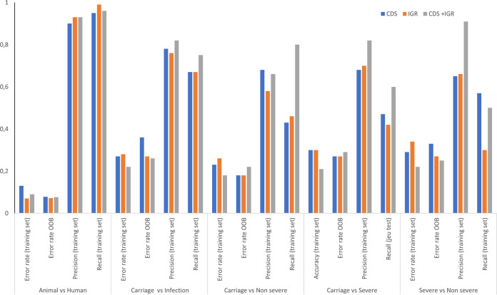
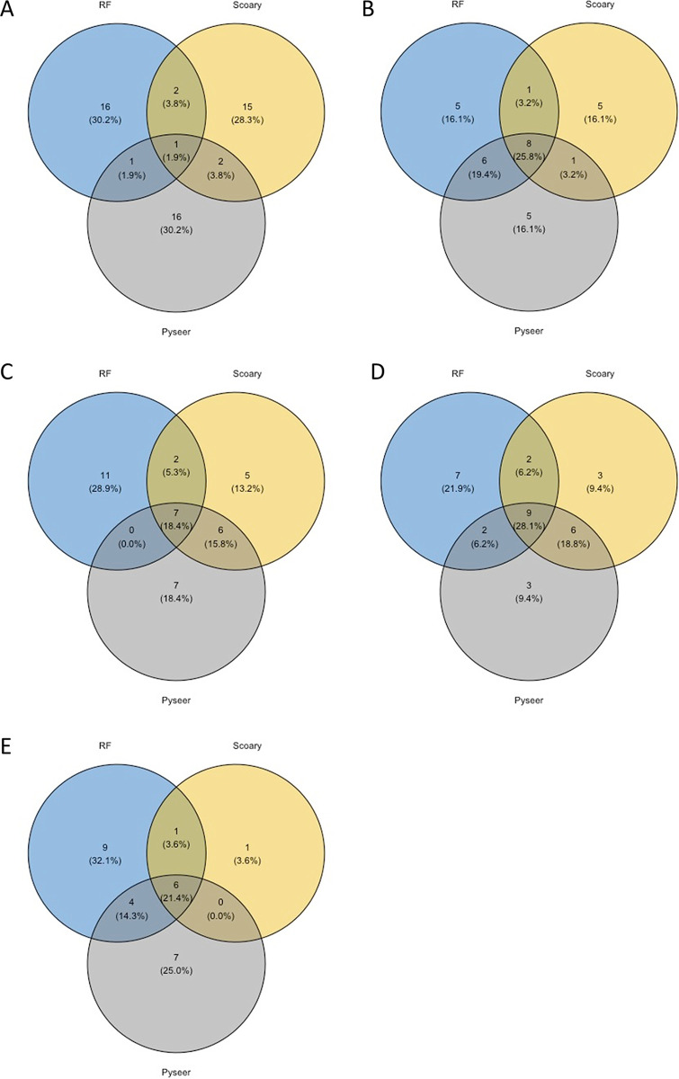
The comparative genomics and GWAS methods employed above produced a large amount of data and revealed significant differences between categories that compose our data set. However, they also show some limitations to perfectly distinguish strains based on their environmental origins. Therefore, by combining all these data, we estimated random forest (RF) classification algorithms known to smartly deal with correlation and interaction among features in genomics. RF is a widely used ensemble machine learning (ML) method that aims to aggregate several classification trees using bagging (38, 39). In order to ensure that the estimation of the prediction error rate was unbiased, two-thirds of the data set was used as a training set while, in a second step, the remaining third was used as a testing set. We first took advantage of the out-of-bag error as an estimate of the prediction error based on the training set. Then, we used three different types of genomic matrices as inputs: CDS, IGRs, and CDS plus IGRs (CDS+IGRs). sRNAs were discarded since they were less discriminant after GWAS analyses, which was also confirmed using CART (classification and regression trees) as preliminary inquiries (see supplemental material at https://biochpharma.univ-rennes1.fr/supplemental-data). Trees were generated (i) to distinguish animal from human strains or (ii) to differentiate human isolates based on the clinical status of isolation, namely, colonization versus infections, colonization versus nonsevere infections, colonization versus severe infections, and nonsevere versus severe infections. Overall, the accuracy was high (error rate of the training set, ~10%) in separating strains isolated from animals from strains isolated from humans, reaching 93% (error rate of 7%) when using only IGRs (Fig. 4). However, the error rate ranged from 26% to 33% as a function of the input used for comparisons based on human clinical and carriage status. Although no strong differences were observed regarding the input used, an overall increase of accuracy was measurable when CDS and IGRs were combined. To decipher whether some specific genes or IGRs were critical to build the trees, we took advantage of the variable importance measure of RF and report the five most important genes in Table 1. Interestingly, scn was the variable of most importance to segregate animal from human isolates. Group 3413, which was the second most important variable, was also retrieved with the pyseer approach and ranked 15th (Table S4), whereas the three other genes were not previously identified through GWAS. Among a total of 6,638 CDSs used to build trees, group 3780, group 3965, sak, and recT identified as significant by GWAS were ranked 21st, 35th, 37th, and 54th, respectively. This suggests good correlation between those approaches. Among the 15 CDSs identified with Scoary to discern carriage from infection, the first three (mecA, maoC, and ugpQ_1) were ranked 4th, 5th, and 2nd, respectively. With the IGR matrices, an even better correlation was observed with clusters 15862, 15817, 24735, and 16230 (Table S3), being among the top five variables of importance among a set of 8,045 clusters needed to generate the trees (Table 1; Table S7). Similarly, cluster 25463, identified with pyseer (Table S4), was ranked first and second to distinguish colonization versus infection strains and colonization versus severe infection strains, respectively. To better compare our findings, we extracted the first 20 hits obtained from Scoary, pyseer, and RF measures of importance from RF and illustrate the data by using Venn diagrams (Fig. 5). Overall, Fig. 5 shows that better correlation was obtained using IGRs than using CDSs for distinguishing both animal versus human isolates and colonization versus infection. Noteworthy, up to 9 IGRs were in common using the three approaches using IGRs while comparing colonization and infection (Fig. 5C).
FIG 4.
Host preference or clinical status prediction within the data set of 356 S. aureus strains assessed by random forest analysis. OOB, out of bag.
TABLE 1.
Top five variables of most importance for random forest
| Parameter | Variables of most importance |
||
|---|---|---|---|
| Animal vs human | Colonization vs infection | Colonization vs severe | |
| CDS | scn, group_3413, group_1916, lukD, hlb | group_5661”, ugpQ_1, yezG_2, mecA, maoC | NAb |
| IGRa | Cluster_15817, cluster_15862, cluster_24735, cluster_22030, cluster_16230 | Cluster_25463, cluster_21327, cluster_5558, cluster_21209, cluster_1153 | Cluster_21327, cluster_25463, cluster_1153, cluster_19750, cluster_8836 |
Flanking genes are accessible in Table S7 in the supplemental material.
NA, not applicable.
FIG 5.
Comparison and correlation between GWAS and random forest approaches. Venn diagrams were produced after extraction of the first 20 hits obtained from random forest (blue), from Scoary (brown), or from pyseer (purple). (A to E) Comparisons using CDS for animal versus human isolates (A), CDS for colonization versus infection isolates (B), IGR for animal versus human isolates (C), IGR for colonization versus infection isolates (D), and IGR for colonization versus severe infection isolates (E).
List of genes flanking IGRs that belong to the top five variables of most importance for random forest. Download Table S7, XLSX file, 0.01 MB (11.8KB, xlsx) .
Copyright © 2022 Sassi et al.
This content is distributed under the terms of the Creative Commons Attribution 4.0 International license.
To verify the robustness of the RF classification algorithms estimated on the training set, we calculated error rates using the testing set (out of bag). As for the training set, the algorithm was more powerful distinguishing animal strains from human strains than distinguishing strains in the other categories (Fig. 4). An ~7 to 8% error rate was observed to separate these strains using CDS, IGRs, or CDS+IGRs as inputs. Although predictions were less reliable when human isolates were compared based on their clinical status, the error rate did not exceed 18% in discriminating colonization from nonsevere infections when the combination of CDS and IGRs was used while reaching 27% in discriminating colonization versus severe infections, indicating that this comparison is probably the most difficult to make. Also, the use of IGRs or CDS plus IGRs improved the prediction to distinguish colonization versus infection and severe versus nonsevere infections but not for the two other categories. Finally, to verify that errors did not cluster in a particular clade, we plotted predicted and actual phenotypes on a phylogenetic tree, which showed that they were mostly random (see Fig. S10 and S11 at https://biochpharma.univ-rennes1.fr/supplemental-data).
Together, these results indicate that the RF methodology is efficient for classifying strains regarding their environmental isolation status and revealed that the use of IGRs is important to improve discrimination. Additionally, the out-of-bag error rates indicate that RF is suitable for a good prediction accuracy in distinguishing animal from human isolates.
Close-up on ST8 to search for discriminant genomic markers.
We tested whether putting the focus on a more clonal set of strains is relevant to improving the discrimination of isolates based on genomic features. To that aim, we selected ST8 isolates, as they belong to one of the most prevalent clonal complexes (CC8) worldwide and also because we already had 80 ST8 genomes in our main data set. They were classified into three subgroups according to clinical data: colonization, nonsevere infections, and severe infections (Table S1). The 80 ST8 isolates yielded a CDS pangenome size of 4,275 orthologs. The ST8 core genome contains 2,180 CDSs, representing ~79% of the entire genomes, compared with only ~21% when the 356 genomes from the 30 CCs are analyzed (Fig. S2). The accessory genome of the ST8 strains reached a total of 782 CDSs, while 1,313 singletons were found, corresponding to around 16 unique genes per isolate. Gene function annotations (COG databases) revealed that, as for the whole pangenome analysis (n = 356), ~43% of the core genome is devoted to metabolism. The ST8 pan-sRNome was composed of 613 sRNA genes. Five hundred six belong to the core genome (80% of those listed in the SRD database [27]), while 92 sRNAs composed the accessory genome and 15 sRNAs were found as singletons (all in JKD6008 strains). Finally, 19 sRNAs reported in the SRD database were missing in all the ST8 isolates. RF was applied to search for genomic markers in the ST8 set (Table 2). Surprisingly, the accuracy fell dramatically, especially when CDSs were used as the sole input. Conversely, validation using the out-of-bag error rates showed that error rates were generally lower after using the training set with CDSs, except for prediction of severe or nonsevere infections, where the use of IGRs substantially improved predictions. These data suggest that the reduction of the set’s size is detrimental to forecasting the outcome.
TABLE 2.
Use of random forest to predict host preference or clinical status within the ST8
| Matrix | Comparison | Error rate Out of bag (OOB) | Accuracy (training set) | Precision (training set) | Recall (training set) |
|---|---|---|---|---|---|
| CDS | Colonization vs infection | 0.26 | 0.62 | 0.71 | 0.71 |
| Colonization vs nonsevere | 0.46 | 0.59 | 0.60 | 0.38 | |
| Colonization vs severe | 0.41 | 0.59 | 0.60 | 0.38 | |
| Severe vs nonsevere | 0.50 | 0.69 | 0.64 | 0.88 | |
| IGRs | Colonization vs infection | 0.37 | 0 0.73 | 0.71 | 1 |
| Colonization vs nonsevere | 0.51 | 0.76 | 0.83 | 0.63 | |
| Colonization vs severe | 0.54 | 0.88 | 1 | 0.75 | |
| Severe vs nonsevere | 0.28 | 0.81 | 0.86 | 0.75 | |
| CDS+IGRs | Colonization vs infection | 0.37 | 0.73 | 0.71 | 1 |
| Colonization vs nonsevere | 0.46 | 0.71 | 0.67 | 0.75 | |
| Colonization vs severe | 0.51 | 0.76 | 1 | 0.5 | |
| Severe vs nonsevere | 0.30 | 0.81 | 0.86 | 0.75 |
Use of RNA-seq under conditions mimicking nasal colonization and bacteremia to identify potent transcriptomic markers in ST8. (i) Bacteremia medium versus SNM3 medium.
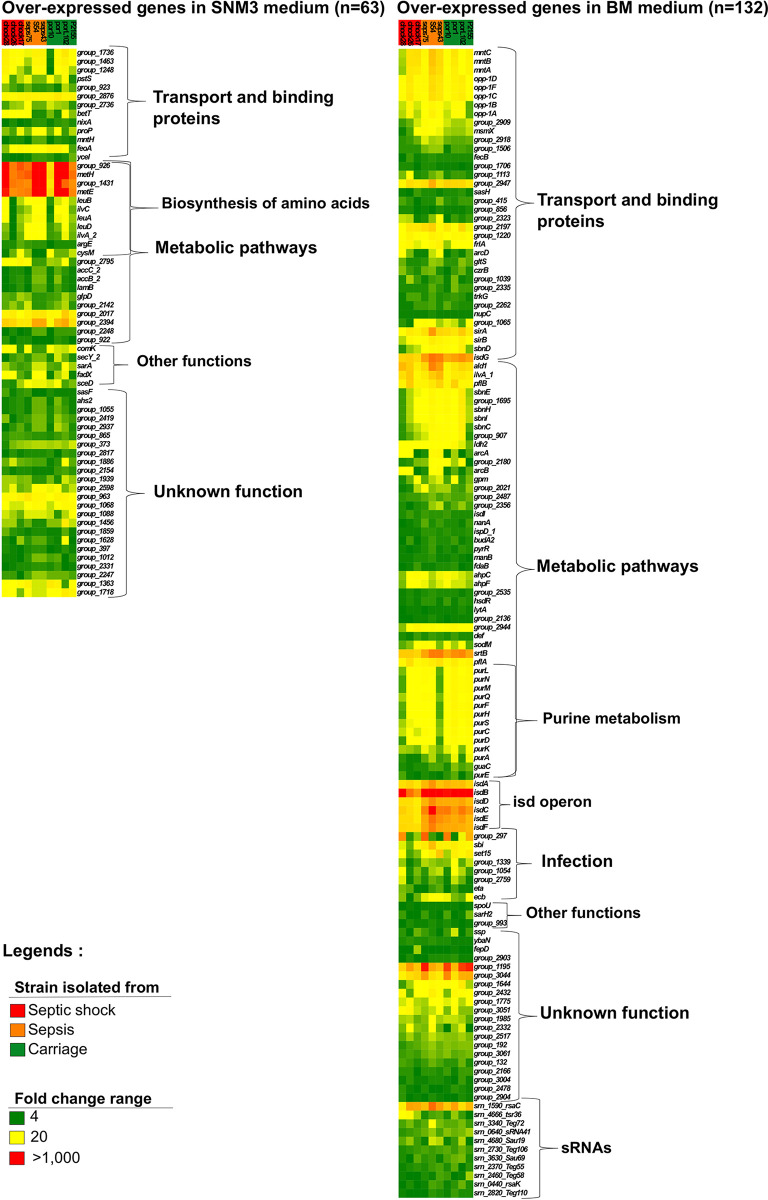
Although promising, GWAS or RF performed on the multiple-CC data set or on ST8 illustrated some limitations in discriminating or predicting strain origins and phenotypic outcomes. To further widen our approach, we investigated whole-gene expression by RNA-seq. We selected 10 ST8 isolates from our ST8 data set (Fig. 6; Table S1). Four were isolated from asymptomatic carriers and six from patients with severe infections treated in our hospital. Within the severe infection strains, we distinguished strains that yielded bacteremia without organ failure (n = 3) from the one that yielded septic shock (n = 3) according to the Third International Consensus Definitions for Sepsis and Septic Shock (40). Cells were grown in medium that mimics nasal colonization (SNM3) and then transferred to bacteremia mimicking medium (BMM), which mimics some aspect of a bacteremia. Overall, 19.5% of genes (including sRNA genes) were differentially expressed between the two conditions, ranging from 15% for shock strain 28 to 22% for colonization strain P1 (Table S7). All of the septic shock strains presented an overall expression variation lower than that of the seven other strains. In BMM, the isd operon, involved in iron binding and transport, was overexpressed, with fold changes ranging from 200 to 1,000 as a function of the gene and strain considered (Fig. 6; Table S7). sirA, also involved in iron acquisition, was significantly induced (around 150 times). Among genes involved in virulence, srtB (41) was more than 300 times overexpressed in BMM while sbi and spa, encoding immune evasion factors, increased 200 and 80 times, respectively. Regarding sRNAs, a nearly 300-fold increase was observed for Srn_1590_RsaC, an sRNA that modulates the oxidative stress response during manganese starvation (42) and that is known to be overexpressed during acute in vivo infection (43). Apart from Srn_1590_RsaC, few sRNAs were strongly induced under these bacteremia-mimicking conditions. On the other hand, genes overexpressed in SNM3 were mostly involved in amino acid transport and metabolism, consistent with nutrient limitation in the human nose (44) (Fig. 6; Table S7). To summarize, based on the overall set of genes overexpressed in SNM3 and BMM, these data indicate that the in vitro culture media used in this study indeed mimic conditions that can occur during colonization and severe infections.
FIG 6.
Transcriptome profile of ST8 isolates under synthetic nasal conditions (SNM3) and bacteremia mimicking conditions (BMM). Heat maps display the intensity of genes overexpressed in SNM3 (left panel) and BMM (right panel). Strain names are indicated above each column, and their clinical metadata are indicated as follows: green, colonization; orange, sepsis; red, septic shock. The intensity range of overexpression within the heat maps is represented as follows: green, fold change up to 4; yellow, fold change up to 20; red, fold change of more than 1,000.
(ii) Search for discriminant transcriptomic markers.
Based on the conditions used above, we searched for gene expression variations that could discriminate the clinical origin of the studied isolates. We pooled appropriate RNA-seq data in the categories of colonization, infections, bacteremia without organ failure, and septic shock. Additionally, we applied a DESeq cutoff of 2 and a P of <0.01 to identify the most significant variations (Table S7). First, we compared colonization and strains that yielded infections. In BMM, the expression of norB, encoding a quinolone resistance protein but also a participant in bacterial fitness within abscesses (36), was increased by more than 2-fold in infectious strains. In SNM3, the expression of lrgA and lrgB was lowered by around 3-fold in these strains, indicating that they could be used as biomarkers to forecast staphylococcal infections. Then, we compared strains isolated from healthy carriers with either bacteremia without organ failure or with septic shock strains. The comparison between bacteremia without organ failure and colonization showed that although no significant differences could be depicted in SNM3, the transcript levels of radC and ald1 were modified in BMM by 0.4- and 2.4-fold, respectively (Table S8). More differences were detected between septic shock and colonization, with six and seven genes differentially expressed in BMM and SNM3, respectively. In BMM, all were mRNAs, either up- or downregulated, while in SMN3, one belonged to sRNAs (Srn_4470; i.e., SSR42, known to be involved in virulence [45–47]). Finally, we performed comparisons between strains isolated from bacteremia without organ failure and strains isolated from bacteremia with septic shock (Table S8). In BMM, 12 genes were differentially expressed. These included 10 mRNAs and 2 sRNAs. In SNM3, more differences could be observed, since 26 mRNAs and 3 sRNAs were differentially expressed. Among the mRNAs, the isd genes, encoding coagulase and several hemolysins, were upregulated in septic shock strains, indicating that some of the virulence equipment is already active during colonization-mimicking conditions. Additionally, study of the sRNAs revealed that RNAIII (Srn_3910) was also overexpressed in septic shock strains. Together, this transcriptomic analysis performed under nose-mimicking and blood infection-mimicking conditions showed that some genes are differentially expressed between the different categories and that septic shock strains exhibited higher transcript levels of a few virulence factors in SNM3. lrgA and lrgB are promising targets that could be used as primary tools to discriminate strains and, therefore, ultimately predict infections.
List of genes differentially expressed based on culture conditions (SNM3 versus BMM) and list of potential RNA biomarkers as a function of clinical status of strains that belong to ST8. Download Table S8, XLSX file, 0.2 MB (185.5KB, xlsx) .
Copyright © 2022 Sassi et al.
This content is distributed under the terms of the Creative Commons Attribution 4.0 International license.
DISCUSSION
S. aureus mortality is impacted by a variety of host-related factors, such as age and comorbidities (9), but the transition from colonization to infection, in both animals and humans, is much less understood. Also, the virulence of the pathogen has a significant influence in the outcome of bacterial colonizations, which can lead to mild or severe infections. Combining genomic and transcriptomic approaches for pathogens might enhance our understanding of bacterial infections at the molecular level (48). Among bacterial pathogens, the identification of molecular signatures at DNA and/or RNA levels, which could prognosticate the issue of a colonization or an infection, would lead to the holy grail of clinicians, a step toward personalized medicine and infectious disease management.
Comparative genomic and transcriptomic analyses of multiple strains within a species could be a powerful means to uncover pathoadaptive genetic acquisitions and specific gene expression patterns. To date, several studies have focused on the genomic exploration of clinical isolates for both staphylococci and other bacterial pathogens. For instance, these include the identification of natural mutations associated with virulence (46), the evolutionary genomics study of host specificity (49), and the characterization of molecular signatures of persistent bacteremia in S. aureus (50). In this last study, comparative genomics revealed that isolates from persistent bacteremia have a low mutation frequency, but most are nonsilent mutations, suggesting that the S. aureus genome sequence can influence the clinical onset of infected hosts. Besides staphylococci, a recent study in Streptococcus agalactiae revealed the identification of CC-specific genes associated with virulence and conclude that this may explain differences in terms of virulence potential for certain CCs (51). Conversely, few studies performed whole-transcriptome comparisons between S. aureus strains to show that gene sets can be related to phenotypic features, such as bacterial invasion and host adaptation (52).
Here, we compiled a multicountry S. aureus genome data set containing 356 strains from livestock and humans with reliable clinical information responsible for colonization and nonsevere or severe infections. MLST representation of our S. aureus isolate data set indicates that our collection is a robust representation of the worldwide clonal diversity. Furthermore, our study fits with the reported prevalence of MRSA strains at a global scale (25, 53). The set of mobile genetic elements, some from phage origin, varies significantly between livestock and human isolates and also between colonization and infection strains, implying that analyzing the S. aureus mobilome gene content and expression is highly instructive. GWAS revealed that two genes, sak and scn, are absent in all livestock-specific strains but are detected in all strains infecting humans. Although promising, our findings also indicate that strains that belong to STs that can infect both animals and humans cannot be discriminated using those two markers. This result is congruent with previous findings indicating that all “sak-positive” strains are human associated (11). Staphylokinase (sak) is a virulence factor acting as a thrombolytic agent, which leads to tissue damage and improves bacterial invasiveness. The SAK protein has a dual role in human illnesses, promoting skin infections while, surprisingly, decreasing disease severity (54). The staphylococcal complement inhibitor SCIN (encoded by scn) helps the bacteria escape attack from the host immune system (55). The protein allows complement evasion by inhibiting the central complement convertases, reduces phagocytosis following opsonization, and binds to the alpha-defensins secreted from human neutrophils to counteract their bactericidal properties (56). Thus, SCIN is specific to human infections due to its ability to neutralize both the innate and adaptive immune host responses. Besides the SAK and SCIN genes, other determinants identified by Scoary and pyseer were phage-related functions and hypothetical proteins, suggesting the important role of mobile elements in S. aureus evolutionary history. Additionally, our analyses showed that sRNA genes, which contribute to tight gene expression regulation in bacteria (57), were not discriminant. This result was not surprising since several sRNAs are known to be implicated in staphylococcal virulence (58). Conversely, IGRs, which are often neglected in genomic analyses, provided significant information and consistency using both GWAS approaches, indicating that they might be useful.
However, in the present report, analysis of gene content and mutation rate showed some limitations in discriminating strains regarding the clinical status of their hosts and their origins. Therefore, we implemented RF, a well-established statistical method that has two main advantages in our context: (i) RF can be used to rank the importance of the variables using permutations and out-of-bags, and (ii) RF is not limited to linear association and is flexible enough to catch nonlinear as well as interaction relationships between genes and infection status. Based on GWAS results, we explored CDS and IGR content but not the sRNA content. Again, the use of IGRs in combination or not with CDSs showed that these data are relevant to predict host specificity but can also be useful in increasing prediction of clinical status. Our out-of-bag prediction showed that RF was powerful in determining whether a strain was specific to humans or animals. Interestingly, determination of variables of importance indicated a good correlation between GWAS and RF. sak and scn were again within the short set of the most important variables along with some IGRs that were found significant using Scoary or pyseer (clusters 15862, 15817, 24735, and 16230). This suggests that a small set of genes may be sufficient to distinguish strains based on their genomic sequences. However, predictive assessment showed limitations as soon as the predictions went beyond host specificity. This lack of accuracy seems correlated with the size of the data set. In this retrospective work, a main limitation lies in the absence of a precise description of strain isolation conditions. From a set of thousands of S. aureus genome sequences available from the literature that we analyzed, only 356 met the necessary requirements (Fig. 1) for subsequent analyses. The close-up on strains from ST8 (n = 80) illustrates the importance of a large collection, as the error rate for predictions dramatically increased. An alternative hypothesis to explain more reliable predictions on the species level would be that strains belonging to a single ST (or specifically ST8) do not show strong differences in their propensity to cause invasive infection. However, from a positive standpoint, our data suggest that RF models should be implemented as additional information for clinician practitioners and that the collection of an extensive data set may lead to the development of dedicated ML programs based on the presence/absence of a defined set of genes.
Inadequacy of genome-based predictions performed on a limited number of isolates (i.e., the ST8 data set) was counterbalanced by RNA sequencing data. First, the use of SNM3 (44) and BMM (our study) revealed that these media can mimic colonization and bacteremia under laboratory conditions. A large set of genes associated with virulence and iron acquisition were overexpressed in blood, whereas S. aureus mostly expressed genes implicated in amino acid transport and metabolism in SNM3, consistent with the poor nutritional content of this medium mimicking human nose conditions. Among the virulence factors induced in BMM, the isd operon (59), immune evasion system genes sbi and spa (60), and srtB, which encodes a surface-anchored protein (61), are the most overexpressed genes. The comparison of strains based on their clinical descriptions allowed the identification of putative RNA markers with increased or decreased transcripts in subpopulations. Strains that led to infections had lower expression of the lrgAB operon in the nasal colonization medium SNM3, while norB increased under bacteremia conditions. The lrgAB operon encodes a murein hydrolase, with expression controlled by the LytSR two-component system (62), and the norB quinolone efflux pump (63) is known to participate in virulence in abscesses (36). Although the identification of norB can be interpreted as a bias due to quinolone treatment, an in-depth study of metadata from these strains, all isolated in our facility, indicated that the patients were not treated with this antibiotic. Therefore, this result should not be considered an artifact. Additionally, when we deciphered transcriptomes from strains that actually progressed to infections and compared infection severity, a second set of genes, larger than the previous one, was found significant and therefore could be used to forecast infection severity. To date, our study includes only 356 clinical isolates, but future broader investigations will be set to collect clones from asymptomatic carriers or from infected patients to build a larger collection to identify discriminant RNA markers.
To conclude, the use of comparative genomics, GWAS, RF analyses, and transcriptomics studies within a single pilot study identified genomic combinations that distinguish strain host specificity and enabled estimation, through ML algorithms, of whether a colonized individual will be at higher risk to have a severe staphylococcal infection. We also highlight the importance of IGRs when considering the genomic elements. Furthermore, the expression pattern of a gene set may act as a biomarker to forecast strain clinical outcomes. The data from this study contribute to a better understanding of the critical factors that allow a pathogenic strain to establish a staphylococcal infection. In the future, a similar approach focusing on the role of host factors may be useful to provide an extensive prediction of risk associated with S. aureus.
MATERIALS AND METHODS
Construction of representative and harmonized S. aureus data sets.
Two different sets of genomes were generated during this study. The first one included strains that belong to multiple clonal complexes (see Table S1 in the supplemental material), whereas the second was devoted solely to ST8 isolates, which belong to CC8 (Table S1). The first set included genomes sequenced from strains of both animal and human origins. Most genomes were obtained from a literature survey. We used the terms “Staphylococcus aureus” AND “genomes” to search the PubMed database for studies published from July 2014 to June 2015, which corresponded with the beginning of the present study. Two authors (M. Sassi and Y. Augagneur) independently screened titles and abstracts to identify relevant studies. The search was supplemented by seeking the references of all eligible studies. Human studies were considered eligible when their authors (i) reported extractable and complete data on S. aureus genomes and (ii) documented sufficiently accurate clinical information. To prevent confounding factors, studies dealing with immunodeficient patients were excluded. A restriction on English and French literature was also imposed. To complete our data sets, we obtained strains from the infectious diseases and intensive care units located in our health care facility (Rennes University Hospital, France), from the Victoria Hospital (Australia), and from the Lausanne Hospital (Switzerland). Strains from asymptomatic individuals were also recovered from these facilities. Other strains were obtained from the National Reference Laboratory for Staphylococci in Lyon, France. All strains available in our laboratory were confirmed as being S. aureus by using multilocus sequence typing (64) and S. aureus protein A typing (Table S1). Then, two clinicians (P.-Y. Donnio and M. Revest) independently classified the clinical characteristics of patients from whom the bacterial strains were extracted. Patients were classified into three groups: those with severe infections, those with nonsevere infections, and those that were colonized. Strains for which both investigators could not agree were excluded from the analysis. Infections were considered severe if they led to bacteremia, infective endocarditis, pneumonia, or severe abscesses requiring surgery. Nonsevere infections were superficial infections treated on an outpatient basis. All other type of infections were excluded from the analysis. Patients with a superficial sample positive for S. aureus but without clinical symptoms were considered colonized. Additionally, S. aureus genomes from animal strains were kindly provided by R. Fitzgerald (Edinburgh, Scotland). The second set contained all the isolates typed as ST8 during the construction of the first set, to which we added 10 novel ST8 isolates for transcriptomic studies.
Genome sequencing and assembly of additional S. aureus isolates.
S. aureus strains sequenced in Rennes facilities were grown in brain heart infusion (BHI) broth (Oxoid) at 37°C and under agitation (160 rpm). Genomic DNA was isolated using the Wizard genomic DNA purification kit (Promega) in the presence of lytic enzymes (lysozyme and lysostaphin) in accordance with the manufacturer’s recommendations for Gram-positive bacteria. Subsequently, genomic DNA was precipitated with sodium acetate and washed two times with 70% (vol/vol) ethanol. DNA was sheared using a Covaris M220 focused ultrasonicator to generate an average fragment size of 600 bp. Unwanted smaller and larger fragments were removed by size selection using AMPure XP beads (Beckman Coulter). A DNA library was prepared using the NEBNext Ultra DNA library prep kit for Illumina (NEB) and sequenced as paired-end reads using an Illumina MiSeq platform and a MiSeq reagent kit v3 (600 cycles) (Illumina, Inc., San Diego, CA). Illumina reads for 27 strains (Table S1) were (i) trimmed using Trimmomatic (65), (ii) filtered based on quality using the Fastx-toolkit (http://hannonlab.cshl.edu/fastx_toolkit/) and, (iii) assembled using the SPAdes software (66, 67). SIS and GapFiller version 1.10 (68, 69) were used to improve the initial set of contigs, and the closest complete genome was used as the reference to order and orient the contigs.
S. aureus genomic annotation and pangenomic analyses.
To normalize coding sequence predictions, genomes were annotated using Prokka (70). Genes from each strain were functionally annotated using COG scripts (https://github.com/transcript/COG) and the COG databases (31). The number of genes encoding each COG family function were used to investigate pangenome associations with host and clinical phenotypes. We used R software to calculate the mean, the standard deviation, and P values (t test). The difference between COG families was considered significant when P was <0.01. Figures were generated using ggplot package in R software. sRNA genes were predicted after extracting all sRNA sequences available in the SRD database (27) (http://srd.genouest.org/) and were subsequently aligned with each genome using BLAST and a homemade script (https://github.com/mosassi/Staphylococcus_annotation.git). The pangenome is the sum of the core genome (the set of genes present in all genomes) and the dispensable, accessory genome containing genes present in some but not all the strains, as well as strain-specific genes (71). The pangenomic analysis was performed on CDSs, sRNA genes, and intergenic regions (IGRs). Gene homologies between strains were assessed using Panaroo (26) with a >95% nucleotide identity cutoff and a refinding step with at least 50% of the sequence within a radius of 1,000 nucleotides to identify genes that were missed during annotation. The sRNA homologies between the strains were evaluated using BLAST and with >90% nucleotide identity and >70% sequence coverage. The IGR homologies between the strains were estimated using Piggy software (72) with >80% nucleotide identity and >60% sequence coverage cutoffs. Core genome alignments were performed with PRANK (73) and with single nucleotide polymorphisms (SNPs) extracted. Antibiotic resistance and virulence genes were annotated using BLAST, ResFinder (74), and VirulenceFinder (75) databases.
Minimum spanning trees and phylogenetic reconstructions.
Multilocus sequence typing (MLST) was performed on all genomes, and the results are presented in Table S1. In order to define the relationships among strains at the microevolutionary level, we performed allelic profile-based comparisons using a minimum spanning tree (MST) analysis with BioNumerics v7.6 software (Applied-Maths, Sint-Martens-Latem, Belgium). MST analysis links profiles so that the sum of the distances (number of distinct alleles between two STs) is minimized. Strains were grouped into clonal complexes (clonal families), defined as groups of profiles differing by no more than one gene from at least one other profile of the group. Accordingly, singletons were defined as STs having at least two allelic mismatches with all other STs.
A phylogenetic tree was constructed by considering the 26,577 polymorphic sites retained in the core genome. A transversion substitution model was selected on the basis of the Akaike’s information criterion with jModelTest2 (76). Maximum likelihood phylogeny was constructed using PhyML with 1,000 nonparametric bootstrap iterations (77). A phylogenetic tree was visualized using Figtree (http://tree.bio.ed.ac.uk/software/figtree/).
Selection signatures.
To infer homoplastic SNPs that might be driven by convergent and positive selection, we analyzed the global SNP matrix with the HomoplasyFinder algorithm (33).
GWAS.
Two genome-wide association study (GWAS) analyses (Scoary and pyseer) were performed to investigate pangenome associations with host and clinical phenotypes. The input for both approaches were CDS and IGR presence/absence and SNP matrices (78). Using Scoary, genetic elements that matched our inclusion criteria (Bonferroni’s P < 10−4; best pairwise comparison P value of <0.01) were considered significant. Due to the strong population structure of our complete collection and the dominance of the ST8 type, an in-depth analysis of genetic elements associated with a host or with a severity outcome was also performed using pyseer (v.1.3.6) (79). To do this, a phylogenetic distance file was calculated by using the phylogeny_distance.py included in pyseer on the maximum likelihood phylogeny. The distance, trait, gene, IGR, and sRNA presence and absence matrices and SNP files were used to run pyseer as described in the pyseer tutorial (https://pyseer.readthedocs.io/en/master/tutorial.html). A genome-wide association was considered statistically significant if the likelihood ratio test (lrt) P was <0.00001.
RF.
To build a classifier of the infection status of patients with respect to their genome, we used the well-known ensemble learning method called random forest (RF) (80). RF aims at building a forest of individual decision trees (obtained by applying the classification and regression tree algorithm), combined with randomized node optimization and bagging. We applied a random forest algorithm to our data set by using the software R (81) (function randomForest from the package randomForest maintained by A. Liaw and M. Wiener [82]). We used a total of 500 trees for each forest (ntree = 500) and a number of variables randomly sampled at each split given by srqt(p), where p is the number of explanatory variables in a matrix of predictors (mtry = sqrt(p)).
Transcriptomic analysis. (i) Growth and RNA extraction.
Only ST8 colonization isolates and ST8 strains recovered from bacteremia were used at this stage. S. aureus was precultured in tryptone soya broth (TSB; Oxoid) diluted 5 times in RPMI 1640 (Life Technologies) under agitation at 37°C. Overnight cultures were washed twice in synthetic nasal medium SNM3 (44) and then transferred in fresh SNM3 medium at an optical density at 600 nm (OD600) of 0.1. Cells were grown overnight and monitored by measuring the OD600. Appropriate volumes of cells were centrifuged and transferred either to fresh SNM3 or to fresh bacteremia-mimicking medium (BMM) at an OD600 of 0.1. BMM was prepared from 50% human AB serum (Etablissement Français du Sang), 44% RPMI 1640, and 6% packed red blood cells (washed in RPMI 1640 and set to a hematocrit of 50). Then, S. aureus was incubated for 4 h at 37°C under agitation. Cells were harvested by centrifugation at 4,500 rpm for 8 min, and pellets were washed with 5 mL of H2O, allowing lysis of red blood cells, and centrifuged again at 4,500 rpm for 8 min, and pellets were frozen prior to RNA extraction. Dried frozen pellets were resuspended in 500 μL of cold lysis buffer (20 mM sodium acetate, 1 mM EDTA, 0.5% SDS, pH 5.5). Total RNAs were extracted by phenol (pH 4) using a FP120 FastPrep cell disruptor (MP Biomedicals), as described previously (83).
(ii) cDNA library synthesis and Illumina RNA sequencing.
Up to 10 μg of total RNA was doubled treated with amplification-grade DNase I (Invitrogen) to remove genomic contaminations. The absence of DNA was checked by quantitative PCR (qPCR) with primers targeting S. aureus hup in an Applied Biosystems 7500 instrument. RNA integrity was verified on a bioanalyzer (Agilent). rRNAs were depleted using a Ribo-Zero magnetic kit (Epicentre), and the remaining RNAs were purified by ethanol precipitation according to the manufacturer’s recommendations. Efficiency of depletion was estimated on a bioanalyzer (Agilent). Stranded cDNA libraries were prepared using the NEBNext Ultra directional RNA library prep kit for Illumina (New England Biolabs) following an initial fragmentation step of 13 min at 95°C resulting in cDNA libraries of ~330 bp. The concentration, quality, and purity of the libraries were determined using a bioanalyzer, a Qubit fluorometer (Invitrogen), and a NanoDrop spectrophotometer (Thermo Scientific). Indexed libraries were equimolarly mixed in two pools and sequenced on an Illumina HiSeq 1500 system (high output, 200 cycles, paired end), as described in the manufacturer’s instructions.
(iii) Read mapping and differential expression analysis.
All genomes from strains used for transcriptome analysis were annotated using Prokka (70). Genes from each strain were functionally annotated using the KEGG database (84). Two annotation files (in GFF format) were generated per strain. One contained all CDSs predicted from Prokka, and the other was composed of all sRNA genes identified based on sequence similarity with the SRD database (27). To allow comparisons between all strains sequenced, orthologous genes were clustered by sequence homologies and unique identifiers were generated based on pangenomic analyses as described above. Quality control of RNA-seq reads was done as described previously (27). Illumina reads were trimmed using the FASTX-Toolkit (http://hannonlab.cshl.edu/fastx_toolkit/) and then mapped onto their respective genomes. SAM files were filtered on bitwise flag values (85) to keep only properly paired reads and counted by HTSeq count (86) for stranded library with the intersection nonempty mode. Fragments per kilobase per million (FPKM) normalization was calculated to remove weakly expressed transcripts. To do so, we removed all transcripts which led to an FPKM lower than 10 in each strain and condition tested. Then, differential expression analyses were calculated using DESeq with the per-condition method and the parametric fit type (threshold P = 0.05; fold change threshold = 4; baseMean > 15) (87) for SNM3 medium versus BMM or with the pooled method and the parametric fit type (threshold P = 0.01; fold change threshold = 2; baseMean > 15) to search for transcriptomic markers.
Data availability.
The genomes sequenced during this study are available under the following BioProject accession numbers: PRJNA273632, PRJNA280933, and PRJNA290551. The RNA sequences have been submitted to the NCBI SRA database under BioProject accession number PRJNA647528.
Supplementary Material
ACKNOWLEDGMENTS
We thank Ben Howden and Ross Fitzgerald for kindly providing human and animal strains. We also thank the Roscoff Bioinformatics platform ABiMS (http://abims.sb-roscoff.fr) for providing computational resources and the BioGenouest GEH facility (https://geh.univ-rennes1.fr/) for technical support.
We declare that we have no competing interests.
This work was supported by Inserm, University of Rennes1, and BIOSIT. M.S. was supported by Region Bretagne grant no. SAD SARS 8254 and SARS_2 9181 (to Y.A.). J.B. was a recipient of a fellowship from the Direction Générale pour l’Armement (Agence Innovation Défense) and the Conseil Régional de Bretagne.
The funders had no role in study design, data collection and interpretation, or the decision to submit the work for publication.
Contributor Information
Thierry Wirth, Email: thierry.wirth@mnhn.fr.
Yoann Augagneur, Email: yoann.augagneur@univ-rennes1.fr.
Benjamin E. Wolfe, Tufts University
Nicole Wheeler, University of Birmingham.
REFERENCES
- 1.van Belkum A, Melles DC, Nouwen J, van Leeuwen WB, van Wamel W, Vos MC, Wertheim HFL, Verbrugh HA. 2009. Co-evolutionary aspects of human colonisation and infection by Staphylococcus aureus. Infect Genet Evol 9:32–47. doi: 10.1016/j.meegid.2008.09.012. [DOI] [PubMed] [Google Scholar]
- 2.Bierowiec K, Płoneczka-Janeczko K, Rypuła K. 2016. Is the colonisation of Staphylococcus aureus in pets associated with their close contact with owners? PLoS One 11:e0156052. doi: 10.1371/journal.pone.0156052. [DOI] [PMC free article] [PubMed] [Google Scholar]
- 3.Lowy FD. 1998. Staphylococcus aureus infections. N Engl J Med 339:520–532. doi: 10.1056/NEJM199808203390806. [DOI] [PubMed] [Google Scholar]
- 4.Naber CK. 2009. Staphylococcus aureus bacteremia: epidemiology, pathophysiology, and management strategies. Clin Infect Dis 48(Suppl 4):S231–S237. doi: 10.1086/598189. [DOI] [PubMed] [Google Scholar]
- 5.Tong SY, Davis JS, Eichenberger E, Holland TL, Fowler VG, Jr. 2015. Staphylococcus aureus infections: epidemiology, pathophysiology, clinical manifestations, and management. Clin Microbiol Rev 28:603–661. doi: 10.1128/CMR.00134-14. [DOI] [PMC free article] [PubMed] [Google Scholar]
- 6.Kluytmans J, van Belkum A, Verbrugh H. 1997. Nasal carriage of Staphylococcus aureus: epidemiology, underlying mechanisms, and associated risks. Clin Microbiol Rev 10:505–520. doi: 10.1128/CMR.10.3.505. [DOI] [PMC free article] [PubMed] [Google Scholar]
- 7.Wertheim HF, Melles DC, Vos MC, van Leeuwen W, van Belkum A, Verbrugh HA, Nouwen JL. 2005. The role of nasal carriage in Staphylococcus aureus infections. Lancet Infect Dis 5:751–762. doi: 10.1016/S1473-3099(05)70295-4. [DOI] [PubMed] [Google Scholar]
- 8.Laabei M, Recker M, Rudkin JK, Aldeljawi M, Gulay Z, Sloan TJ, Williams P, Endres JL, Bayles KW, Fey PD, Yajjala VK, Widhelm T, Hawkins E, Lewis K, Parfett S, Scowen L, Peacock SJ, Holden M, Wilson D, Read TD, van den Elsen J, Priest NK, Feil EJ, Hurst LD, Josefsson E, Massey RC. 2014. Predicting the virulence of MRSA from its genome sequence. Genome Res 24:839–849. doi: 10.1101/gr.165415.113. [DOI] [PMC free article] [PubMed] [Google Scholar]
- 9.Recker M, Laabei M, Toleman MS, Reuter S, Saunderson RB, Blane B, Török ME, Ouadi K, Stevens E, Yokoyama M, Steventon J, Thompson L, Milne G, Bayliss S, Bacon L, Peacock SJ, Massey RC. 2017. Clonal differences in Staphylococcus aureus bacteraemia-associated mortality. Nat Microbiol 2:1381–1388. doi: 10.1038/s41564-017-0001-x. [DOI] [PubMed] [Google Scholar]
- 10.Bouchiat C, Moreau K, Devillard S, Rasigade J-P, Mosnier A, Geissmann T, Bes M, Tristan A, Lina G, Laurent F, Piroth L, Aissa N, Duval X, Le Moing V, Vandenesch F, Chirouze C, Curlier E, Descottes-Genon C, Hoen B, Patry I, Vettoretti L, Chavanet P, Eicher J-C, Gohier-Treuvelot S, Greusard M-C, Neuwirth C, Péchinot A, Piroth L, Célard M, Cornu C, Delahaye F, Hadid M, Rausch P, Coma A, Galtier F, Géraud P, Jean-Pierre H, Le Moing V, Sportouch C, Reynes J, Aissa N, Doco-Lecompte T, Goehringer F, Keil N, Letranchant L, Malela H, May T, Selton-Suty C, Bedos N, Lavigne J-P, et al. 2015. Staphylococcus aureus infective endocarditis versus bacteremia strains: subtle genetic differences at stake. Infect Genet Evol 36:524–530. doi: 10.1016/j.meegid.2015.08.029. [DOI] [PubMed] [Google Scholar]
- 11.Bosi E, Monk JM, Aziz RK, Fondi M, Nizet V, Palsson BØ. 2016. Comparative genome-scale modelling of Staphylococcus aureus strains identifies strain-specific metabolic capabilities linked to pathogenicity. Proc Natl Acad Sci USA 113:E3801–E3809. doi: 10.1073/pnas.1523199113. [DOI] [PMC free article] [PubMed] [Google Scholar]
- 12.Wirth T, Wong V, Vandenesch F, Rasigade J-P. 2020. Applied phyloepidemiology: detecting drivers of pathogen transmission from genomic signatures using density measures. Evol Appl 13:1513–1525. doi: 10.1111/eva.12991. [DOI] [PMC free article] [PubMed] [Google Scholar]
- 13.Young BC, Wu C-H, Charlesworth J, Earle S, Price JR, Gordon NC, Cole K, Dunn L, Liu E, Oakley S, Godwin H, Fung R, Miller R, Knox K, Votintseva A, Quan TP, Tilley R, Scarborough M, Crook DW, Peto TE, Walker AS, Llewelyn MJ, Wilson DJY. 2021. Antimicrobial resistance determinants are associated with Staphylococcus aureus bacteraemia and adaptation to the healthcare environment: a bacterial genome-wide association study. Microb Genom 7:e000700. doi: 10.1099/mgen.0.000700. [DOI] [PMC free article] [PubMed] [Google Scholar]
- 14.Wirth T, Bergot M, Rasigade J-P, Pichon B, Barbier M, Martins-Simoes P, Jacob L, Pike R, Tissieres P, Picaud J-C, Kearns A, Supply P, Butin M, Laurent F, International Consortium for Staphylococcus capitis neonatal sepsis, ESGS Study Group of ESCMID . 2020. Niche specialization and spread of Staphylococcus capitis involved in neonatal sepsis. Nat Microbiol 5:735–745. doi: 10.1038/s41564-020-0676-2. [DOI] [PubMed] [Google Scholar]
- 15.Eichner H, Karlsson J, Loh E. 1 April 2022. The emerging role of bacterial regulatory RNAs in disease. Trends Microbiol doi: 10.1016/j.tim.2022.03.007. [DOI] [PubMed] [Google Scholar]
- 16.Wang B, Muir TW. 2016. Regulation of virulence in Staphylococcus aureus: molecular mechanisms and remaining puzzles. Cell Chem Biol 23:214–224. doi: 10.1016/j.chembiol.2016.01.004. [DOI] [PMC free article] [PubMed] [Google Scholar]
- 17.Shen P, Zhou K, Wang Y, Song J, Liu Y, Zhou Y, Xiao Y. 2019. High prevalence of a globally disseminated hypervirulent clone, Staphylococcus aureus CC121, with reduced vancomycin susceptibility in community settings in China. J Antimicrob Chemother 74:2537–2543. doi: 10.1093/jac/dkz232. [DOI] [PubMed] [Google Scholar]
- 18.Stinear TP, Holt KE, Chua K, Stepnell J, Tuck KL, Coombs G, Harrison PF, Seemann T, Howden BP. 2014. Adaptive change inferred from genomic population analysis of the ST93 epidemic clone of community-associated methicillin-resistant Staphylococcus aureus. Genome Biol Evol 6:366–378. doi: 10.1093/gbe/evu022. [DOI] [PMC free article] [PubMed] [Google Scholar]
- 19.Laabei M, Massey R. 2016. Using functional genomics to decipher the complexity of microbial pathogenicity. Curr Genet 62:523–525. doi: 10.1007/s00294-016-0576-4. [DOI] [PMC free article] [PubMed] [Google Scholar]
- 20.Young BC, Golubchik T, Batty EM, Fung R, Larner-Svensson H, Votintseva AA, Miller RR, Godwin H, Knox K, Everitt RG, Iqbal Z, Rimmer AJ, Cule M, Ip CL, Didelot X, Harding RM, Donnelly P, Peto TE, Crook DW, Bowden R, Wilson DJ. 2012. Evolutionary dynamics of Staphylococcus aureus during progression from carriage to disease. Proc Natl Acad Sci USA 109:4550–4555. doi: 10.1073/pnas.1113219109. [DOI] [PMC free article] [PubMed] [Google Scholar]
- 21.Gallegos MT, Schleif R, Bairoch A, Hofmann K, Ramos JL. 1997. Arac/XylS family of transcriptional regulators. Microbiol Mol Biol Rev 61:393–410. doi: 10.1128/mmbr.61.4.393-410.1997. [DOI] [PMC free article] [PubMed] [Google Scholar]
- 22.Laabei M, Uhlemann A-C, Lowy FD, Austin ED, Yokoyama M, Ouadi K, Feil E, Thorpe HA, Williams B, Perkins M, Peacock SJ, Clarke SR, Dordel J, Holden M, Votintseva AA, Bowden R, Crook DW, Young BC, Wilson DJ, Recker M, Massey RC. 2015. Evolutionary trade-offs underlie the multi-faceted virulence of Staphylococcus aureus. PLoS Biol 13:e1002229. doi: 10.1371/journal.pbio.1002229. [DOI] [PMC free article] [PubMed] [Google Scholar]
- 23.Gordon NC, Pichon B, Golubchik T, Wilson DJ, Paul J, Blanc DS, Cole K, Collins J, Cortes N, Cubbon M, Gould FK, Jenks PJ, Llewelyn M, Nash JQ, Orendi JM, Paranthaman K, Price JR, Senn L, Thomas HL, Wyllie S, Crook DW, Peto TEA, Walker AS, Kearns AM. 2017. Whole-genome sequencing reveals the contribution of long-term carriers in Staphylococcus aureus outbreak investigation. J Clin Microbiol 55:2188–2197. doi: 10.1128/JCM.00363-17. [DOI] [PMC free article] [PubMed] [Google Scholar]
- 24.Grundmann H, Schouls LM, Aanensen DM, Pluister GN, Tami A, Chlebowicz M, Glasner C, Sabat AJ, Weist K, Heuer O, Friedrich AW, ESCMID Study Group on Molecular Epidemiological Markers, European Staphylococcal Reference Laboratory Working Group . 2014. The dynamic changes of dominant clones of Staphylococcus aureus causing bloodstream infections in the European region: results of a second structured survey. Euro Surveill 19:20987. doi: 10.2807/1560-7917.es2014.19.49.20987. [DOI] [PubMed] [Google Scholar]
- 25.Voss A, Doebbeling BN. 1995. The worldwide prevalence of methicillin-resistant Staphylococcus aureus. Int J Antimicrob Agents 5:101–106. doi: 10.1016/0924-8579(94)00036-t. [DOI] [PubMed] [Google Scholar]
- 26.Tonkin-Hill G, MacAlasdair N, Ruis C, Weimann A, Horesh G, Lees JA, Gladstone RA, Lo S, Beaudoin C, Floto RA, Frost SDW, Corander J, Bentley SD, Parkhill J. 2020. Producing polished prokaryotic pangenomes with the Panaroo pipeline. Genome Biol 21:180. doi: 10.1186/s13059-020-02090-4. [DOI] [PMC free article] [PubMed] [Google Scholar]
- 27.Sassi M, Augagneur Y, Mauro T, Ivain L, Chabelskaya S, Hallier M, Sallou O, Felden B. 2015. SRD: a Staphylococcus regulatory RNA database. RNA 21:1005–1017. doi: 10.1261/rna.049346.114. [DOI] [PMC free article] [PubMed] [Google Scholar]
- 28.Liu W, Rochat T, Toffano-Nioche C, Le Lam TN, Bouloc P, Morvan C. 2018. Assessment of bona fide sRNAs in Staphylococcus aureus. Front Microbiol 9:228. doi: 10.3389/fmicb.2018.00228. [DOI] [PMC free article] [PubMed] [Google Scholar]
- 29.Pichon C, Felden B. 2005. Small RNA genes expressed from Staphylococcus aureus genomic and pathogenicity islands with specific expression among pathogenic strains. Proc Natl Acad Sci USA 102:14249–14254. doi: 10.1073/pnas.0503838102. [DOI] [PMC free article] [PubMed] [Google Scholar]
- 30.Howden BP, Beaume M, Harrison PF, Hernandez D, Schrenzel J, Seemann T, Francois P, Stinear TP. 2013. Analysis of the small RNA transcriptional response in multidrug-resistant Staphylococcus aureus after antimicrobial exposure. Antimicrob Agents Chemother 57:3864–3874. doi: 10.1128/AAC.00263-13. [DOI] [PMC free article] [PubMed] [Google Scholar]
- 31.Tatusov RL, Galperin MY, Natale DA, Koonin EV. 2000. The COG database: a tool for genome-scale analysis of protein functions and evolution. Nucleic Acids Res 28:33–36. doi: 10.1093/nar/28.1.33. [DOI] [PMC free article] [PubMed] [Google Scholar]
- 32.Redfern J, Wallace J, van Belkum A, Jaillard M, Whittard E, Ragupathy R, Verran J, Kelly P, Enright MC. 2021. Biofilm associated genotypes of multiple antibiotic resistant Pseudomonas aeruginosa. BMC Genomics 22:572. doi: 10.1186/s12864-021-07818-5. [DOI] [PMC free article] [PubMed] [Google Scholar]
- 33.Crispell J, Balaz D, Gordon SV. 2019. HomoplasyFinder: a simple tool to identify homoplasies on a phylogeny. Microb Genom 5:e000245. doi: 10.1099/mgen.0.000245. [DOI] [PMC free article] [PubMed] [Google Scholar]
- 34.Baker J, Sengupta M, Jayaswal RK, Morrissey JA. 2011. The Staphylococcus aureus CsoR regulates both chromosomal and plasmid-encoded copper resistance mechanisms. Environ Microbiol 13:2495–2507. doi: 10.1111/j.1462-2920.2011.02522.x. [DOI] [PubMed] [Google Scholar]
- 35.Meehl M, Herbert S, Götz F, Cheung A. 2007. Interaction of the GraRS two-component system with the VraFG ABC transporter to support vancomycin-intermediate resistance in Staphylococcus aureus. Antimicrob Agents Chemother 51:2679–2689. doi: 10.1128/AAC.00209-07. [DOI] [PMC free article] [PubMed] [Google Scholar]
- 36.Ding Y, Onodera Y, Lee JC, Hooper DC. 2008. NorB, an efflux pump in Staphylococcus aureus strain MW2, contributes to bacterial fitness in abscesses. J Bacteriol 190:7123–7129. doi: 10.1128/JB.00655-08. [DOI] [PMC free article] [PubMed] [Google Scholar]
- 37.Hassanzadeh S, Ganjloo S, Pourmand MR, Mashhadi R, Ghazvini K. 2020. Epidemiology of efflux pumps genes mediating resistance among Staphylococcus aureus; a systematic review. Microb Pathog 139:103850. doi: 10.1016/j.micpath.2019.103850. [DOI] [PubMed] [Google Scholar]
- 38.Chen X, Ishwaran H. 2012. Random forests for genomic data analysis. Genomics 99:323–329. doi: 10.1016/j.ygeno.2012.04.003. [DOI] [PMC free article] [PubMed] [Google Scholar]
- 39.Acharjee A, Larkman J, Xu Y, Cardoso VR, Gkoutos GV. 2020. A random forest based biomarker discovery and power analysis framework for diagnostics research. BMC Med Genomics 13:178. doi: 10.1186/s12920-020-00826-6. [DOI] [PMC free article] [PubMed] [Google Scholar]
- 40.Shankar-Hari M, Phillips GS, Levy ML, Seymour CW, Liu VX, Deutschman CS, Angus DC, Rubenfeld GD, Singer M, Sepsis Definitions Task Force . 2016. Developing a new definition and assessing new clinical criteria for septic shock: for the Third International Consensus Definitions for Sepsis and Septic Shock (Sepsis-3). JAMA 315:775–787. doi: 10.1001/jama.2016.0289. [DOI] [PMC free article] [PubMed] [Google Scholar]
- 41.Jonsson I-M, Mazmanian SK, Schneewind O, Bremell T, Tarkowski A. 2003. The role of Staphylococcus aureus sortase A and sortase B in murine arthritis. Microbes Infect 5:775–780. doi: 10.1016/s1286-4579(03)00143-6. [DOI] [PubMed] [Google Scholar]
- 42.Lalaouna D, Baude J, Wu Z, Tomasini A, Chicher J, Marzi S, Vandenesch F, Romby P, Caldelari I, Moreau K. 2019. RsaC sRNA modulates the oxidative stress response of Staphylococcus aureus during manganese starvation. Nucleic Acids Res 47:9871–9887. doi: 10.1093/nar/gkz728. [DOI] [PMC free article] [PubMed] [Google Scholar]
- 43.Szafranska AK, Oxley APA, Chaves-Moreno D, Horst SA, Roßlenbroich S, Peters G, Goldmann O, Rohde M, Sinha B, Pieper DH, Löffler B, Jauregui R, Wos-Oxley ML, Medina E. 2014. High-resolution transcriptomic analysis of the adaptive response of Staphylococcus aureus during acute and chronic phases of osteomyelitis. mBio 5:e01775-14. doi: 10.1128/mBio.01775-14. [DOI] [PMC free article] [PubMed] [Google Scholar]
- 44.Krismer B, Liebeke M, Janek D, Nega M, Rautenberg M, Hornig G, Unger C, Weidenmaier C, Lalk M, Peschel A. 2014. Nutrient limitation governs Staphylococcus aureus metabolism and niche adaptation in the human nose. PLoS Pathog 10:e1003862. doi: 10.1371/journal.ppat.1003862. [DOI] [PMC free article] [PubMed] [Google Scholar]
- 45.Morrison JM, Miller EW, Benson MA, Alonzo F, III, Yoong P, Torres VJ, Hinrichs SH, Dunman PM. 2012. Characterization of SSR42, a novel virulence factor regulatory RNA that contributes to the pathogenesis of a Staphylococcus aureus USA300 representative. J Bacteriol 194:2924–2938. doi: 10.1128/JB.06708-11. [DOI] [PMC free article] [PubMed] [Google Scholar]
- 46.Das S, Lindemann C, Young BC, Muller J, Osterreich B, Ternette N, Winkler AC, Paprotka K, Reinhardt R, Forstner KU, Allen E, Flaxman A, Yamaguchi Y, Rollier CS, van Diemen P, Blattner S, Remmele CW, Selle M, Dittrich M, Muller T, Vogel J, Ohlsen K, Crook DW, Massey R, Wilson DJ, Rudel T, Wyllie DH, Fraunholz MJ. 2016. Natural mutations in a Staphylococcus aureus virulence regulator attenuate cytotoxicity but permit bacteremia and abscess formation. Proc Natl Acad Sci USA 113:E3101-10. [DOI] [PMC free article] [PubMed] [Google Scholar]
- 47.Horn J, Klepsch M, Manger M, Wolz C, Rudel T, Fraunholz M. 2018. Long noncoding RNA SSR42 controls Staphylococcus aureus alpha-toxin transcription in response to environmental stimuli. J Bacteriol 200:e00252-18. doi: 10.1128/JB.00252-18. [DOI] [PMC free article] [PubMed] [Google Scholar]
- 48.McAdam PR, Richardson EJ, Fitzgerald JR. 2014. High-throughput sequencing for the study of bacterial pathogen biology. Curr Opin Microbiol 19:106–113. doi: 10.1016/j.mib.2014.06.002. [DOI] [PMC free article] [PubMed] [Google Scholar]
- 49.Matuszewska M, Murray GGR, Harrison EM, Holmes MA, Weinert LA. 2020. The evolutionary genomics of host specificity in Staphylococcus aureus. Trends Microbiol 28:465–477. doi: 10.1016/j.tim.2019.12.007. [DOI] [PubMed] [Google Scholar]
- 50.Giulieri SG, Baines SL, Guerillot R, Seemann T, Gonçalves da Silva A, Schultz M, Massey RC, Holmes NE, Stinear TP, Howden BP. 2018. Genomic exploration of sequential clinical isolates reveals a distinctive molecular signature of persistent Staphylococcus aureus bacteraemia. Genome Med 10:65. doi: 10.1186/s13073-018-0574-x. [DOI] [PMC free article] [PubMed] [Google Scholar]
- 51.Gori A, Harrison OB, Mlia E, Nishihara Y, Chan JM, Msefula J, Mallewa M, Dube Q, Swarthout TD, Nobbs AH, Maiden MCJ, French N, Heyderman RS. 2020. Pan-GWAS of Streptococcus agalactiae highlights lineage-specific genes associated with virulence and niche adaptation. mBio 11:e00728-20. doi: 10.1128/mBio.00728-20. [DOI] [PMC free article] [PubMed] [Google Scholar]
- 52.Capra E, Cremonesi P, Pietrelli A, Puccio S, Luini M, Stella A, Castiglioni B. 2017. Genomic and transcriptomic comparison between Staphylococcus aureus strains associated with high and low within herd prevalence of intra-mammary infection. BMC Microbiol 17:21. doi: 10.1186/s12866-017-0931-8. [DOI] [PMC free article] [PubMed] [Google Scholar]
- 53.Tsering DC, Pal R, Kar S. 2011. Methicillin-resistant Staphylococcus aureus: prevalence and current susceptibility pattern in Sikkim. J Glob Infect Dis 3:9–13. doi: 10.4103/0974-777X.77289. [DOI] [PMC free article] [PubMed] [Google Scholar]
- 54.Kwiecinski J, Jacobsson G, Karlsson M, Zhu X, Wang W, Bremell T, Josefsson E, Jin T. 2013. Staphylokinase promotes the establishment of Staphylococcus aureus skin infections while decreasing disease severity. J Infect Dis 208:990–999. doi: 10.1093/infdis/jit288. [DOI] [PubMed] [Google Scholar]
- 55.Ricklin D, Tzekou A, Garcia BL, Hammel M, McWhorter WJ, Sfyroera G, Wu Y-Q, Holers VM, Herbert AP, Barlow PN, Geisbrecht BV, Lambris JD. 2009. A molecular insight into complement evasion by the staphylococcal complement inhibitor protein family. J Immunol 183:2565–2574. doi: 10.4049/jimmunol.0901443. [DOI] [PMC free article] [PubMed] [Google Scholar]
- 56.Rooijakkers SHM, van Wamel WJB, Ruyken M, van Kessel KPM, van Strijp J A G. 2005. Anti-opsonic properties of staphylokinase. Microbes Infect 7:476–484. doi: 10.1016/j.micinf.2004.12.014. [DOI] [PubMed] [Google Scholar]
- 57.Quereda JJ, Cossart P. 2017. Regulating bacterial virulence with RNA. Annu Rev Microbiol 71:263–280. doi: 10.1146/annurev-micro-030117-020335. [DOI] [PubMed] [Google Scholar]
- 58.Felden B, Vandenesch F, Bouloc P, Romby P. 2011. The Staphylococcus aureus RNome and its commitment to virulence. PLoS Pathog 7:e1002006. doi: 10.1371/journal.ppat.1002006. [DOI] [PMC free article] [PubMed] [Google Scholar]
- 59.Mazmanian SK, Skaar EP, Gaspar AH, Humayun M, Gornicki P, Jelenska J, Joachmiak A, Missiakas DM, Schneewind O. 2003. Passage of heme-iron across the envelope of Staphylococcus aureus. Science 299:906–909. doi: 10.1126/science.1081147. [DOI] [PubMed] [Google Scholar]
- 60.Atkins KL, Burman JD, Chamberlain ES, Cooper JE, Poutrel B, Bagby S, Jenkins ATA, Feil EJ, van den Elsen JMH. 2008. S. aureus IgG-binding proteins SpA and Sbi: host specificity and mechanisms of immune complex formation. Mol Immunol 45:1600–1611. doi: 10.1016/j.molimm.2007.10.021. [DOI] [PubMed] [Google Scholar]
- 61.Weiss WJ, Lenoy E, Murphy T, Tardio L, Burgio P, Projan SJ, Schneewind O, Alksne L. 2004. Effect of srtA and srtB gene expression on the virulence of Staphylococcus aureus in animal models of infection. J Antimicrob Chemother 53:480–486. doi: 10.1093/jac/dkh078. [DOI] [PubMed] [Google Scholar]
- 62.Groicher KH, Firek BA, Fujimoto DF, Bayles KW. 2000. The Staphylococcus aureus lrgAB operon modulates murein hydrolase activity and penicillin tolerance. J Bacteriol 182:1794–1801. doi: 10.1128/JB.182.7.1794-1801.2000. [DOI] [PMC free article] [PubMed] [Google Scholar]
- 63.Truong-Bolduc QC, Hooper DC. 2007. The transcriptional regulators NorG and MgrA modulate resistance to both quinolones and beta-lactams in Staphylococcus aureus. J Bacteriol 189:2996–3005. doi: 10.1128/JB.01819-06. [DOI] [PMC free article] [PubMed] [Google Scholar]
- 64.Enright MC, Day NP, Davies CE, Peacock SJ, Spratt BG. 2000. Multilocus sequence typing for characterization of methicillin-resistant and methicillin-susceptible clones of Staphylococcus aureus. J Clin Microbiol 38:1008–1015. doi: 10.1128/JCM.38.3.1008-1015.2000. [DOI] [PMC free article] [PubMed] [Google Scholar]
- 65.Bolger AM, Lohse M, Usadel B. 2014. Trimmomatic: a flexible trimmer for Illumina sequence data. Bioinformatics 30:2114–2120. doi: 10.1093/bioinformatics/btu170. [DOI] [PMC free article] [PubMed] [Google Scholar]
- 66.Bankevich A, Nurk S, Antipov D, Gurevich AA, Dvorkin M, Kulikov AS, Lesin VM, Nikolenko SI, Pham S, Prjibelski AD, Pyshkin AV, Sirotkin AV, Vyahhi N, Tesler G, Alekseyev MA, Pevzner PA. 2012. SPAdes: a new genome assembly algorithm and its applications to single-cell sequencing. J Comput Biol 19:455–477. doi: 10.1089/cmb.2012.0021. [DOI] [PMC free article] [PubMed] [Google Scholar]
- 67.Nurk S, Bankevich A, Antipov D, Gurevich AA, Korobeynikov A, Lapidus A, Prjibelski AD, Pyshkin A, Sirotkin A, Sirotkin Y, Stepanauskas R, Clingenpeel SR, Woyke T, McLean JS, Lasken R, Tesler G, Alekseyev MA, Pevzner PA. 2013. Assembling single-cell genomes and mini-metagenomes from chimeric MDA products. J Comput Biol 20:714–737. doi: 10.1089/cmb.2013.0084. [DOI] [PMC free article] [PubMed] [Google Scholar]
- 68.Boetzer M, Pirovano W. 2012. Toward almost closed genomes with GapFiller. Genome Biol 13:R56. doi: 10.1186/gb-2012-13-6-r56. [DOI] [PMC free article] [PubMed] [Google Scholar]
- 69.Nadalin F, Vezzi F, Policriti A. 2012. GapFiller: a de novo assembly approach to fill the gap within paired reads. BMC Bioinformatics 13(Suppl 14):S8. doi: 10.1186/1471-2105-13-S14-S8. [DOI] [PMC free article] [PubMed] [Google Scholar]
- 70.Seemann T. 2014. Prokka: rapid prokaryotic genome annotation. Bioinformatics 30:2068–2069. doi: 10.1093/bioinformatics/btu153. [DOI] [PubMed] [Google Scholar]
- 71.Caputo A, Fournier P-E, Raoult D. 2019. Genome and pan-genome analysis to classify emerging bacteria. Biol Direct 14:5. doi: 10.1186/s13062-019-0234-0. [DOI] [PMC free article] [PubMed] [Google Scholar]
- 72.Thorpe HA, Bayliss SC, Sheppard SK, Feil EJ. 2018. Piggy: a rapid, large-scale pan-genome analysis tool for intergenic regions in bacteria. Gigascience 7:1–11. doi: 10.1093/gigascience/giy015. [DOI] [PMC free article] [PubMed] [Google Scholar]
- 73.Löytynoja A, Goldman N. 2010. webPRANK: a phylogeny-aware multiple sequence aligner with interactive alignment browser. BMC Bioinformatics 11:579. doi: 10.1186/1471-2105-11-579. [DOI] [PMC free article] [PubMed] [Google Scholar]
- 74.Zankari E, Hasman H, Cosentino S, Vestergaard M, Rasmussen S, Lund O, Aarestrup FM, Larsen MV. 2012. Identification of acquired antimicrobial resistance genes. J Antimicrob Chemother 67:2640–2644. doi: 10.1093/jac/dks261. [DOI] [PMC free article] [PubMed] [Google Scholar]
- 75.Joensen KG, Scheutz F, Lund O, Hasman H, Kaas RS, Nielsen EM, Aarestrup FM. 2014. Real-time whole-genome sequencing for routine typing, surveillance, and outbreak detection of verotoxigenic Escherichia coli. J Clin Microbiol 52:1501–1510. doi: 10.1128/JCM.03617-13. [DOI] [PMC free article] [PubMed] [Google Scholar]
- 76.Darriba D, Taboada GL, Doallo R, Posada D. 2012. jModelTest 2: more models, new heuristics and parallel computing. Nat Methods 9:772. doi: 10.1038/nmeth.2109. [DOI] [PMC free article] [PubMed] [Google Scholar]
- 77.Guindon S, Dufayard JF, Lefort V, Anisimova M, Hordijk W, Gascuel O. 2010. New algorithms and methods to estimate maximum-likelihood phylogenies: assessing the performance of PhyML 3.0. Syst Biol 59:307–321. doi: 10.1093/sysbio/syq010. [DOI] [PubMed] [Google Scholar]
- 78.Brynildsrud O, Bohlin J, Scheffer L, Eldholm V. 2016. Rapid scoring of genes in microbial pan-genome-wide association studies with Scoary. Genome Biol 17:238. doi: 10.1186/s13059-016-1108-8. [DOI] [PMC free article] [PubMed] [Google Scholar]
- 79.Lees JA, Galardini M, Bentley SD, Weiser JN, Corander J. 2018. pyseer: a comprehensive tool for microbial pangenome-wide association studies. Bioinformatics 34:4310–4312. doi: 10.1093/bioinformatics/bty539. [DOI] [PMC free article] [PubMed] [Google Scholar]
- 80.Breiman L. 2001. Random forests. Machine Learning 45:5–32. doi: 10.1023/A:1010933404324. [DOI] [Google Scholar]
- 81.R Core Team. R: the R project for statistical computing. https://www.r-project.org/. Accessed 21 July 2021.
- 82.Liaw A, Wiener M. 2001. Classification and regression by RandomForest. Forest 23. [Google Scholar]
- 83.Bronsard J, Pascreau G, Sassi M, Mauro T, Augagneur Y, Felden B. 2017. sRNA and cis-antisense sRNA identification in Staphylococcus aureus highlights an unusual sRNA gene cluster with one encoding a secreted peptide. Sci Rep 7:4565. doi: 10.1038/s41598-017-04786-3. [DOI] [PMC free article] [PubMed] [Google Scholar]
- 84.Kanehisa M, Furumichi M, Tanabe M, Sato Y, Morishima K. 2017. KEGG: new perspectives on genomes, pathways, diseases and drugs. Nucleic Acids Res 45:D353–D361. doi: 10.1093/nar/gkw1092. [DOI] [PMC free article] [PubMed] [Google Scholar]
- 85.Li H, Handsaker B, Wysoker A, Fennell T, Ruan J, Homer N, Marth G, Abecasis G, Durbin R, 1000 Genome Project Data Processing Subgroup. 2009. The Sequence Alignment/Map format and SAMtools. Bioinformatics 25:2078–2079. doi: 10.1093/bioinformatics/btp352. [DOI] [PMC free article] [PubMed] [Google Scholar]
- 86.Anders S, Pyl PT, Huber W. 2015. HTSeq—a Python framework to work with high-throughput sequencing data. Bioinformatics 31:166–169. doi: 10.1093/bioinformatics/btu638. [DOI] [PMC free article] [PubMed] [Google Scholar]
- 87.Anders S, Huber W. 2010. Differential expression analysis for sequence count data. Genome Biol 11:R106. doi: 10.1186/gb-2010-11-10-r106. [DOI] [PMC free article] [PubMed] [Google Scholar]
Associated Data
This section collects any data citations, data availability statements, or supplementary materials included in this article.
Supplementary Materials
Geographical location of the 356 S. aureus sequenced strains with reliable metadata. The color code is indicated in the right panel. Dot sizes reflects the number of strains per country. Download FIG S1, PDF file, 0.4 MB (427.5KB, pdf) .
Copyright © 2022 Sassi et al.
This content is distributed under the terms of the Creative Commons Attribution 4.0 International license.
Overview of the S. aureus strains used in this study. Abbreviations: CC, clonal complex; MRSA, methicillin-resistant Staphylococcus aureus. Download Table S1, XLSX file, 0.04 MB (40KB, xlsx) .
Copyright © 2022 Sassi et al.
This content is distributed under the terms of the Creative Commons Attribution 4.0 International license.
Minimum spanning tree representing the repartition and diversity of our data set. The size of the circles is a function of the number of clonal isolates. Colors indicate the clonal complex diversity of the data set. Download FIG S2, PDF file, 0.1 MB (76KB, pdf) .
Copyright © 2022 Sassi et al.
This content is distributed under the terms of the Creative Commons Attribution 4.0 International license.
Analysis of the pangenome using Panaroo. Download Table S2, XLSX file, 10.5 MB (10.8MB, xlsx) .
Copyright © 2022 Sassi et al.
This content is distributed under the terms of the Creative Commons Attribution 4.0 International license.
Identification of significant differences based on host preference or clinical status using Scoary. Download Table S3, XLSX file, 0.04 MB (46.1KB, xlsx) .
Copyright © 2022 Sassi et al.
This content is distributed under the terms of the Creative Commons Attribution 4.0 International license.
Identification of significant differences based on host preference or clinical status using pyseer. Download Table S4, XLSX file, 0.2 MB (251.5KB, xlsx) .
Copyright © 2022 Sassi et al.
This content is distributed under the terms of the Creative Commons Attribution 4.0 International license.
Intergenic region matrix of the data set to identify and locate differences. Download Table S5, XLSX file, 13.6 MB (13.9MB, xlsx) .
Copyright © 2022 Sassi et al.
This content is distributed under the terms of the Creative Commons Attribution 4.0 International license.
List of homoplastic single nucleotide polymorphisms of the data set. Download Table S6, XLSX file, 0.2 MB (140.4KB, xlsx) .
Copyright © 2022 Sassi et al.
This content is distributed under the terms of the Creative Commons Attribution 4.0 International license.
List of genes flanking IGRs that belong to the top five variables of most importance for random forest. Download Table S7, XLSX file, 0.01 MB (11.8KB, xlsx) .
Copyright © 2022 Sassi et al.
This content is distributed under the terms of the Creative Commons Attribution 4.0 International license.
List of genes differentially expressed based on culture conditions (SNM3 versus BMM) and list of potential RNA biomarkers as a function of clinical status of strains that belong to ST8. Download Table S8, XLSX file, 0.2 MB (185.5KB, xlsx) .
Copyright © 2022 Sassi et al.
This content is distributed under the terms of the Creative Commons Attribution 4.0 International license.
Data Availability Statement
The genomes sequenced during this study are available under the following BioProject accession numbers: PRJNA273632, PRJNA280933, and PRJNA290551. The RNA sequences have been submitted to the NCBI SRA database under BioProject accession number PRJNA647528.


