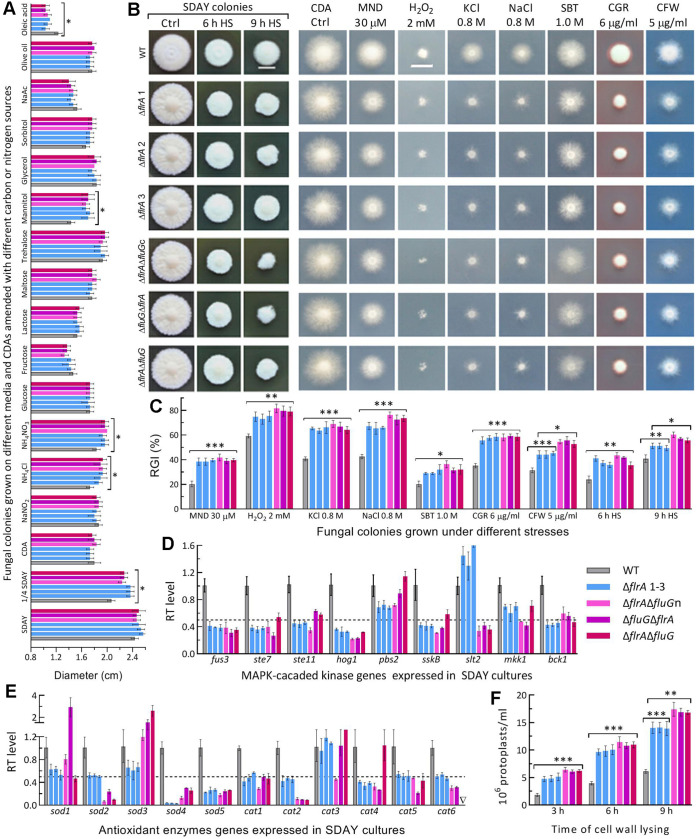
FIG 2.
Radial growth rates of B. bassiana in the absence of flrA and of both flrA and fluG. (A) Diameters of fungal colonies grown at the optimal regime of 25°C and L:D 12:12 for 7 days on the plates of rich medium SDAY, 1/4 SDAY, minimal medium CDA, and CDAs amended with different carbon/nitrogen sources. (B, C) Images (scale bar: 10 mm) and relative growth inhibition (RGI) percentages of fungal colonies incubated at 25°C for 7 days on CDA plates supplemented with indicated concentrations of menadione (MND), H2O2, KCl, NaCl, sorbitol (SBT), Congo red (CGR) and calcofluor white (CFW), and of SDAY colonies incubated at 25°C for 5-day growth recovery after 2-day-old colonies were exposed to a 42°C heat shock (HS) for 6 h and 9 h, respectively. Each colony was initiated by spotting 1 μL of a 106 conidia/mL suspension. (D, E) Relative transcript (RT) levels of MAPK-cascaded and antioxidant enzyme genes in the 3-day-old SDAY cultures of mutants with respect to the WT standard. The dashed line denotes a significant level of 1-fold downregulation. (F) Concentrations of protoplasts released from cell suspensions after 6 h and 9 h of cell wall lysing with enzymes in 1 M NaCl at 37°C. P < 0.05*; 0.01**; or 0.001*** in Tukey’s HSD tests. Error bars: SDs of the means from three independent replicates.

