PLOS ONE logoLink to PLOS ONE
. 2022 Aug 30;17(8):e0269316. doi: 10.1371/journal.pone.0269316

Bioaffinity-based surface immobilization of antibodies to capture endothelial colony-forming cells

Mariève D Boulanger 1,, Hugo A Level 1,, Mohamed A Elkhodiry 1,, Omar S Bashth 1, Pascale Chevallier 2,3, Gaétan Laroche 2,3, Corinne A Hoesli 1,*
Editor: Oksana Lockridge4
PMCID: PMC9426933  PMID: 36040884

Abstract

Maximizing the re-endothelialization of vascular implants such as prostheses or stents has the potential to significantly improve their long-term performance. Endothelial progenitor cell capture stents with surface-immobilized antibodies show significantly improved endothelialization in the clinic. However, most current antibody-based stent surface modification strategies rely on antibody adsorption or direct conjugation via amino or carboxyl groups which leads to poor control over antibody surface concentration and/or molecular orientation, and ultimately bioavailability for cell capture. Here, we assess the utility of a bioaffinity-based surface modification strategy to immobilize antibodies targeting endothelial cell surface antigens. A cysteine-tagged truncated protein G polypeptide containing three Fc-binding domains was conjugated onto aminated polystyrene substrates via a bi-functional linking arm, followed by antibody immobilization. Different IgG antibodies were successfully immobilized on the protein G-modified surfaces. Covalent grafting of the protein G polypeptide was more effective than surface adsorption in immobilizing antibodies at high density based on fluorophore-labeled secondary antibody detection, as well as endothelial colony-forming cell capture through anti-CD144 antibodies. This work presents a potential avenue for enhancing the performance of cell capture strategies by using covalent grafting of protein G polypeptides to immobilize IgG antibodies.

1. Introduction

Despite decades of continuous improvement in the overall performance of vascular implants, biocompatibility challenges remain a major area of concern [1, 2]. A healthy endothelium can modulate protein deposition, platelet activation, and proliferation of the underlying smooth muscle layer which are critical in reducing the risk of re-stenosis and thrombosis [3]. A promising approach to enhance stent endothelialization and reduce the risks of implant failure is to capture circulating endothelial progenitor cells (EPCs) [4]. Capturing EPCs, particularly their functional subtype [5] endothelial colony forming cells (ECFCs), accelerates the formation of a neo-endothelium due to their high proliferative potential and clonal expansion [68].

One way to promote ECFC capture on the surface of blood contacting devices is to modify the surface with antibodies that target ECFC surface antigens [9]. The surface-immobilized antibodies mimic the function of glycoproteins present on the vessel lining such as selectins and intercellular adhesion molecule-1 in the endogenous recruitment of circulating ECFCs to tissues undergoing vascular regeneration [1012]. This antibody-based strategy was commercially adopted to create the Genous stent (Orbusneich, USA), an anti-CD34 antibody surface-modified metal stent. In a pilot study with 193 patients, the Genous stent was shown to be as safe as drug-eluting stents (the control group) with no observed difference in adverse cardiac effects. The study also revealed a promising reduction in the rate of in-stent thrombosis in patients with the Genous stent compared to the drug-eluting stent group despite an increased rate of re-stenosis [13].

Since the first generation of EPC capture stents, potential areas of improvement in stent design were investigated. For example, the choice of CD34 as a target antigen has been under scrutiny due to its presence on the surface of hematopoietic progenitors that can exacerbate intimal hyperplasia [14]. Stents modified with anti-vascular endothelial cadherin (CD144) were shown to be more effective in accelerating endothelialization in animal models [15]. Furthermore, available antibody-modified implants mostly utilize passive adsorption or covalent conjugation to immobilize antibodies on the surface. Using passive adsorption, non-covalent interactions between the antibody and the surface dictates the strength and longevity of the surface modification often leading to lack of durability and lack of control over antibody orientation [16, 17]. Using covalent conjugation, most strategies exploit reactive groups introduced on surfaces to conjugate antibodies via free amine or carboxyl groups present in the antibody sequence [18, 19]. The abundance of these functional groups per antibody results in random antibody orientations on the surface and could lead to changes in conformation affecting its antigen binding efficacy [20, 21]. Unwanted reactions between amino acids in the hypervariable region of the antibody and the cross-linking reagents used for covalent immobilization could also directly affect the antigen-binding capacity of the antibody.

More recently, improved bio-affinity-based antibody immobilization techniques emerged as an alternative that can enhance the potential of next-generation ECFC-capture stents [2224]. Li et al. demonstrated that stent surfaces modified with anti-CD34 antibodies immobilized via the Fc-binding protein A improved in vivo stent endothelialization compared to unmodified controls [25]. However, due to the layer-by-layer coating strategy applied in this study, protein A molecules can take different conformations on the surface which reduces the availability of sites available for antibody immobilization and hence antibody surface density. Recombinant DNA technology can be used to introduce cysteine residues in the sequence of Fc-binding proteins which can then be conjugated onto surfaces via the thiol group. Gold substrates modified with cysteine-tagged protein G increased antibody surface density compared to adsorption controls while maintaining optimal antibody orientation leading to enhanced antigen binding capacity. This technique has been successfully applied to design immune-assays on a chip [26, 27]. Oriented surface immobilization of antibodies via covalent grafting of cysteine-tagged protein G remains untested for in vivo cell capture applications, particularly of ECFCs.

Here, we grafted a cysteine-tagged protein G polypeptide containing three Fc-binding domains on polystyrene, the most commonly-used tissue culture plastic, in order to immobilize antibodies which recognize endothelial surface markers such as CD31 and CD144. We hypothesized that our modified surfaces would increase ECFC capture compared to conventional antibody immobilization via passive adsorption. To test this hypothesis, a parallel plate flow chamber was used to study how ECFCs—derived from human donors—are captured under dynamic flow conditions.

2. Materials and methods

2.1 Surface modification and antibody immobilization

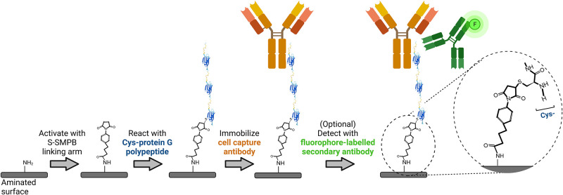
The bioaffinity-based antibody immobilization strategy relied on a 3-step process consisting of (1) activation of an aminated surface with an amine to sulfhydryl heterobifunctional linker, (2) covalent attachment of a cysteine-tagged protein G sequence containing three Fc-binding domains through sulfhydryl/maleimide reaction and (3) addition of IgG antibodies (Fig 1). First, aminated surfaces (BD PureCoat Amine Culture Dishes, BD Biosciences, now a part of Corning®, VWR) were reacted for 2 h with 150 μL/cm2 of a 3 mg/mL suspension of sulfo-succinimidyl-4-(p-maleimidophenyl)-butyrate (S-SMPB, #BC24, G-Biosciences) in phosphate buffered saline solution (PBS, #21600010, Thermo Fisher Scientific). Next, the cysteine-tagged protein G sequence was attached to the linking arm by adding 150 μL/cm2 of a 5.5 μM recombinant cysteine-tagged protein G polypeptide (henceforth termed “protein G polypeptide”—a recombinant non-glycosylated polypeptide chain produced in E. coli containing an N-terminal Cys followed by amino acids 298–497 of the streptococcal protein G sequence, #PRO-1328, Prospec-Tany Technogene Ltd) solution in PBS for 1 h. Finally, primary antibodies targeting cell surface antigens (mouse anti-human CD31 antibody #303101; mouse anti-human CD105 #323202; mouse anti-human CD144 #348502; and mouse anti-human CD14, anti-CD14, #367102; all from Biolegend, San Diego, US) were immobilized on the protein-G modified surfaces by adding 150 μL/cm2 of antibody solution at 5 μg/mL in PBS for 1 h. Surfaces were then rinsed twice with PBS, once with a 1% SDS-TRIS pH 11 solution (5% v/v of 20% sodium dodecyl sulfate #05030, from Sigma Aldrich and 2.4% w/v TRIS base PBP151-500 from Fisher Scientific in reverse osmosis water, pH adjusted to 11 with 2N NaOH solution) to remove adsorbed molecules, twice with PBS, and finally rinsed with reverse osmosis (RO) filtered water. The surfaces were then air-dried and stored for at most 1 week at room temperature before use. Adsorption controls followed the same surface modification scheme, except for the omission of S-SMPB prior to incubation with cysteine-tagged protein G polypeptide.

Fig 1. Schematic representation of the antibody immobilization process followed by a fluorescence-based antibody detection step.

Fig 1

For cell capture experiments under flow, surfaces were obtained by cutting aminated polystyrene Petri dishes (BD Purecoat Amine #354732, BD Biosciences, San Jose, USA) into 3.0 cm × 2.5 cm slides using a Micro Mill (Datron Neo 3-axis CNC Mill, Cell imaging and analysis network, McGill University, Canada). The circumference of these cut samples was lined with Teflon tape (#3213–103, polytetrafluoroethylene (PTFE) thread sealant) to maintain solutions on surfaces during the reaction steps. All other surface modifications were performed directly in well plates. All incubation steps were performed in the dark with 90 rpm agitation on a rotary shaker (Ecotron, Infors HT) at room temperature. After each reaction step, solutions containing reactants were removed, and surfaces were rinsed twice with 0.2 μm-filtered PBS. Collagen-coated surfaces were used as a native protein control and were prepared by adding 0.15 mL/cm2 of 50 μg/mL type 1 rat-tail collagen (Thermo Fisher Scientific) in 0.02 N acetic acid (Thermo Fisher Scientific). All surfaces were sterilized by incubation in 95% ethanol.

2.2 X-Ray Photoelectron Spectroscopy (XPS)

The chemical composition of the aminated surfaces, before and after functionalization was investigated by XPS using a PHI 5600-ci spectrometer (Physical Electronics, Eden Prairie, MN). The main XPS chamber was maintained at a base pressure of < 8×10⁻⁹ Torr. A standard aluminum X-ray (Al Kα = 1486.6 eV) source was used at 300 W to record survey spectra with charge neutralization, while C1s high resolution spectra were recorded with a standard magnesium X-ray source without neutralization. The detection angle was set at 45º with respect to the normal of the surface and the analyzed area was 0.5 mm2. The spectrometer work function was adjusted to 285.0 eV for the main C (1s) peak. Curve fitting of high-resolution peaks were determined by means of the least squares minimization procedure employing Gaussian-Lorentzian functions and a Shirley-type background.

2.3 Enzyme-Linked Immunosorbent Assay (ELISA) for protein G

A direct ELISA was developed to detect and quantify protein G polypeptide surface concentrations. Purecoat 96 well plates (BD Purecoat Amine # 356717, BD Biosciences) were functionalized as described above (S-SMPB(+)) or by omitting the S-SMPB activation step (S-SMPB(-)). To block further protein adsorption, 200 μL/well of 1% BSA solution in PBS was introduced and left to incubate for 90 min at 37ºC on a rotary shaker at 90 RPM. Wells were rinsed twice with wash buffer consisting of 0.05% Tween-20 (#P1379, Sigma-Aldrich) solution in PBS. To detect protein G polypeptide, a chicken immunoglobulin Y (IgY) anti-protein G was used. This antibody was selected due to the absence of affinity between protein G and the Fc fragment of IgY antibodies [28]: only the antigen binding fragment of the IgY anti-protein G can interact with protein G, which should facilitate quantification of surface ligands [29]. A volume of 100 μL of horseradish peroxidase (HRP)-conjugated IgY anti-protein G secondary antibody (HRP anti-protein G, OAIA00498, Aviva systems biology) solution (0.02 μg/mL of HRP anti-protein G diluted in rinsing solution with 1% BSA) was incubated in each well for 2 h at room temperature. Wells were immediately rinsed once with 1% SDS-TRIS solution at pH 11 and twice with wash buffer. To detect HRP, 100 μL of Slow TMB-ELISA substrate solution (#34024, Thermo Fisher) was added per well. After 25 min of incubation without agitation at room temperature, 100 μL/well of 1M sulfuric acid solution was added to stop the reaction, and absorbance measurements were immediately taken at 450 nm on a Benchmark plate reader (Bio-Rad, Berkeley, USA).

2.4 Immobilized antibody detection and quantification

Purecoat amine surfaces were modified as described above, but only certain regions of test surfaces were treated with protein G polypeptide by adding spots of 0.5 μL cysteine-tagged protein G polypeptide solution at concentrations ranging between 0.055 μM and 55 μM. To assess the effect of adsorption on surface amounts of protein G polypeptide, the spots were deposited on surfaces with (S-SMPB (+)) or without (S-SMPB (-)) S-SMPB treatment (previously treated with 3 mg/mL for 2 hours unless otherwise mentioned). After 1 h incubation, surfaces were rinsed with PBS and covered (spot and surrounding region) with primary antibody solution for 1 h as described above. After two washes in PBS, surfaces were covered with Alexa Fluor 488-conjugated goat anti-mouse secondary antibody solution at 20 μg/mL. After 1 h of incubation at room temperature, surfaces were rinsed twice with 1% SDS-TRIS solution, twice with PBS and twice with RO water before air drying. Spots were then imaged using a laser scanning confocal microscope (Zeiss LSM 5 Exciter, Germany) at 10X with an argon laser (488 nm). A total of 10 images per spot were taken to obtain the mean fluorescence intensity of one spot along with the associated standard deviation value. A total of 3 spots per replicate were studied to obtain the mean fluorescence intensity of each condition.

2.5 ECFC capture under flow

Peripheral blood mononuclear cells (PBMCs) were isolated from fresh adult human peripheral blood and ECFCs were expanded as previously described [30]. Fresh blood samples were collected from adult donors (N = 4: 2 females and 2 males; mean age 25.5 years) under informed consent following a protocol (Study No. A06-M33-15A) approved by the Ethics Institutional Review Board at McGill University. To study cell capture by antibody-modified surfaces under flow, functionalized surfaces were assembled into a custom parallel-plate flow chamber system with 4 independent chambers and flow paths, as previously described [31]. The flow chamber was ethanol-sterilized and then assembled inside an incubator with humidified air maintained at 5% CO2 and 37 ºC. Each chamber was connected to a reservoir that was pre-filled with 15 mL of warm serum-free EGM-2 (endothelial cell growth medium-2 without serum added from the kit, Lonza). ECFCs were harvested and resuspended in serum-free EGM-2 and added to the reservoirs to reach an overall cell density of 125,000 cells/mL. A peristaltic pump (Masterflex RK-7543-02 with Masterflex L/S two channels Easy Load II pump head using L/S 13 BPT tubing) was used to circulate the cell suspension in the system at a flow rate of 0.18 mL/s to obtain 1.5 dyn/cm2 wall shear stress. After 1 h of circulation, cells were fixed using a 4% paraformaldehyde solution (VWR) for 10 min, rinsed once in PBS and stored in PBS for immunocytochemistry.

2.6 Immunohistochemistry and microscopy

Fixed cells were permeabilized for 15 min with 0.1% Triton X (VWR) in PBS. Nuclei were stained with 1 μg/mL DAPI (Sigma) diluted in RO water for 10 min. Slides were then rinsed with RO water and stored in PBS before being imaged on an inverted fluorescent microscope (Olympus IX81). Images were acquired at 10X in phase contrast and fluorescence. At least 40 phase contrast images were acquired on each slide per cell capture experiment. Captured cells were enumerated using the “analyze particles command” of ImageJ from the 40 acquired DAPI images.

2.7 Statistical methods

Statistical analysis was performed with JMP Pro 13 software (SAS Institute, Cary, NC). Unless otherwise stated, data represent the average ± the standard error of the mean of 3 independent experiments. The criteria for statistically significant differences was selected to be p < 0.05. The Shapiro-Wilk normality test was applied prior to performing parametric tests. Student’s t-test was used for comparisons between two sample groups and comparisons between multiple groups were performed using two-way analysis of variance (ANOVA) followed by Tukey-Kramer HSD post hoc test. For ECFC capture experiments, each of the 4 replicas was conducted with ECFCs derived from a different donor.

3. Results

To develop a suitable antibody screening platform for cell capture, the proposed surface modification steps were first characterized, followed by testing the effect of different immobilized antibodies on ECFC capture under laminar flow.

3.1 Characterization of the Purecoat substrate and S-SMPB activation of the surface

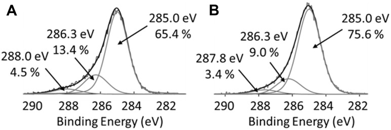
Cysteine-tagged protein G polypeptide was conjugated onto commercially-available aminated polystyrene surfaces via the amine-to-sulfhydryl linking arm S-SMPB (Fig 1). The surface compositon at each step was assessed by XPS (Table 1). As expected, the nitrogen content decreased after S-SMPB treatment when compared to the initial aminated substrates. The surface density of amino groups also decreased after the S-SMPB activation step based on the concentration of surface-bound Orange II (Fig A1 in S1 Appendix). The carbon content increased, and oxygen content decreased after the S-SMPB reaction due to the elemental composition of the linking arm. The increase in water static contact angles observed after S-SMPB treatment (Fig A1 in S1 Appendix) is consistent with the reduction in O and N content observed by XPS and addition of hydrophobic moieties present in the S-SMPB structure. All experiments were conducted at 3 mg/mL S-SMPB in this study, but this concentration could potentially be reduced based on the Orange II and water contact angle results. Subsequent protein G polypeptide grafting led to an increase in the O and N content related to the presence of these atoms in amino acid side chains and C terminus. The penetration depth of XPS analysis is ~5 nm which is inferior to the size of antibodies and on the same range as the size of a ~22 kDa polypeptide (~2 nm expected size) such as the protein G sequence used here. Atomic ratios therefore provide a useful metric to determine whether protein G polypeptide and antibody immobilization steps were successful. The O/C and N/C ratios decreased when anti-CD144 antibodies were immobilized on the surface. This observation suggests that anti-CD144 antibodies have higher C-rich amino acid content than the protein G polypeptide. This observation was corroborated by C1s high-resolution spectra (Fig 2): the peak at 285.0 eV, associated to C-C/C-H bonds, reached 76% for CD144 surface whereas it was 65% on Cys-Protein G.

Table 1. Surface atomic composition assessed by XPS survey analyses*.

Reaction step after which XPS analysis was conducted Atomic percentage Atomic ratios
%C %O %N N/C O/C
Initial pureCoat amine polystyrene surfaces 63 ± 2 17.2 ± 0.9 18.8 ± 0.6 0.30 ± 0.02 0.26 ± 0.02
After S-SMPB 86 ± 1 7.6 ± 0.9 5.0 ± 0.5 0.057 ± 0.006 0.09 ± 0.01
After Cys-protein G 83.3 ± 0.9 9.7 ± 0.7 7.0 ± 0.4 0.084 ± 0.006 0.116 ± 0.009
After antibody immobilization 85.8 ± 0.9 8.5 ± 0.9 4.7 ± 1.1 0.05 ± 0.01 0.09 ± 0.01

* Error estimates represent the standard deviation of areas analyzed on one sample to assess the grafting homogeneity.

Fig 2. High resolution C1s XPS spectra.

Fig 2

(A) Surfaces analyzed after the protein G polypeptide graftin step. (B) Surfaces after the antibody immobilization step using anti-CD144 antibodies.

3.2 Analysis of protein G polypeptide grafting efficiency

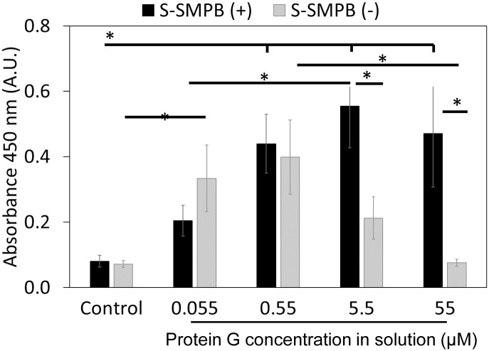
Next, cysteine-tagged protein G polypeptide grafting was evaluated with a direct ELISA developed to detect and confirm protein G polypeptide presence on surfaces (Fig 3). As shown in Fig 3, a positive correlation between protein G polypeptide concentration and absorbance signal was observed under covalent conjugation conditions. This trend was reproduced on other commercially-available aminated polystyrene substrates (Fig A2 in S1 Appendix). This correlation was not observed for adsorbed protein G polypeptide in the absence of the linking arm. This suggests that the covalent conjugation method via S-SMPB activation improved control over the amount of protein G polypeptide present on surface compared to adsorption. Together, the results shown in Table 1, Figs 2, and 3 highlight the effectiveness of this covalent conjugation strategy in grafting protein G polypeptide in controlled amounts on amine-functionalized surfaces.

Fig 3. Direct ELISA detection of surface-immobilized protein G polypeptide.

Fig 3

Conditions tested include covalent conjugation (S-SMPB(+) or adsorption (S-SMPB omitted during the first reaction step shown in Fig 1, S-SMPB(-)) of cysteine-tagged protein G polypeptide applied at different concentrations in solution. Control: no protein G polypeptide added. *P < 0.05 with N = 3. Raw data for this Fig 3 can be found in S2 Appendix.

3.3 Antibodies interact specifically with protein G modified surfaces

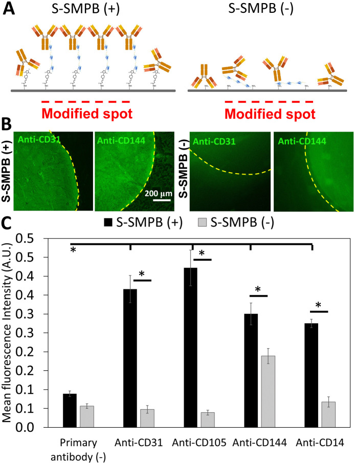
After covalent conjugation of protein G, the next step was to immobilize IgG antibodies onto the functionalized surfaces. As shown in Fig 4B, anti-CD31 antibodies were successfully immobilized on surfaces functionalized with protein G polypeptide based on fluorescent secondary antibody detection. The fluorescence intensity in the region where protein G polypeptide was deposited was significantly higher when applying at least 5.5 μM of cysteine-tagged protein G polypeptide as compared to 0.55 μM or to the surrounding region without polypeptide (Fig A3 in S1 Appendix). This was not observed on surfaces with adsorbed protein G polypeptide (S-SMPB(-)). A concentration 5.5 μM of protein G was selected for further studies given the higher background signal observed when applying 55 μM protein G polypeptide on S-SMPB(-) surfaces (Fig A3 in S1 Appendix). These experiments were repeated with four different IgG antibodies targeting endothelial (anti-CD31, anti-CD105, anti-CD144) or macrophage/monocyte (anti-CD14) surface markers which were successfully immobilized on surfaces using the same strategy (Fig 4C). Atomic force microscopy imaging performed on the modified surfaces also shows high density of immobilized antibodies. Immobilization via the protein G polypeptide created features of 10 nm ~ 15 nm height on surfaces (Fig A4 in S1 Appendix), as would be expected for oriented antibodies immobilized via Fc domains [26].

Fig 4. Fluorescence-based detection of immobilized IgG antibodies on conjugated protein G polypeptide spots.

Fig 4

(A) Schematic representation of antibody immobilization on protein G spots with (S-SMPB(+)) or without (S-SMPB(-)) covalent grafting of protein G polypeptide. (B) Spotted anti-CD31 or anti-CD144 antibodies detected through fluorophore-labelled anti-mouse antibodies as described in the last step of Fig 1. The protein G polypeptide concentration applied was 5.5 μM. (C) Successful immobilization of different primary antibodies on conjugated protein G polypeptide (S-SMPB (+)) based on the detection of fluorophore-labeled secondary antibodies added after protein G polypeptide and primary antibody immobilization. Surfaces without S-SMPB and/or without primary antibodies were used as negative controls. *P < 0.05 with N = 3. Raw data for Fig 4C can be found in S3 Appendix.

3.4 Antibody-functionalized surfaces can capture ECFCs

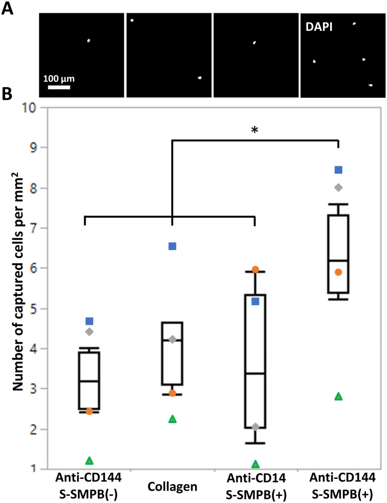
ECFCs were injected into a flow loop with a parallel plate chamber and circulated for 1 h at 1.5 dyn/cm2 wall-shear stress to determine whether antibody-modified surfaces can mediate cell capture. This wall shear stress is at the lower end of the physiological range and was selected to allow quantification of cell capture in vitro [32, 33]. Surfaces with (1) adsorbed anti-CD144 (S-SMPB (-)), (2) collagen-coated surfaces, (3) immobilized anti-CD14 on conjugated protein G polypeptide (S-SMPB (+)) and (4) immobilized anti-CD144 on conjugated protein G polypeptide (S-SMPB (+)) were tested in the flow system. Out of the four conditions, only the immobilized anti-CD144 on the conjugated protein G polypeptide had a significant effect in enhancing ECFC capture under flow (Fig 5). Surfaces with adsorbed anti-CD144 showed no significant difference in the number of captured ECFCs compared with surfaces modified with anti-CD14, a surface antigen that is not expressed by ECFCs [30]. Surfaces coated with rat tail collagen, a commonly used ECFC substrate, also had a significantly lower number of captured cells compared to the immobilized anti-CD144 on the conjugated protein G polypeptide. As a control, PBMCs, rich in CD14+ cells (>45%) but with low or undetectable CD144+ cell populations (<0.1%), were separately circulated in the same conditions over the same surfaces. In this arrangement, a significantly higher number of captured cells were observed on surfaces with immobilized anti-CD14 compared to surfaces with anti-CD144, confirming the specificity of the cell capturing strategy (S1 Appendix).

Fig 5. ECFC capture from laminar flow conditions on functionalized surfaces.

Fig 5

Conditions tested include anti-C144 (present on ECFCs) on adsorbed or conjugated protein G polypeptide, anti-CD14 (not present on ECFCs) on conjugated protein G polypeptide (negative control), or collagen. (A) Fluorescence images of ECFC nuclei on modified surfaces after 1 h of exposure to cell suspension under flow conditions. (B) Quantification of number of cells per mm2 on the modified surfaces at the end of the 1 h of flow. Each symbol represents data collected using ECFCs from a separate donor. *P < 0.05 with N = 4.

4. Discussion

To our knowledge, this study is the first demonstration of the utility of the covalent conjugation of polypeptides containing the Fc-binding domains of protein G for antibody immobilization towards cell capture applications. This strategy was selected to maximize the immunoaffinity of the antibodies compared to adsorption or direct covalent conjugation methods which can result in random orientation and reduced availability of antigen-binding sites [3436]. The mild conditions (physiological pH, room temperature, aqueous conditions) of this surface modification strategy assures compatibility with a wide variety of cell culture substrates and biomaterials. The covalent conjugation of the protein G polypeptide via its N-terminal cysteine tag enhances the ability to control the orientation of the protein G and of the immobilized antibody [26]. Different IgG antibodies were successfully immobilized on conjugated protein G polypeptide, achieving better control over protein G polypeptide surface density compared with adsorption (omission of the linking arm). The surfaces with grafted protein G polypeptide and immobilized anti-CD144 successfully captured circulating ECFCs at 1.5 dyn/cm2 wall shear stress, contrary to surfaces where the linking arm was omitted from the surface treatment. The grafted protein G polypeptide anti-CD144 surfaces also captured significantly higher circulating ECFC numbers compared with surfaces with antibodies which do not target ECFCs (anti-CD14). Compared to a native extracellular matrix protein such as collagen, the anti-CD144 antibodies immobilized via grafted protein G polypeptide resulted in significantly higher levels of ECFC capture demonstrating the advantage of targeting specific cell-surface antigens. These promising findings highlight the value of our proposed surface modification strategy for the design of EPC capture vascular biomaterials.

S-SMPB is a versatile linking arm which has been applied to vascular biomaterials such as aminated polytetrafluoroethylene (PTFE) [37], poly (L-lactide (PLLA), poly (ɛ-caprolactone) (PCL)[38] and other aminated model surfaces [39]. The maleimide functional group of the S-SMPB reacts with the sulfhydryl group present solely in the cysteine tag of the protein G polypeptide, thus creating a selective oriented conjugation strategy. Compared to protein G polypeptide adsorption, conjugation via S-SMPB led to a better control over protein G polypeptide surface density. With covalent protein G polypeptide grafting, surface concentration of protein G polypeptide followed an expected saturation profile (Fig 3). Conversely, the maximum achievable protein G polypeptide surface concentration achieved through adsorption was lower and was followed by a decreased in surface densities at higher concentrations. A possible explanation for the adsorption profiles at high polypeptide concentrations in solution is the ability of free cysteines to interact in solution to form disulfide bonds which can produce dimers reducing effective interaction with the detection antibodies of the ELISA. High concentrations may also lead to multilayers on surfaces with changes in conformation leading to reduced secondary antibody detection. Therefore, the conjugation scheme with the S-SMPB linking arm allowed better control over protein G surface density.

The protein G polypeptide used in this study comprises amino acids 298–497 of the full streptococcal protein G sequence, which includes the three Fc-binding domains of this protein. Streptococcal protein G interacts with immunoglobulin G antibodies of most mammalian species [40, 41]. This interaction can be mediated both through Fc and Fab regions [42], although the association constant is one order of magnitude higher for Fc fragments [43] with the exception of mouse IgG1 where Fab plays a more significant role [44, 45]. A polypeptide fragment containing the sequence used in the current study was previously shown to interact specifically with the Fc and not the Fab [42, 46] region of human IgG antibodies. All antibodies in the current work were of IgG1 subclass except anti-CD144, of IgG2a subclass. It is possible that the difference in subclass led to different antibody interactions with adsorbed vs conjugated protein G observed in Fig 5. Theoretically, the higher ratio between Fc:Fab protein G binding affinity expected for IgG2 antibodies would lead to improved orientation on surfaces. However, even for antibodies where Fab interactions with protein G are significant, the hypervariable region is expected to remain available for antigen recognition [47].

We have recently shown that antibodies immobilized through surface-grafted RRGW peptides designed to interact with the Fc region of IgG2a antibodies can selectively capture ECFCs from a mixture of cells under dynamic flow conditions [23]. A major obstacle hampering the use of protein G on implanted biomaterials such as stents is its unknown immunogenic profile and the possibility that it can provoke undesirable host immune responses [48]. Fc-binding peptides such as RRGW, on the other hand, can pose a lower risk of triggering an immune response due to its small chemically defined structure and the absence of endotoxins due to chemical synthesis. Larger polypeptides can also be more susceptible to enzymatic degradation and thermal denaturation compared to smaller Fc-binding peptides. A potential advantage of the protein G polypeptide strategy over the previously proposed RRGW peptide are the existing protein G supply chains allowing its use in cGMP bioprocessing plants, which may facilitate large-scale use in other biomedical and clinical applications [49]. Furthermore, the recombinant cysteine-tagged protein G polypeptide has 25 times the molecular weight of the RRGW peptide which can lead to 2 to 3 nm additional spacing between the antigen-binding site and the modified surface, reducing steric hindrance [27]. There are three Fc fragment binding sites available on each protein G polypeptide compared to the RRGW peptide’s single antibody binding capacity, potentially increasing the density of antibodies which can be immobilized on protein G polypeptide modified surfaces. Further development of antibody immobilization strategies via protein G polypeptide and RRGW for in vivo use will require side-by-side comparison of the hemocompatibility, immunogenicity, and stability of both molecules. Given the promising results obtained via protein G-mediated antibody immobilization for cell capture, other methods that aim to orient antibodies on surfaces [50] could also show significant promise in cell capture applications. Comparison to other antibody-based biocoatings [5153] which lead to varying degrees of antibody orientation on surfaces could help elucidate the effect of antibody conformation on cell capture efficiency.

Given the versatility of the proposed bioaffinity-based antibody immobilization strategy as demonstrated through immobilization of IgGs targeting different cell antigens, it would be interesting to study selective capture of different immune cell subsets from peripheral blood. The proliferation of ECFC post-capture should also be assessed. Combination of Fc-binding peptides or polypeptides with integrin-binding peptides shows significant promise in this regard [54]. This bi-functionnal coating could also be applied on various substrates to efficiently isolate ECFC from peripheral blood in vitro, paving the way to the development of novel cell-culture materials. All in all, the proposed bioaffinity-based antibody immobilization strategy shows promise towards engineering clinically successful EPC-capture biomaterials.

5. Conclusion

This study presents a 3-step surface functionalization strategy to immobilize antibodies on aminated surfaces via bioaffinity interactions with a protein G polypeptide containing its three Fc-binding domains. This technology can be applied to engineer endothelial progenitor cell capture stents and other cell separation devices. Model aminated polystyrene surfaces were first reacted with an amine to sulfhydryl linking arm. The linking arm was then used to conjugate protein G to the surface through a cysteine tag maximizing its antibody immobilization capacity. Different IgG antibodies were successfully immobilized on the surface and detected using a simple fluorescence-based approach. Finally, surfaces modified with anti-CD144 via our protein G polypeptide-based approach displayed superior ability in capturing human derived ECFCs from flow compared to surfaces modified through passive adsorption. Our work highlights the potential of grafted protein G-based surface functionalization strategies in enhancing the potential of ECFC capture on the surface of vascular implants. Bioaffinity-based antibody immobilization on EPC capture stents may accelerate the endothelialization process essential in vascular regeneration and homeostasis.

Supporting information

S1 Appendix. Supplementary methods and figures: Orange II assay, static water contact angle, atomic force microscopy and peripheral blood mononuclear cell capture on anti-CD144 and anti-CD14 surfaces.

(PDF)

S2 Appendix. Spreadsheet comprising the raw ELISA readouts used to generate Fig 3.

(XLSX)

S3 Appendix. Spreadsheet comprising the raw fluorescence intensity data used to generate Fig 4C.

(XLSX)

Acknowledgments

The authors thank Ariane Beland, Stéphanie Vanslambrouck, Lisa Danielczak, Ranjan Roy, Frank Caporuscio, Natalie Fekete and Gad Sabbatier for technical support and Raymond Tran for his help in reviewing the manuscript.

Data Availability

All relevant data are within the manuscript and its Supporting information files.

Funding Statement

This study was financially supported by the Canadian Institutes of Health Research (CIHR, MOP 142285; CAH and GL) and the Canadian Foundation for Innovation (CFI, project 35507; CAH). This research was undertaken, in part, thanks to funding from the Canada Research Chairs Program (CAH). This work was supported via travel awards and networking opportunities offered by ThéCell (The Quebec Network for Cell, Tissue and Gene Therapy; MDB, MAE, OSB, CH), the Quebec Center for Advanced Materials (QCAM; MDB, MAE, OSB, GL, CH), PROTEO (The Quebec Network for Research on Protein Function; MDB, MAE, OSB, CH), CMDO (the Cardiometabolic Health, Diabetes, and Obesity Research Network; MDB, MAE, OSB, CH), and the MRM (McGill Regenerative Medicine; MDB, MAE, OSB, CH) network. MDB received a Graduate Excellence Fellowship from the Faculty of Engineering of McGill University. MAE received a scholarship from the Fonds de recherche du Québec - Nature et technologies (Programme de bourses d’excellence pour étudiants étrangers 262907). OSB received a 2017 Hadhramout Establishment for Human Development scholarship. The funders had no role in study design, data collection and analysis, decision to publish, or preparation of the manuscript.

References

  • 1.Jaganathan SK, Supriyanto E, Murugesan S, Balaji A, Asokan MK. Biomaterials in cardiovascular research: applications and clinical implications. BioMed research international. 2014;2014:459465-. Epub 05/08. doi: 10.1155/2014/459465 . [DOI] [PMC free article] [PubMed] [Google Scholar]
  • 2.Byrne RA, Joner M, Kastrati A. Stent thrombosis and restenosis: what have we learned and where are we going? The Andreas Grüntzig Lecture ESC 2014. Eur Heart J. 2015;36(47):3320–31. Epub 09/28. doi: 10.1093/eurheartj/ehv511 . [DOI] [PMC free article] [PubMed] [Google Scholar]
  • 3.Ravindranath RR, Romaschin A, Thompson M. In vitro and in vivo cell-capture strategies using cardiac stent technology—A review. Clin Biochem. 2016;49(1):186–91. doi: 10.1016/j.clinbiochem.2015.09.012 [DOI] [PubMed] [Google Scholar]
  • 4.Avci-Adali M, Ziemer G, Wendel HP. Induction of EPC homing on biofunctionalized vascular grafts for rapid in vivo self-endothelialization—A review of current strategies. Biotechnol Adv. 2010;28(1):119–29. doi: 10.1016/j.biotechadv.2009.10.005 [DOI] [PubMed] [Google Scholar]
  • 5.Medina RJ, Barber CL, Sabatier F, Dignat-George F, Melero-Martin JM, Khosrotehrani K, et al. Endothelial Progenitors: A Consensus Statement on Nomenclature. Stem Cells Transl Med. 2017;6(5):1316–20. Epub 2017/03/16. doi: 10.1002/sctm.16-0360 . [DOI] [PMC free article] [PubMed] [Google Scholar]
  • 6.Kipshidze N, Dangas G, Tsapenko M, Moses J, Leon MB, Kutryk M, et al. Role of the endothelium in modulating neointimal formation: vasculoprotective approaches to attenuate restenosis after percutaneous coronary interventions. J Am Coll Cardiol. 2004;44(4):733–9. doi: 10.1016/j.jacc.2004.04.048 [DOI] [PubMed] [Google Scholar]
  • 7.Liu T, Liu S, Zhang K, Chen J, Huang N. Endothelialization of implanted cardiovascular biomaterial surfaces: the development from in vitro to in vivo. J Biomed Mater Res A. 2014;102(10):3754–72. Epub 2013/11/19. doi: 10.1002/jbm.a.35025 . [DOI] [PubMed] [Google Scholar]
  • 8.Ingram DA, Mead LE, Tanaka H, Meade V, Fenoglio A, Mortell K, et al. Identification of a novel hierarchy of endothelial progenitor cells using human peripheral and umbilical cord blood. Blood. 2004;104(9):2752–60. Epub 2004/07/01. doi: 10.1182/blood-2004-04-1396 . [DOI] [PubMed] [Google Scholar]
  • 9.Sethi R, Lee CH. Endothelial progenitor cell capture stent: safety and effectiveness. Journal of interventional cardiology. 2012;25(5):493–500. Epub 2012/05/23. doi: 10.1111/j.1540-8183.2012.00740.x . [DOI] [PubMed] [Google Scholar]
  • 10.Lewis DM, Abaci HE, Xu Y, Gerecht S. Endothelial progenitor cell recruitment in a microfluidic vascular model. Biofabrication. 2015;7(4):045010. Epub 2015/12/24. doi: 10.1088/1758-5090/7/4/045010 . [DOI] [PubMed] [Google Scholar]
  • 11.Liu Z-J, Tian R, Li Y, Zhang L, Shao H, Yang C, et al. SDF-1α-induced dual pairs of E-selectin/ligand mediate endothelial progenitor cell homing to critical ischemia. Sci Rep. 2016;6(1):34416. doi: 10.1038/srep34416 [DOI] [PMC free article] [PubMed] [Google Scholar]
  • 12.Foubert P, Silvestre JS, Souttou B, Barateau V, Martin C, Ebrahimian TG, et al. PSGL-1-mediated activation of EphB4 increases the proangiogenic potential of endothelial progenitor cells. J Clin Invest. 2007;117(6):1527–37. doi: 10.1172/JCI28338 . [DOI] [PMC free article] [PubMed] [Google Scholar]
  • 13.Beijk MA, Klomp M, van Geloven N, Koch KT, Henriques JP, Baan J, et al. Two-year follow-up of the Genous endothelial progenitor cell capturing stent versus the Taxus Liberte stent in patients with de novo coronary artery lesions with a high-risk of restenosis: a randomized, single-center, pilot study. Catheter Cardiovasc Interv. 2011;78(2):189–95. Epub 2011/05/05. doi: 10.1002/ccd.23143 . [DOI] [PubMed] [Google Scholar]
  • 14.Leopold JA. Prohealing endothelial progenitor cell capture stents: do the cells captured explain the clinical outcomes? Circulation Cardiovascular interventions. 2013;6(5):494–5. doi: 10.1161/CIRCINTERVENTIONS.113.000812 . [DOI] [PMC free article] [PubMed] [Google Scholar]
  • 15.Lee JM, Choe W, Kim BK, Seo WW, Lim WH, Kang CK, et al. Comparison of endothelialization and neointimal formation with stents coated with antibodies against CD34 and vascular endothelial-cadherin. Biomaterials. 2012;33(35):8917–27. doi: 10.1016/j.biomaterials.2012.08.066 [DOI] [PubMed] [Google Scholar]
  • 16.Shen M, Rusling J, Dixit CK. Site-selective orientated immobilization of antibodies and conjugates for immunodiagnostics development. Methods. 2017;116:95–111. Epub 2016/11/24. doi: 10.1016/j.ymeth.2016.11.010 . [DOI] [PMC free article] [PubMed] [Google Scholar]
  • 17.Butler JE, Ni L, Brown WR, Joshi KS, Chang J, Rosenberg B, et al. The immunochemistry of sandwich ELISAs—VI. Greater than 90% of monoclonal and 75% of polyclonal anti-fluorescyl capture antibodies (CAbs) are denatured by passive adsorption. Mol Immunol. 1993;30(13):1165–75. Epub 1993/09/01. doi: 10.1016/0161-5890(93)90135-x . [DOI] [PubMed] [Google Scholar]
  • 18.Eswaramoorthy H. CHEMICAL CROSSLINKING OF PROTEINS: A REVIEW. Journal of Pharmaceutical and Scientific Innovation. 2012. [Google Scholar]
  • 19.Chiadò A, Palmara G, Ricciardi S, Frascella F, Castellino M, Tortello M, et al. Optimization and characterization of a homogeneous carboxylic surface functionalization for silicon-based biosensing. Colloids Surf B Biointerfaces. 2016;143:252–9. doi: 10.1016/j.colsurfb.2016.03.048 [DOI] [PubMed] [Google Scholar]
  • 20.Nimse SB, Song K, Sonawane MD, Sayyed DR, Kim T. Immobilization techniques for microarray: challenges and applications. Sensors (Basel). 2014;14(12):22208–29. Epub 2014/11/28. doi: 10.3390/s141222208 . [DOI] [PMC free article] [PubMed] [Google Scholar]
  • 21.Sharma S, Byrne H, O’Kennedy RJ. Antibodies and antibody-derived analytical biosensors. Essays Biochem. 2016;60(1):9–18. doi: 10.1042/EBC20150002 . [DOI] [PMC free article] [PubMed] [Google Scholar]
  • 22.Qi P, Chen S, Liu T, Chen J, Yang Z, Weng Y, et al. New strategies for developing cardiovascular stent surfaces with novel functions (Review). Biointerphases. 2014;9(2):029017. doi: 10.1116/1.4878719 [DOI] [PubMed] [Google Scholar]
  • 23.Bashth OS, Elkhodiry MA, Laroche G, Hoesli CA. Surface grafting of Fc-binding peptides as a simple platform to immobilize and identify antibodies that selectively capture circulating endothelial progenitor cells. Biomaterials Science. 2020. doi: 10.1039/d0bm00650e [DOI] [PubMed] [Google Scholar]
  • 24.Yu Q, Wang Q, Li B, Lin Q, Duan Y. Technological Development of Antibody Immobilization for Optical Immunoassays: Progress and Prospects. Crit Rev Anal Chem. 2015;45(1):62–75. doi: 10.1080/10408347.2014.881249 [DOI] [Google Scholar]
  • 25.Li Q-L, Huang N, Chen C, Chen J-L, Xiong K-Q, Chen J-Y, et al. Oriented immobilization of anti-CD34 antibody on titanium surface for self-endothelialization induction. Journal of Biomedical Materials Research Part A. 2010;94A(4):1283–93. doi: 10.1002/jbm.a.32812 [DOI] [PubMed] [Google Scholar]
  • 26.Kim ES, Shim CK, Lee JW, Park JW, Choi KY. Synergistic effect of orientation and lateral spacing of protein G on an on-chip immunoassay. Analyst. 2012;137(10):2421–30. Epub 2012/04/06. doi: 10.1039/c2an16137k . [DOI] [PubMed] [Google Scholar]
  • 27.Lee JM, Park HK, Jung Y, Kim JK, Jung SO, Chung BH. Direct immobilization of protein g variants with various numbers of cysteine residues on a gold surface. Anal Chem. 2007;79(7):2680–7. Epub 2007/03/08. doi: 10.1021/ac0619231 . [DOI] [PubMed] [Google Scholar]
  • 28.Lee W, Atif AS, Tan SC, Leow CH. Insights into the chicken IgY with emphasis on the generation and applications of chicken recombinant monoclonal antibodies. Journal of Immunological Methods. 2017;447:71–85. doi: 10.1016/j.jim.2017.05.001 [DOI] [PubMed] [Google Scholar]
  • 29.Schade R, Staak C, Hendriksen C, Erhard M, Hugl H, Koch G, et al. The production of avian (egg yolk) antibodies: IgY—The report and recommendations of ECVAM Workshop 21. Atla-Altern Lab Anim. 1996;24(6):925–34. [Google Scholar]
  • 30.Elkhodiry MA, Boulanger MD, Bashth O, Tanguay J-F, Laroche G, Hoesli CA. Isolating and expanding endothelial progenitor cells from peripheral blood on peptide-functionalized polystyrene surfaces. Biotechnol Bioeng. 2019;116(10):2598–609. doi: 10.1002/bit.27107 [DOI] [PubMed] [Google Scholar]
  • 31.Hoesli CA, Tremblay C, Juneau P-M, Boulanger MD, Beland AV, Ling SD, et al. Dynamics of Endothelial Cell Responses to Laminar Shear Stress on Surfaces Functionalized with Fibronectin-Derived Peptides. ACS Biomaterials Science & Engineering. 2018. doi: 10.1021/acsbiomaterials.8b00774 [DOI] [PubMed] [Google Scholar]
  • 32.Green JV, Murthy SK. Microfluidic enrichment of a target cell type from a heterogenous suspension by adhesion-based negative selection. Lab Chip. 2009;9(15):2245–8. Epub 2009/07/17. doi: 10.1039/b906768j . [DOI] [PubMed] [Google Scholar]
  • 33.Plouffe BD, Njoka DN, Harris J, Liao J, Horick NK, Radisic M, et al. Peptide-mediated selective adhesion of smooth muscle and endothelial cells in microfluidic shear flow. Langmuir: the ACS journal of surfaces and colloids. 2007;23(9):5050–5. doi: 10.1021/la0700220 [DOI] [PubMed] [Google Scholar]
  • 34.Markway BD, McCarty OJ, Marzec UM, Courtman DW, Hanson SR, Hinds MT. Capture of flowing endothelial cells using surface-immobilized anti-kinase insert domain receptor antibody. Tissue engineering Part C, Methods. 2008;14(2):97–105. doi: 10.1089/ten.tec.2007.0300 [DOI] [PubMed] [Google Scholar]
  • 35.Vashist SK, Dixit CK, MacCraith BD, O’Kennedy R. Effect of antibody immobilization strategies on the analytical performance of a surface plasmon resonance-based immunoassay. The Analyst. 2011;136(21):4431–6. doi: 10.1039/c1an15325k [DOI] [PubMed] [Google Scholar]
  • 36.Makaraviciute A, Ramanaviciene A. Site-directed antibody immobilization techniques for immunosensors. Biosensors and Bioelectronics. 2013;50:460–71. doi: 10.1016/j.bios.2013.06.060 [DOI] [PubMed] [Google Scholar]
  • 37.Vallières K, Petitclerc É, Laroche G. Covalent Grafting of Fibronectin onto Plasma-Treated PTFE: Influence of the Conjugation Strategy on Fibronectin Biological Activity. Macromolecular Bioscience. 2007;7(5):738–45. doi: 10.1002/mabi.200600267 [DOI] [PubMed] [Google Scholar]
  • 38.Larrañaga A, Guay-Bégin A-A, Chevallier P, Sabbatier G, Fernández J, Laroche G, et al. Grafting of a model protein on lactide and caprolactone based biodegradable films for biomedical applications. Biomatter. 2014;4:e27979. doi: 10.4161/biom.27979 [DOI] [PMC free article] [PubMed] [Google Scholar]
  • 39.Hoesli CA, Garnier A, Juneau P-M, Chevallier P, Duchesne C, Laroche G. A fluorophore-tagged RGD peptide to control endothelial cell adhesion to micropatterned surfaces. JBMT Biomaterials. 2014;35(3):879–90. doi: 10.1016/j.biomaterials.2013.09.076 [DOI] [PubMed] [Google Scholar]
  • 40.Bjorck L, Kronvall G. Purification and some properties of streptococcal protein G, a novel IgG-binding reagent. J Immunol. 1984;133(2):969–74. Epub 1984/08/01. . [PubMed] [Google Scholar]
  • 41.Boyle MD. CHAPTER 1—Introduction to bacterial immunoglobulin-binding proteins,. In: Boyle MD, editor. Bacterial Immunoglobulin-Binding Proteins: Academic Press; 1990. p. 1–21. [Google Scholar]
  • 42.Erntell M, Myhre EB, Sjobring U, Bjorck L. Streptococcal protein G has affinity for both Fab- and Fc-fragments of human IgG. Mol Immunol. 1988;25(2):121–6. Epub 1988/02/01. doi: 10.1016/0161-5890(88)90059-4 . [DOI] [PubMed] [Google Scholar]
  • 43.Stone GC, Sjobring U, Bjorck L, Sjoquist J, Barber CV, Nardella FA. The Fc binding site for streptococcal protein G is in the C gamma 2-C gamma 3 interface region of IgG and is related to the sites that bind staphylococcal protein A and human rheumatoid factors. J Immunol. 1989;143(2):565–70. Epub 1989/07/15. . [PubMed] [Google Scholar]
  • 44.Derrick JP, Wigley DB. The third IgG-binding domain from streptococcal protein G. An analysis by X-ray crystallography of the structure alone and in a complex with Fab. J Mol Biol. 1994;243(5):906–18. Epub 1994/11/11. doi: 10.1006/jmbi.1994.1691 . [DOI] [PubMed] [Google Scholar]
  • 45.Kato K, Lian LY, Barsukov IL, Derrick JP, Kim H, Tanaka R, et al. Model for the complex between protein G and an antibody Fc fragment in solution. Structure. 1995;3(1):79–85. Epub 1995/01/15. doi: 10.1016/s0969-2126(01)00136-8 . [DOI] [PubMed] [Google Scholar]
  • 46.Derrick JP, Wigley DB. Crystal structure of a streptococcal protein G domain bound to an Fab fragment. Nature. 1992;359(6397):752–4. Epub 1992/10/22. doi: 10.1038/359752a0 . [DOI] [PubMed] [Google Scholar]
  • 47.Erntell M, Myhre EB, Kronvall G. Alternative non-immune F(ab’)2-mediated immunoglobulin binding to group C and G streptococci. Scand J Immunol. 1983;17(3):201–9. Epub 1983/03/01. doi: 10.1111/j.1365-3083.1983.tb00783.x . [DOI] [PubMed] [Google Scholar]
  • 48.Choe W, Durgannavar TA, Chung SJ. Fc-Binding Ligands of Immunoglobulin G: An Overview of High Affinity Proteins and Peptides. Materials. 2016;9(12):994. doi: 10.3390/ma9120994 [DOI] [PMC free article] [PubMed] [Google Scholar]
  • 49.Roque ACA, Silva CSO, Taipa MÂ. Affinity-based methodologies and ligands for antibody purification: Advances and perspectives. J Chromatogr. 2007;1160(1):44–55. doi: 10.1016/j.chroma.2007.05.109 [DOI] [PubMed] [Google Scholar]
  • 50.Welch NG, Scoble JA, Muir BW, Pigram PJ. Orientation and characterization of immobilized antibodies for improved immunoassays (Review). Biointerphases. 2017;12(2):02D301. Epub 2017/03/18. doi: 10.1116/1.4978435 . [DOI] [PubMed] [Google Scholar]
  • 51.Su H, Xue G, Ye C, Wang Y, Zhao A, Huang N, et al. The effect of anti-CD133/fucoidan bio-coatings on hemocompatibility and EPC capture. J Biomater Sci Polym Ed. 2017;28(17):2066–81. Epub 2017/09/01. doi: 10.1080/09205063.2017.1373989 . [DOI] [PubMed] [Google Scholar]
  • 52.Park KS, Kang SN, Kim DH, Kim HB, Im KS, Park W, et al. Late endothelial progenitor cell-capture stents with CD146 antibody and nanostructure reduce in-stent restenosis and thrombosis. Acta Biomater. 2020;111:91–101. Epub 2020/05/21. doi: 10.1016/j.actbio.2020.05.011 . [DOI] [PubMed] [Google Scholar]
  • 53.Wu X, Yin T, Tian J, Tang C, Huang J, Zhao Y, et al. Distinctive effects of CD34- and CD133-specific antibody-coated stents on re-endothelialization and in-stent restenosis at the early phase of vascular injury. Regen Biomater. 2015;2(2):87–96. Epub 2016/01/28. doi: 10.1093/rb/rbv007 . [DOI] [PMC free article] [PubMed] [Google Scholar]
  • 54.Hoesli CA, Bashth O, Elkhodiry MA, Boulanger M, Laroche G, inventorsDual function surface for cell capture and spreading2021 2020-07-14 (USPTO), 2021-07-14 (PCT).

Decision Letter 0

Oksana Lockridge

2 Nov 2021

PONE-D-21-26288Bioaffinity-based surface immobilization of antibodies to capture endothelial colony-forming cellsPLOS ONE

Dear Dr. Hoesli,

Thank you for submitting your manuscript to PLOS ONE. After careful consideration, we feel that it has merit but does not fully meet PLOS ONE’s publication criteria as it currently stands. Therefore, we invite you to submit a revised version of the manuscript that addresses the points raised during the review process.

Please submit your revised manuscript by Dec 16 2021 11:59PM. If you will need more time than this to complete your revisions, please reply to this message or contact the journal office at plosone@plos.org. When you're ready to submit your revision, log on to https://www.editorialmanager.com/pone/ and select the 'Submissions Needing Revision' folder to locate your manuscript file.

Please include the following items when submitting your revised manuscript:

  • A rebuttal letter that responds to each point raised by the academic editor and reviewer(s). You should upload this letter as a separate file labeled 'Response to Reviewers'.

  • A marked-up copy of your manuscript that highlights changes made to the original version. You should upload this as a separate file labeled 'Revised Manuscript with Track Changes'.

  • An unmarked version of your revised paper without tracked changes. You should upload this as a separate file labeled 'Manuscript'.

If you would like to make changes to your financial disclosure, please include your updated statement in your cover letter. Guidelines for resubmitting your figure files are available below the reviewer comments at the end of this letter.

If applicable, we recommend that you deposit your laboratory protocols in protocols.io to enhance the reproducibility of your results. Protocols.io assigns your protocol its own identifier (DOI) so that it can be cited independently in the future. For instructions see: https://journals.plos.org/plosone/s/submission-guidelines#loc-laboratory-protocols. Additionally, PLOS ONE offers an option for publishing peer-reviewed Lab Protocol articles, which describe protocols hosted on protocols.io. Read more information on sharing protocols at https://plos.org/protocols?utm_medium=editorial-email&utm_source=authorletters&utm_campaign=protocols.

We look forward to receiving your revised manuscript.

Kind regards,

Oksana Lockridge, Ph.D.

Academic Editor

PLOS ONE

Journal Requirements:

When submitting your revision, we need you to address these additional requirements.

1. Please ensure that your manuscript meets PLOS ONE's style requirements, including those for file naming. The PLOS ONE style templates can be found at 

https://journals.plos.org/plosone/s/file?id=wjVg/PLOSOne_formatting_sample_main_body.pdf and 

https://journals.plos.org/plosone/s/file?id=ba62/PLOSOne_formatting_sample_title_authors_affiliations.pdf

2. Please amend your current ethics statement to address the following concerns:

a) Did participants provide their written or verbal informed consent to participate in this study?

b) If consent was verbal, please explain i) why written consent was not obtained, ii) how you documented participant consent, and iii) whether the ethics committees/IRB approved this consent procedure.

3. Thank you for stating the following in the Acknowledgments Section of your manuscript: 

This study was financially supported by the Canadian Institutes of Health Research (CIHR, MOP 142285; CAH and GL) and the Canadian Foundation for Innovation (CFI, project 35507; CAH). This research was undertaken, in part, thanks to funding from the Canada Research Chairs Program (CAH). This work was supported via travel awards and networking opportunities offered by ThéCell (The Quebec Network for Cell, Tissue and Gene Therapy; MDB, MAE, OSB, CH), the Quebec Center for Advanced Materials (QCAM; MDB, MAE, OSB, GL, CH), PROTEO (The Quebec Network for Research on Protein Function; MDB, MAE, OSB, CH), CMDO (the Cardiometabolic Health, Diabetes, and Obesity Research Network; MDB, MAE, OSB, CH), and the MRM (McGill Regenerative Medicine; MDB, MAE, OSB, CH) network. MDB received a Graduate Excellence Fellowship from the Faculty of Engineering of McGill University. MAE received a scholarship from the Fonds de recherche du Québec - Nature et technologies (Programme de bourses d'excellence pour étudiants étrangers 262907). OSB received a 2017 Hadhramout Establishment for Human Development scholarship. The funders had no role in study design, data collection and analysis, decision to publish, or preparation of the manuscript.

Please note that funding information should not appear in the Acknowledgments section or other areas of your manuscript. We will only publish funding information present in the Funding Statement section of the online submission form. 

Please remove any funding-related text from the manuscript and let us know how you would like to update your Funding Statement. Currently, your Funding Statement reads as follows: 

This study was financially supported by the Canadian Institutes of Health Research (CIHR, MOP 142285; CAH and GL) and the Canadian Foundation for Innovation (CFI, project 35507; CAH). This research was undertaken, in part, thanks to funding from the Canada Research Chairs Program (CAH). This work was supported via travel awards and networking opportunities offered by ThéCell (The Quebec Network for Cell, Tissue and Gene Therapy; MDB, MAE, OSB, CH), the Quebec Center for Advanced Materials (QCAM; MDB, MAE, OSB, GL, CH), PROTEO (The Quebec Network for Research on Protein Function; MDB, MAE, OSB, CH), CMDO (the Cardiometabolic Health, Diabetes, and Obesity Research Network; MDB, MAE, OSB, CH), and the MRM (McGill Regenerative Medicine; MDB, MAE, OSB, CH) network. MDB received a Graduate Excellence Fellowship from the Faculty of Engineering of McGill University. MAE received a scholarship from the Fonds de recherche du Québec - Nature et technologies (Programme de bourses d'excellence pour étudiants étrangers 262907). OSB received a 2017 Hadhramout Establishment for Human Development scholarship. The funders had no role in study design, data collection and analysis, decision to publish, or preparation of the manuscript.

Please include your amended statements within your cover letter; we will change the online submission form on your behalf. 

[Note: HTML markup is below. Please do not edit.]

Reviewers' comments:

Reviewer's Responses to Questions

Comments to the Author

1. Is the manuscript technically sound, and do the data support the conclusions?

The manuscript must describe a technically sound piece of scientific research with data that supports the conclusions. Experiments must have been conducted rigorously, with appropriate controls, replication, and sample sizes. The conclusions must be drawn appropriately based on the data presented.

Reviewer #1: Yes

Reviewer #2: Yes

**********

2. Has the statistical analysis been performed appropriately and rigorously?

Reviewer #1: Yes

Reviewer #2: No

**********

3. Have the authors made all data underlying the findings in their manuscript fully available?

The PLOS Data policy requires authors to make all data underlying the findings described in their manuscript fully available without restriction, with rare exception (please refer to the Data Availability Statement in the manuscript PDF file). The data should be provided as part of the manuscript or its supporting information, or deposited to a public repository. For example, in addition to summary statistics, the data points behind means, medians and variance measures should be available. If there are restrictions on publicly sharing data—e.g. participant privacy or use of data from a third party—those must be specified.

Reviewer #1: Yes

Reviewer #2: Yes

**********

4. Is the manuscript presented in an intelligible fashion and written in standard English?

PLOS ONE does not copyedit accepted manuscripts, so the language in submitted articles must be clear, correct, and unambiguous. Any typographical or grammatical errors should be corrected at revision, so please note any specific errors here.

Reviewer #1: Yes

Reviewer #2: Yes

**********

5. Review Comments to the Author

Please use the space provided to explain your answers to the questions above. You may also include additional comments for the author, including concerns about dual publication, research ethics, or publication ethics. (Please upload your review as an attachment if it exceeds 20,000 characters)

Reviewer #1: The authors investigate the immobilization of EPC capture antibodies on cysteine-tagged protein G grafted polystyrene surface, as well as the ECFCs capture capability of modified surface under dynamic flow conditions. The manuscript was well prepared and some interesting outcomes were obtained. However, more advanced experiments should be included so that the ECFCs capture mechanism of the modified surface can be better clarified. This paper is not recommended for publication until it is properly revised.

1. The orientation of the grafted antibodies was demonstrated playing a major role in ECFCs capture. However, no evidence was provided to verify the orientation of antibodies on the polystyrene surface, which hardly makes the outcomes and corresponding analysis convincing. Therefore, the orientation of antibodies on different modified surface MUST be supplemented.

2. The success of antibodies grafting cannot be confirmed by the content changes of C, N and O element. The high-resolution XPS results should be provided. Also, more advanced characterization should be considered.

3. The ECFCs capture capability of the modified surface should be further studied. The authors can refer to the evaluation methods in the following reference: “Bio-clickable and Mussel Adhesive Peptide Mimics for Engineering Vascular Stent Surfaces”.

Reviewer #2: Overall Opinion

• The paper is written to a high grade and is an eloquently designed study with a detailed methodology and results section.

Overall Comments

• Could you use this technology be adapted to isolate ECFCs from peripheral blood for in vitro expansion as this is often a problem due to the low frequency of ECFCs in PBMCs?

• Is there any evidence to support the idea that ECFCs can proliferate successfully on this novel surface?

• There is a lot of review articles references, the inclusion of a higher number of primary articles is needed.

• It may be worth having a chemical diagram to demonstrate the process + binding (even if it is part of the supplemental material) such as that featured in the following paper, figure 1, which was previously published by your lab. It would diagrammatically demonstrate the binding to the aminated surfaces. https://pubs.rsc.org/en/content/articlepdf/2020/bm/d0bm00650e?page=search Bashth, O.S., Elkhodiry, M.A., Laroche, G. and Hoesli, C.A., 2020. Surface grafting of Fc-binding peptides as a simple platform to immobilize and identify antibodies that selectively capture circulating endothelial progenitor cells. Biomaterials Science, 8(19), pp.5465-5475.

Abstract:

• Authors should mention that their strategy facilitates improved ECFC binding due to improved orientation of capture antibodies.

Introduction:

• Background well described. Each point is explained and referenced before making the next point. However many of the references used are review articles and not primary articles, it would be beneficial to include more primary references for points made if possible

• Line 41: ‘Capturing EPCs, … ‘ should include primary references such as: https://ashpublications.org/blood/article/104/9/2752/19414/Identification-of-a-novel-hierarchy-of-endothelial Ingram, D.A., Mead, L.E., Tanaka, H., Meade, V., Fenoglio, A., Mortell, K., Pollok, K., Ferkowicz, M.J., Gilley, D. and Yoder, M.C., 2004. Identification of a novel hierarchy of endothelial progenitor cells using human peripheral and umbilical cord blood. Blood, 104(9), pp.2752-2760.

• Line 64: What do the authors mean by ‘partial denaturation’ and is this the appropriate term in this case?

• Line 60: ‘Using passive adsorption, …’ may benefit from referring to a primary article along with the already referenced to better support the point being made

• Line 80, 82: Due to the confusion associated with EPCs vs ECFCs that exists in the literature I think it important to keep it consistent after introducing ECFCs, therefore I suggest using ECFCs throughout the manuscript.

Methods:

• Methods section is very well described with detailed description of step by step procedures

• Line 104: minor typo of ‘2N’ where I assume it should be ‘2M’

• Line 106: The authors mention that the antibody coated plates were left to dry for up to one week, is there any idea as to whether there is any degree of antibody denaturation or reduced cell capture with time?

• Line 159: How did the authors confirm that only the FAB part of the chick antibody binds to the protein G?

• Section 2.6: Why was the surface not rinsed with BSA to prevent antibody non-specific binding? There seems to be quite a bit of background fluorescence in the images outside the drop area where there should be no protein G to the point where there is more fluorescence in the background than the lowest dose of protein G.

• There is no evidence that the data was tested for normality.

• For improved reproducibility/comparibility it would be helpful if the authors provided the ECFC donors mean age, N number and genders

Results

• Figure 2)

o Why were only the 1mg/ml and the 3mg/ml included in part B (Orange II concentration) and not 0.5 and 1.5mg/ml treated surfaces?

o There is no explanation as to why 1mg/ml and 3mg/ml were used going forward. Or which one was used for future studies

• Paragraph starting on line 251: Is it possible that S-SMPB is binding more than 1 protein G which is facilitating the higher absorbance?

• Figure 4)

o What S-SMPB concentration was used for this experiment?

• Figure 5)

o Is there more protein G binding outside the drop in the 0.55uM concentration? (Figure Bi) Can the authors suggest a reason for why it would be higher?

o The inclusion of a S-SMPB(-) fluorescence image here as a negative control would also benefit to highlight the improvement in fluorescent signal

o Why continue with CD144 and not CD31? Since the authors decided to use CD144 for the remainder of the study then the images in figure 5B should be of CD144 and not CD31.

� Additionally, the authors should include fluorescent images of the other antibodies mentioned in section 3.3 in the supplementary figures section (i.e. anti-CD105, anti-CD144, CD14).

o There are some ‘defects’ in binding in some areas of the droplet resulting in non-homogenous staining, especially in the higher concentrations, and therefore would indicate inhomogeneity in ECFC binding later. Would this affect endothelialisation of a graft with ECFCs if there are regions of non-binding?

o What concentration of protein G was used to generate the graph in part C? and why was this concentration chosen over the others?

• Section 3.4:

o The authors have not explained their reasoning for selecting CD144 over CD31 and CD105 as a capture antibody?

o While I see that using ECFCs at a low shear stress demonstrates an important initial proof of concept of cell capture, I believe an important experiment that would need consideration is passing PBMCs over the treated surface to determine ECFC capture from a mixed cell population and at rate at a higher and more physiological shear stress for longer periods of time. i.e. to better mimic in vivo conditions and to demonstrate proof of concept in a more relevant in vitro model. This would also better facilitate moving into an animal model. Additionally, due to the potential inflammatory effect of the protein G this experiment could also double up to demonstrate immune cell activation.

• Figure 6)

o Interesting that the ECFCs indicated by the green triangle seem to have lower binding than the others in all conditions. It might be worthwhile measuring CD144 expression on the ECFCs of that donor relative to the others.

• Figure S1

o Was there any endothelial specific marker (not CD144) used to identify any ECFC binding to the surface? It will be problematic if only immune cells are binding to the surface, this may prevent any ECFCs binding to the surface.

Discussion

• While the discussion discusses the feasibility of the strategy used in this paper and compares it to a past paper providing a balanced discussion, it does not compare it to other modification strategies used by other papers/groups. An improved literature comparison would benefit this section.

• Line 324: Due to the confusion that remains in the field it may be best to consistently use the term ‘ECFC’ instead of ‘EPC’ as EPC can be thought of as a cell with immune cell properties.

• Line 355: Change EPC to ECFC

Conclusion

• Line 360: Change ‘endothelial progenitor cell’ to ‘ECFC’

• Line 369: Change ‘EPC’ to ‘ECFC’

**********

6. PLOS authors have the option to publish the peer review history of their article (what does this mean?). If published, this will include your full peer review and any attached files.

If you choose “no”, your identity will remain anonymous but your review may still be made public.

Do you want your identity to be public for this peer review? For information about this choice, including consent withdrawal, please see our Privacy Policy.

Reviewer #1: No

Reviewer #2: Yes: Caomhán J. Lyons

[NOTE: If reviewer comments were submitted as an attachment file, they will be attached to this email and accessible via the submission site. Please log into your account, locate the manuscript record, and check for the action link "View Attachments". If this link does not appear, there are no attachment files.]

While revising your submission, please upload your figure files to the Preflight Analysis and Conversion Engine (PACE) digital diagnostic tool, https://pacev2.apexcovantage.com/. PACE helps ensure that figures meet PLOS requirements. To use PACE, you must first register as a user. Registration is free. Then, login and navigate to the UPLOAD tab, where you will find detailed instructions on how to use the tool. If you encounter any issues or have any questions when using PACE, please email PLOS at figures@plos.org. Please note that Supporting Information files do not need this step.

Attachment

Submitted filename: Paper Review_ECFC Capture with antibody coated surfaces.docx

PLoS One. 2022 Aug 30;17(8):e0269316. doi: 10.1371/journal.pone.0269316.r002

Author response to Decision Letter 0


9 May 2022

Please refer to the attached document which contains image that are not pasted below.

----------------

Dear PLOS ONE editor and reviewers,

We thank you for the time you invested reviewing our manuscript titled “Bioaffinity-based surface-immobilization of antibodies to capture endothelial colony-forming cells”. We appreciate your patience in receiving this revision caused by the aftershocks of the pandemic, graduation of all students (Elkhodiry, Bashth & Boulanger) that previously performed the work, and stock shortage (over 4 months lead time) in the PureCoat® Amine plates we used for previous experiments.

We appreciated insightful comments on our claims related to oriented immobilization of antibodies and interpretation of XPS results which led to a significant revision of the content of this manuscript. The revised manuscript has been significantly modified and improved through the main changes listed below:

1. Addition of high-resolution XPS data (new Figure 2)

2. Successful Protein G immobilization on another aminated polystyrene substrate which could address future supply issues in PureCoat® Amine plates, which as been a long-standing issue in our experience (new Figure A2 in Appendix S1)

3. More cautious and rigorous assessment of our claim that antibodies are oriented on surfaces to focus rather on affinity-mediated interactions vs other immobilization methods. We also provide proof-of-concept AFM studies indicating that at least some antibodies are indeed oriented on surfaces (new Figure A4 in Appendix S1).

4. Modification of most figures, including Figure 1 (improved graphical representation), the Striking Image (as requested by Reviewer 2) and panel B in Figure 4 (anti-CD144 added, S-SMPB(-) controls added in new Figure A3 in the appendix).

Specific response to comments by Reviewer 1

Overall: “The authors investigate the immobilization of EPC capture antibodies on cysteine-tagged protein G grafted polystyrene surface, as well as the ECFCs capture capability of modified surface under dynamic flow conditions. The manuscript was well prepared and some interesting outcomes were obtained. However, more advanced experiments should be included so that the ECFCs capture mechanism of the modified surface can be better clarified. This paper is not recommended for publication until it is properly revised.”

Response: We thank Reviewer 1 for noting that our manuscript was carefully prepared. We believe that our additional studies (AFM, high-resolution XPS data, demonstration that protein G can be immobilized on other commercial aminated polystyrene substrates) help clarify how antibodies are immobilized and interact with ECFC surface antigens.

Reviewer 1, comment 1: “The orientation of the grafted antibodies was demonstrated playing a major role in ECFCs capture. However, no evidence was provided to verify the orientation of antibodies on the polystyrene surface, which hardly makes the outcomes and corresponding analysis convincing. Therefore, the orientation of antibodies on different modified surface MUST be supplemented.”

Response: We thank the reviewers for raising this question, which is at the core of the hypothesis tested in this work. Determining which regions of antibodies interact with different domains of protein G is not trivial and has been the object of numerous previous studies since the 1980s (1-9). Streptococcal protein G has a high promiscuity towards mammalian IgG Fc fragment binding. The Fc-binding domains of protein G can also interact with Fab regions of IgGs of certain species or antibody subtypes, typically with lower affinity than the strong Fc interactions.

The Fc-mediated interactions reportedly allow oriented antibody immbolization on surfaces (10-12). A study by Young Min Bae et al. (10) demonstrated that surface grafting of antibodies through the Fc-binding regions of protein G leads to a higher immobilized antibody concentration when compared to a control surface (i.e. without protein G), which corroborates our results. This study also proposed an immobilization profile of antibodies based on surface plasmon resonance (SPR) and atomic force microscopy (AFM) experiments.

We performed a proof-of-concept AFM study of our different surfaces. The methods and results of this experiment are shown below and were added to the Supplementary Information. We were able to qualitatively observe an increase in the density of features on the surface with protein G immobilization, compared to antibody adsorption (omission of the S-SMPB and protein G polypeptide steps). AFM depth histograms were compared for the different conditions, showing a noticeable shift toward higher relative depth with antibody immobilization on protein G.

We recognize that our experiments, despite the addition of the AFM data, do not directly address antibody orientation on surfaces. To clarify this question, we have added further details on the recombinant protein G polypeptide and its known interactions with different IgG domains.

Added text in the abstract:

A cysteine-tagged truncated protein G polypeptide containing three Fc-binding domains was conjugated onto aminated polystyrene substrates via a bi-functional linking arm, followed by antibody immobilization.

Added text in materials and methods:

Next, the cysteine-tagged protein G sequence was attached to the linking arm by adding 150 µL/cm2 of a 5.5 µM recombinant cysteine-tagged protein G polypeptide (henceforth termed “protein G polypeptide” - a recombinant non-glycosylated polypeptide chain produced in E. coli containing an N-terminal Cys followed by amino acids 298-497 of the streptococcal protein G sequence, #PRO-1328, Prospec-Tany Technogene Ltd) suspension in PBS for 1 h.

Added discussion:

The protein G polypeptide used in this study comprises amino acids 298-497 of the full streptococcal protein G sequence, which includes the three Fc-binding domains of this protein. Streptococcal protein G interacts with immunoglobulin G antibodies of most mammalian species (38, 39). This interaction can be mediated both through Fc and Fab regions (40), although the association constant is 1 order of magnitude higher for Fc fragments (41) with the exception of mouse IgG1 where Fab plays a more significant role (42, 43). A polypeptide fragment containing the sequence used in the current study was previously shown to interact specifically with the Fc and not the Fab (40, 44) region of human IgG antibodies. All antibodies in the current work were of IgG1 subclass except anti-CD144, of IgG2a subclass. It is possible that the difference in subclass led to different antibody interactions with adsorbed vs conjugated protein G observed in Figure 5. Theoretically, the higher ratio between Fc:Fab protein G binding affinity expected for IgG2 antibodies would lead to improved orientation on surfaces. However, even for antibodies where Fab interactions with protein G are significant, the hypervariable region is expected to remain available for antigen recognition (45).

Added supplementary information:

3. Atomic Force Microscopy (AFM) imaging of modified polystyrene

A commercial aminated petri dish was cut into 1 cm*1 cm coupons and then functionalized according to the protocol previously described. Briefly, the coupons were covered with 3mg/mL Sulfo-SMPB for 2 hours, rinsed with PBS and then reacted with 5.5 uM Cys-Protein G. Primary antibodies (anti-CD309 mouse IgG1) were then immobilized on the surface at 5 µg/mL for 1 hour. The surfaces were finally rinsed twice with PBS and RO water before air drying and AFM imaging. An unmodified aminated polystyrene coupon was analyzed as a control. The images were acquired both in ambient conditions using a NanoscopeV-Dimension ICON atomic force microscope (Bruker, Santa Barbara, CA, USA). All Imaging was done in the PeakForce Tapping mode (PeakForce QNM®) using SCANASYST-Fluid aluminum-coated Silicon Nitride probes with tip radius ranging between 2-10 nm and a nominal spring constant of 0.4 N/m. The scanning rate was 1 Hz and for each condition, two images were taken with two different resolutions, 5 µm x 5 µm and 1 µm x 1 µm.

Image treatment and analysis was performed with the NanoScope software (Bruker). The arithmetic roughness average was computed for the 1 um2 images to avoid the surface defects observed at larger scales. The depth distribution of a 500 nm x 500 nm area was also obtained for each coupon.

Figure A4. AFM imaging of modified polystyrene surfaces. (A) 5 um2 images of an unmodified polystyrene coupon (aminated PureCoatTM amine), antibody adsorption (surface functionalization omitting S-SMPB) and anti-CD309 antibody immobilization on protein G (complete functionalization scheme). (B) 1 um2 images of the same conditions. The white squares represent the 500 nm2 areas that were used to obtain the surface depth histograms. (C) Surface roughness (arithmetic average, Ra) obtained from the 1 um2 images to avoid surface defects visible at higher scales. (D) Depth distribution profiles obtained from the raw height data of 500 nm2 areas on each image. The vertical red line at 15 nm marks the typical length of an antibody.

Reviewer 1, comment 2: “The success of antibodies grafting cannot be confirmed by the content changes of C, N and O element. The high-resolution XPS results should be provided. Also, more advanced characterization should be considered.”

Response: We thank the reviewers for this comment. Indeed, in the previous version of the manuscript, we didn’t include the XPS analyses for protein G and antibody modification. Based on this comment, we added the survey data associated with these surface modification steps, as well as high resolution spectra of C1s.

Modifications to materials and methods:

The chemical composition of the aminated surfaces, before and after functionalization was investigated by XPS using a PHI 5600-ci spectrometer (Physical Electronics, Eden Prairie, MN). The main XPS chamber was maintained at a base pressure of < 8×10⁻⁹ Torr. A standard aluminum X-ray (Al Kα = 1486.6 eV) source was used at 300 W to record survey spectra with charge neutralization, while C1s high resolution spectra were recorded with a standard magnesium X-ray source without neutralization. The detection angle was set at 45º with respect to the normal of the surface and the analyzed area was 0.5 mm2. The spectrometer work function was adjusted to 285.0 eV for the main C (1s) peak. Curve fitting of high-resolution peaks were determined by means of the least squares minimization procedure employing Gaussian-Lorentzian functions and a Shirley-type background.

Modifications to the results section:

Subsequent protein G polypeptide grafting led to an increase in the O and N content related to the presence of these atoms in amino acid side chains and C terminus. The penetration depth of XPS analysis is ~5 nm which is inferior to the size of antibodies and on the same range as the size of a ~22 kDa polypeptide (~2 nm expected size) such as the protein G sequence used here. Atomic ratios therefore provide a useful metric to determine whether protein G polypeptide and antibody immobilization steps were successful. The O/C and N/C ratios decreased when anti-CD144 antibodies were immobilized on the surface. This observation suggests that anti-CD144 antibodies have higher C-rich amino acid content than the protein G polypeptide. This observation which was corroborated by C1s high-resolution spectra (Figure 2): the peak at 285.0 eV, associated to C-C/C-H bonds, reached 76% for CD144 surface whereas it was 65% on Cys-Protein G.

Table 1. Surface atomic composition assessed by XPS survey analyses*.

Reaction step after which XPS analysis was conducted Atomic percentage Atomic ratios

%C %O %N N/C O/C

Initial pureCoatTM amine polystyrene surfaces 63 ± 2 17.2 ± 0.9 18.8 ± 0.6 0.30 ± 0.02 0.26 ± 0.02

After S-SMPB 86 ± 1 7.6 ± 0.9 5.0 ± 0.5 0.057 ± 0.006

0.09 ± 0.01

After Cys-protein G 83.3 ± 0.9 9.7 ± 0.7 7.0 ± 0.4 0.084 ± 0.006 0.116 ± 0.009

After antibody immobilization 85.8 ± 0.9 8.5 ± 0.9 4.7 ± 1.1 0.05 ± 0.01 0.09 ± 0.01

* Error estimates represent the standard deviation of areas analyzed on one sample to assess the grafting homogeneity.

Figure 1. High Resolution C1s XPS spectra. (A) Surfaces analyzed after the protein G polypeptide graftin step. (B) Surfaces after the antibody immobilization step using anti-CD144 antibodies.

Reviewer 1 Comment 3: “The ECFCs capture capability of the modified surface should be further studied. The authors can refer to the evaluation methods in the following reference: “Bio-clickable and Mussel Adhesive Peptide Mimics for Engineering Vascular Stent Surfaces”.”

Response: We view this study as a first step towards the development of a variety of cell capture applications using protein G-mediated antibody immobilization. We agree that more in-depth studies of ECFC behaviour on surfaces would be valuable for capture stent or other vascular biomaterials applications. However, our preliminary data suggests that combination of bioaffinity-immobilized antibodies with integrin-binding peptides will be needed to create a suitable environment for ECFC firm adhesion, cell spreading and proliferation (13). We have reported these findings in a patent application and plan to publish this work in the upcoming year, building on the findings published in the current work.

We propose avenues for future development of the reported protein G-based strategy in the following added text in the discussion:

Given the versatility of the proposed bioaffinity-based antibody immobilization strategy as demonstrated through immobilization of IgGs targeting different cell antigens, it would be interesting to study selective capture of different immune cell subsets from peripheral blood. The proliferation of ECFC post-capture should also be assessed. Combination of Fc-binding peptides or polypeptides with integrin-binding peptides shows significant promise in this regard (49).

Specific response to comments by Reviewer 2

Overall opinion: “The paper is written to a high grade and is an eloquently designed study with a detailed methodology and results section.”

We thank the reviewer for their appreciation of our efforts.

Reviewer 2, Comment 1: “Could you use this technology be adapted to isolate ECFCs from peripheral blood for in vitro expansion as this is often a problem due to the low frequency of ECFCs in PBMCs?”

Response: We believe that the bioaffinity-based approaches we presented herein as well as in previous work (14) could be used as a first step towards improving ECFC isolation from blood. Given the sparce nature of peripheral blood ECFCs, we believe that combination with integrin-binding peptides which drive their clonal expansion as we have previously demonstrated (15) could improve primary ECFC isolation yields. We recently filed an international patent application (13) which describes this bi-functional approach. We have not yet tested the idea of using these surfaces for primary colony isolation, but this would be of significant value.

These considerations were added to the “Discussion” section of the manuscript:

Given the versatility of the proposed bioaffinity-based antibody immobilization strategy as demonstrated through immobilization of IgGs targeting different cell antigens, it would be interesting to study selective capture of different immune cell subsets from peripheral blood. The proliferation of ECFC post-capture should also be assessed. Combination of Fc-binding peptides or polypeptides with integrin-binding peptides shows significant promise in this regard (49). This bi-functionnal coating could also be applied on various substrates to efficiently isolate ECFC from peripheral blood in vitro, paving the way to the development of novel cell-culture materials.

Reviewer 2, Comment 2: “Is there any evidence to support the idea that ECFCs can proliferate successfully on this novel surface?”

Response: To our knowledge, there is no evidence of such proliferation activity of ECFC on such substrates – as shown in Figures 14 & 15 of our recent patent application (13). We also believe that there is no obvious reason for immobilized capture antibodies or the Fc-binding protein G fragment to trigger biological effects in ECFC that would enhance their proliferation. In fact, the idea of a bi-functional surface described in our patent application relies on the addition of another molecule (e.g. the RGD peptide), whose role would be to enhance cellular proliferation on the surface. We have drafted a separate manuscript showing most of the results described in the patent application on bi-functional surface modifications which we plan to submit very shortly.

Reviewer 2, Comment 2: “There is a lot of review articles references, the inclusion of a higher number of primary articles is needed.”

Response: Thank you for this comment. We have added the following primary references:

7. Ingram DA, Mead LE, Tanaka H, Meade V, Fenoglio A, Mortell K, et al. Identification of a novel hierarchy of endothelial progenitor cells using human peripheral and umbilical cord blood. Blood. 2004;104(9):2752-60.

16. Butler JE, Ni L, Brown WR, Joshi KS, Chang J, Rosenberg B, et al. The immunochemistry of sandwich ELISAs--VI. Greater than 90% of monoclonal and 75% of polyclonal anti-fluorescyl capture antibodies (CAbs) are denatured by passive adsorption. Mol Immunol. 1993;30(13):1165-75.

42. Bjorck L, Kronvall G. Purification and some properties of streptococcal protein G, a novel IgG-binding reagent. J Immunol. 1984;133(2):969-74.

43. Boyle MD. CHAPTER 1 - Introduction to bacterial immunoglobulin-binding proteins,. In: Boyle MD, editor. Bacterial Immunoglobulin-Binding Proteins: Academic Press; 1990. p. 1-21.

44. Erntell M, Myhre EB, Sjobring U, Bjorck L. Streptococcal protein G has affinity for both Fab- and Fc-fragments of human IgG. Mol Immunol. 1988;25(2):121-6.

45. Stone GC, Sjobring U, Bjorck L, Sjoquist J, Barber CV, Nardella FA. The Fc binding site for streptococcal protein G is in the C gamma 2-C gamma 3 interface region of IgG and is related to the sites that bind staphylococcal protein A and human rheumatoid factors. J Immunol. 1989;143(2):565-70.

46. Derrick JP, Wigley DB. The third IgG-binding domain from streptococcal protein G. An analysis by X-ray crystallography of the structure alone and in a complex with Fab. J Mol Biol. 1994;243(5):906-18.

47. Kato K, Lian LY, Barsukov IL, Derrick JP, Kim H, Tanaka R, et al. Model for the complex between protein G and an antibody Fc fragment in solution. Structure. 1995;3(1):79-85.

48. Derrick JP, Wigley DB. Crystal structure of a streptococcal protein G domain bound to an Fab fragment. Nature. 1992;359(6397):752-4.

49. Erntell M, Myhre EB, Kronvall G. Alternative non-immune F(ab')2-mediated immunoglobulin binding to group C and G streptococci. Scand J Immunol. 1983;17(3):201-9.

Reviewer 2, Comment 2: “It may be worth having a chemical diagram to demonstrate the process + binding (even if it is part of the supplemental material) such as that featured in the following paper, figure 1, which was previously published by your lab. It would diagrammatically demonstrate the binding to the aminated surfaces. https://pubs.rsc.org/en/content/articlepdf/2020/bm/d0bm00650e?page=search

Bashth, O.S., Elkhodiry, M.A., Laroche, G. and Hoesli, C.A., 2020. Surface grafting of Fc-binding peptides as a simple platform to immobilize and identify antibodies that selectively capture circulating endothelial progenitor cells. Biomaterials Science, 8(19), pp.5465-5475.”

Response: We have updated Figure 1 to clarify the surface modification and detection steps. We have also updated the “striking figure” which shows both the surface modification process & cell binding as suggested above. The new figures are shown below.

Figure 1. Schematic representation of the antibody immobilization process followed by a fluorescence-based antibody detection step.

Striking Figure (graphical abstract).

Reviewer 2, Comment on Abstract: “Authors should mention that their strategy facilitates improved ECFC binding due to improved orientation of capture antibodies.”

Response: We thank the reviewers for this comment. Although we expect the Fc-binding interaction to be privileged and therefore to promote a better antibody orientation (as it has been shown elsewhere), the Fc-binding domains of protein G can also interact with the more constant portions of Fab depending on species and antibody subclass as detailed in our response to Reviewer 1, Comment 1. Albeit our AFM data suggests higher fraction of oriented antibodies vs direct adsorption, what is more clear is that ECFC capture was significantly enhanced with our technology. We also achieved a higher antibody coverage than with adsorption (as shown in the revised Figure 4 and in the AFM image in Supplementary Information). We believe that multiple factor in our antibody immobilization strategy can enhance antibody binding and cell capture (concentration, stability, distance created by the protein G…), orientation being only one of those. We were therefore quite cautious to avoid overstating orientation in the abstract and revised manuscript.

Reviewer 2, Comment on Introduction: “Background well described. Each point is explained and referenced before making the next point. However many of the references used are review articles and not primary articles, it would be beneficial to include more primary references for points made if possible.”

Response to Reviewer 2, Comment on Introduction: Thank you for this comment, we added more primary references as described in the response to Comment 2.

Reviewer 2, Comment on Line 41:” ‘Capturing EPCs, … ‘ should include primary references such as:

https://ashpublications.org/blood/article/104/9/2752/19414/Identification-of-a-novel-hierarchy-ofendothelial

Ingram, D.A., Mead, L.E., Tanaka, H., Meade, V., Fenoglio, A., Mortell, K., Pollok, K.,

Ferkowicz, M.J., Gilley, D. and Yoder, M.C., 2004. Identification of a novel hierarchy of endothelial progenitor cells using human peripheral and umbilical cord blood. Blood, 104(9), pp.2752-2760.”

Response: This reference was added as an obvious omission.

Reviewer 2, Comment on Line 64: “What do the authors mean by ‘partial denaturation’ and is this the appropriate term in this case?”

Response: What we meant in this statement was that proteins in contact with surfaces can unfold at least partially due to the so-called hydrophobic effect and interactions with surface functional groups (16). When using crosslinking reagents for direct covalent antibody immobilization (via amine or hydroxyl functional groups), multiple chemical modifications can occur on different amino acids in the antibody sequence, some of which can be near or part of the antigen recognition site, therefore reducing its binding capacity by changing its conformation/structure.

We have rephrased our statement as follows:

The abundance of these functional groups in an antibody results in random antibody orientations on the surface and could lead to changes in conformation affecting its antigen binding efficacy (18, 19). Unwanted reactions between amino acids in hypervariable region of the antibody and the cross-linking reagents used for covalent immobilization could also directly affect the antigen-binding capacity of the antibody.

Reviewer 2, Comment on Line 60: “ ‘Using passive adsorption, …’ may benefit from referring to a primary article along with the already referenced to better support the point being made.”

Response: We have added the following reference:

16. Butler JE, Ni L, Brown WR, Joshi KS, Chang J, Rosenberg B, et al. The immunochemistry of sandwich ELISAs--VI. Greater than 90% of monoclonal and 75% of polyclonal anti-fluorescyl capture antibodies (CAbs) are denatured by passive adsorption. Mol Immunol. 1993;30(13):1165-75.

Reviewer 2, Comment on Lines 80, 82: “Due to the confusion associated with EPCs vs ECFCs that exists in the literature I think it important to keep it consistent after introducing ECFCs, therefore I suggest using ECFCs throughout the manuscript.”

Response: We are aware of the confusion surrounding EPC terminology. However, the literature surrounding stents that use immobilized antibodies to capture cells of endothelial phenotype broadly uses the term “EPC capture stents”. The only clinically-approved endothelial capture stent, commercialized by Orbus-Neich, utilizes this terminology. We believe that the term is appropriate in this context since any given antibody will unlikely be specific only towards ECFCs. The antibody immobilization strategy we use in this manuscript could, in fact, also be used to capture myeloid angiogenic cells which can also express markers such as CD31 or CD144, at least in culture.

Therefore, we have retained the use of “EPC” when referring to capture technologies, but have been very careful in using “ECFCs” when referring to bona fide progenitors, as well as the cell model used in the current study. We also added a reference to the consensus statement on nomenclature in the introduction.

Changes:

Oriented surface immobilization of antibodies via covalent grafting of cysteine tagged protein G remains untested for in vivo cell capture applications, particularly ECFCs.

in order to immobilize antibodies which recognize endothelial surface markers such as CD31 and CD144 (replaced “EPC capture antibodies” in this sentence)

Reviewer 2 Comment on Methods: “Methods section is very well described with detailed description of step by step procedures.”

Response: Thank you for your appreciation.

Reviewer 2 Comment on Line 104: “minor typo of ‘2N’ where I assume it should be ‘2M’ “

Response: “2N” refers to the normality of the solution used (which, in the case of NaOH, would also correspond to the molar concentration). This product was in fact purchased as a “2N sodium hydroxide solution”.

Reviewer 2 Comment on Line 106: “The authors mention that the antibody coated plates were left to dry for up to one week, is there any idea as to whether there is any degree of antibody denaturation or reduced cell capture with time?”

Response: We agree that further investigation of the effect of drying and various storage conditions on changes in antibody conformation, stability, antigen and cell binding capacity would be of high interest. We think that drying the surfaces is the most practical approach for most applications including cell capture stents or culture surfaces. It will be important to study these aspects in future work – we thank the reviewer for this suggestion.

Reviewer 2, Comment on line 159: “How did the authors confirm that only the FAB part of the chick antibody binds to the protein G?”

Response: The affinity of protein G toward the Fc fragment of chicken IgY is considered to be negligible. It has also been reported in the industry that avian egg yolk antibodies cannot be isolated via the conventional protein G immobilization technique (widely used for IgG for instance).

The following references were added to this statement in the manuscript:

30. Lee W, Atif AS, Tan SC, Leow CH. Insights into the chicken IgY with emphasis on the generation and applications of chicken recombinant monoclonal antibodies. Journal of Immunological Methods. 2017;447:71-85.

31. Schade R, Staak C, Hendriksen C, Erhard M, Hugl H, Koch G, et al. The production of avian (egg yolk) antibodies: IgY - The report and recommendations of ECVAM Workshop 21. Atla-Altern Lab Anim. 1996;24(6):925-34.

Reviewer 2 Comment on Section 2.6 (now 2.4): “Why was the surface not rinsed with BSA to prevent antibody non-specific binding? There seems to be quite a bit of background fluorescence in the images outside the drop area where there should be no protein G to the point where there is more fluorescence in the background than the lowest dose of protein G.”

Response: We compared different blocking methods including BSA, Dako Protein Block and no blocking agent added and did not observe any significant differences in signal:noise ratio with these agents on the aminated substrates.

The region where protein G is spotted indeed leads to lower adsorption of secondary antibodies as can be seen in Appendix S1 Figure A3. One likely explanation is that this region was in a sense “blocked” by the protein G polypeptide without providing sufficient surface density for efficient Fc-mediated binding.

Reviewer 2 Comment on Statistics: “There is no evidence that the data was tested for normality.”

Response: The data was tested for normality using the distribution/continuous fit/normal distribution/goodness-of-fit platform in JMP(R) 15.1.0. For all datasets, the Shapiro-Wilk test (as well as the Anderson-Darling test) did not reveal any significant departure from normality. We have added the following text to the Statistical Methods section:

“The Shapiro-Wilk normality test was applied prior to performing parametric tests.”

Reviewer 2 Comment on ECFC donor source: “For improved reproducibility/comparibility it would be helpful if the authors provided the ECFC donors mean age, N number and genders”

Response: Thank you for this suggestion. We believe that the reviewer was referring to biological sex and not gender. The sex, mean age and number of donors have been included in the manuscript (the age of each donor is not available as it could allow identification of individual donors known to our colleagues, and because some donors declined to provide age):

(N=4, 2 females, 2 males, mean age : 25.5)

Reviewer 2 Comment on Results, Figure 2 (now Figure A1 in Appendix S1): “Why were only the 1 mg/ml and the 3mg/ml included in part B (Orange II concentration) and not 0.5 and 1.5mg/ml treated surfaces? There is no explanation as to why 1 mg/ml and 3 mg/ml were used going forward. Or which one was used for future studies.”

Response: Thank you for this relevant question. We initially tested several concentrations of S-SMPB to potentially reduce reagent use compared to the conventional amount we have used in the past (3 mg/mL). In the end, we decided to retain this higher concentration ensure a maximal coverage on the surface.

However, we understand that it may create some confusion and we decided to remove data with S-SMPB concentrations other than 3 mg/mL from the main manuscript – moving this information to Supplementary Information. We believe this could still be useful for users who may want to reduce cost in these studies.

In that context, we replaced the XPS histogram by a table (Table 1) to improve readability and provide error estimates. High resolution XPS spectra were added, as mentioned in our response to Reviewer 1, Comment 2.

We added the following text to the results section:

“All experiments were conducted at 3 mg/mL S-SMPB in this study, but this concentration could potentially be reduced based on the Orange II and water contact angle results.”

Reviewer 2, Comment on paragraph starting on line 251: “Is it possible that S-SMPB is binding more than 1 protein G which is facilitating the higher absorbance?”

Response: It is not possible for a protein-bound Sulfo-SMPB molecule to further react with another thiol group. Once the maleimide functional group reacts with a free thiol (via a Michael addition), it is modified in a way that prevents a second addition (saturated thiosuccinimide). We clarified this in the dotted “inset” in the new version of Figure 1 pasted below.

Reviewer 2, Comment on Figure 4: “What S-SMPB concentration was used for this experiment?”

Response: Please see the answer to the question on Figure 2. A solution of 3 mg/mL of S-SMPB was used for all experiments shown in the main manuscript.

Reviewer 2, Comment on 1 Figure 5 (now Figure A3 in Supplementary Information): “Is there more protein G binding outside the drop in the 0.55uM concentration? (Figure Bi) Can the authors suggest a reason for why it would be higher?”

Response: Thank you for this insightful question. We were also initially puzzled by this phenomenon but in retrospect this is not surprising assuming that the addition of protein G polypeptide changes the surface free energy, and thus changes protein adsorption behaviour.

The experiment design described for this figure implies that no protein G is present outside the drop (which is a 0.55 µM Cys-Protein G drop). The signal you are mentioning therefore comes from adsorption of the primary or of the fluorescent secondary (or both) antibodies onto the substrate. At low protein G polypeptide surface grafting, the blocking effect of the covalently attached protein G may have more negative effects on adsorption than its positive effects on Fc-mediated primary antibody immobilization. At higher protein G polypeptide concentration, the adsorption effects outside the “spotted” area remain unchanged, but the bioaffinity antibody immobilization effect increases.

In short, at low concentrations of protein G polypeptide, we believe that the spotted protein G blocks protein adsorption which is not the case outside the spot area.

Reviewer 2, Comment 2 on Figure 5 (now Figure A3 in Supplementary Information): “The inclusion of a S-SMPB(-) fluorescence image here as a negative control would also benefit to highlight the improvement in fluorescent signal.”

Response: We modified the figure to include a S-SMPB(-) control, as shown below.

Figure A3. Effect of protein G polypeptide concentration in solution on anti-CD31 immobilization via conjugated vs adsorbed protein G. PureCoatTM aminated surfaces were functionalized with protein G polypeptides either via covalent conjugation (S-SMPB(+)) or adsorption (S-SMPB(-)). The protein G polypeptides were applied at different concentrations in solution as indicated in white text on each panel. Mouse IgG1 anti-CD31 antibodies were then added and detected through fluorophore-labelled anti-mouse secondary antibodies. Each condition was applied at least 3 times. Representative spots are shown. Dotted yellow lines represent spot contours.

Reviewer 2, Comment 2 on Figure 5 (now Figure 4 & Figure A3): “Why continue with CD144 and not CD31? Since the authors decided to use CD144 for the remainder of the study then the images in figure 5B should be of CD144 and not CD31. Additionally, the authors should include fluorescent images of the other antibodies mentioned in section 3.3 in the supplementary figures section (i.e. anti-CD105, anti-CD144, CD14).”

Response: We now include both anti-CD31 and anti-CD144 in the main manuscript Figure 4. CD31 is expressed on endothelial cells but also at low levels on platelets, granulocytes, macrophages, dendritic cells and other lymphocytes. CD144, which labels endothelial cell cadherins, is expected to be more selective for cells of endothelial phenotype.

We added the images for anti-CD144 to Figure 4 but decided to keep the CD31 images as well as a point of comparison between the behaviour of IgG1 (e.g. the anti-CD31 used here) and IgG2a (e.g. the anti-CD144 antibody used here) antibodies, given the higher reported Fab binding attributes of IgG1 vs IgG2a. We think adding images for anti-CD105 and anti-CD14 in this figure would be redundant with panel C of the figure (very similar behaviour to anti-CD31). However, if a second revision is needed, these images could be added in Supplementary Information.

In addition to the updated Panel B, we also updated Panel A to reflect the new graphics shown in Figure 1. The modified Figure 4 is shown below:

Reviewer 2, Comment 3 on Figure 5 (now Figure 4 & Figure A3): There are some ‘defects’ in binding in some areas of the droplet resulting in non-homogenous staining, especially in the higher concentrations, and therefore would indicate inhomogeneity in ECFC binding later. Would this affect endothelialisation of a graft with ECFCs if there are regions of non-binding?

Response: We have observed heterogeneous fluorescence when immobilizing not only antibodies via Protein G, but also when grafting TRITC-labeled RGD peptides directly onto the Corning PureCoat(R) Amine substrates (15). We also observed heterogeneity when working with other commercial substrates such as silane coated glass slides (Electron Microscopy Sciences) (17). Much more homogeneous results were obtained when grafting fluorophore-labeled peptides on in-house plasma treated aminated PTFE substrates (18, 19).

Together, our results point towards non-homogeneous distribution of primary amines on many commercially-available substrates, including the PureCoat(R) Amine brand from Corning.

Reviewer 2, Comment 3 on Figure 5 (now Figure 4 & Figure A3): “What concentration of protein G was used to generate the graph in part C? and why was this concentration chosen over the others?”

Response: Analysis of the Protein G grafting as shown that the concentration of protein G is higher on the surface when a solution with a concentration of at least 5.5 μM was used as shown in Figure 3. No significant differences in primary antibody surface concentration as detected by fluorophore-labelled secondaries was noted between 5.5 μM and 55 μM protein G polypeptide concentration. In order to reduce the use of unnecessary protein G, we performed further experiments with 5.5 μM protein G polypeptide concentration.

We added the following text to Section 3.3:

“A concentration 5.5 μM of protein G was selected for further studies given the higher background signal observed when applying 55 µM protein G polypeptide on S-SMPB(-) surfaces (Figure A3 in Appendix S1).”

Reviewer 2, Comment 1 on Section 3.4: “The authors have not explained their reasoning for selecting CD144 over CD31 and CD105 as a capture antibody?”

Response: To target the specific capture of ECFCs on the modified surface, we needed endothelial cell markers that could target specifically this type of cells. ECFCs are known to express endothelial cell markers, such as CD144 (VE-cadherin), and CD31 (PECAM). CD31 is highly expressed by ECFCs, however, it is also expressed by PBMCs and other potentially other blood circulating cells. We have decided to use CD144 because this marker is more specific to ECFCs even if this marker is less expressed than CD31.

Reviewer 2, Comment 2 on Section 3.4: “While we see that using ECFCs at a low shear stress demonstrates an important initial proof of concept of cell capture, I believe an important experiment that would need consideration is passing PBMCs over the treated surface to determine ECFC capture from a mixed cell population and at rate at a higher and more physiological shear stress for longer periods of time. i.e. to better mimic in vivo conditions and to demonstrate proof of concept in a more relevant in vitro model. This would also better facilitate moving into an animal model. Additionally, due to the potential inflammatory effect of the protein G this experiment could also double up to demonstrate immune cell activation.”

Response: We agree that higher wall shear stress would be more physiologically-relevant. Low shear stress studies have been broadly used for in vitro ECFC capture studies to allow quantification of statistically significant cell numbers in time frames that do not lead to ECFC death in circulation loops. We have developed an ex vivo flow loop setup which may be useful in future experiments to improve translation.

In addition, we think that the cell capture conditions applied could be relevant to in vitro cell applications such as diagnostics where the wall shear stress levels applied here would remain relevant.

We do envision studying ECFC adhesion and alignment dynamics under high levels of shear stress on our bi-functional antibody/cell adhesion peptide-modified surfaces in future work, as we previously did with mature endothelial cells (20).

Reviewer 2, Comment 1 on Figure 6: Interesting that the ECFCs indicated by the green triangle seem to have lower binding than the others in all conditions. It might be worthwhile measuring CD144 expression on the ECFCs of that donor relative to the others.

Response: Thank you for this interesting suggestion. We agree that this is a striking and consistent trend, especially since the 4 surfaces were studied in “blocks” (i.e. this is not an experiment-to-experiment difference). From our experience with peripheral blood-derived ECFCs, donor to donor variability clearly plays a major role in the physiology of the cells. However, we have not observed significant donor-to-donor variability in CD31 and CD144 expression percentage. Seeing the results presented in figure 6, it would indeed be appropriate to examine mean fluorescence intensity levels of CD144 in more in-depth flow cytometry studies for these specific donors. However, the overall significant improvement of ECFC adhesion observed in our work didn’t support the need to perform such an experiment in the context of that specific study. We will certainly consider acquiring and reporting MFI values in the future if we observe similar trends.

Reviewer 2, Comment 1 on Figure S1 (now Figure A5 in Appendix S1): “Was there any endothelial specific marker (not CD144) used to identify any ECFC binding to the surface? It will be problematic if only immune cells are binding to the surface, this may prevent any ECFCs binding to the surface.”

Response: Experiment demonstrated in Figure S.1. was performed as a control to confirm the capture specificity of a cellular type. For this experiment a population of PMBCs, rich in CD14+ cells (>45%) and with a low CD144+ cell population (˂0.1%) was flown over a surface with immobilized anti-CD144 and anti-CD14. This experiment demonstrated that PBMCs were captured in a higher number on the surface with immobilized anti-CD14 compared to anti-CD144. As you noted, it would be problematic if only immune cells bind to the surface. First, modified surfaces should immobilize only antibodies that are specific to ECFCs and further work should include an extended cell characterization.

We do not think that the number of ECFCs in this study would be sufficient for detection on surfaces. Future studies could apply mixtures of enriched ECFCs and PBMCs to achieve sufficient detectable ECFC numbers. Pre-labelling the ECFCs could be a promising avenue vs detection through another secondary antibody.

Reviewer 2, Comment 1 on Discussion: “While the discussion discusses the feasibility of the strategy used in this paper and compares it to a past paper providing a balanced discussion, it does not compare it to other modification strategies used by other papers/groups. An improved literature comparison would benefit this section.”

Response: We thank the reviewer for raising that concern. We modified the Discussion section to include the following paragraph with the relevant references:

“Given the promising results obtained via protein G-mediated antibody immobilization for cell capture, other methods that aim to orient antibodies on surfaces (53) could also show significant promise in cell capture applications. Other Comparison to other antibody-based biocoatings (54-56) which lead to varying degrees of antibody orientation on surfaces could help elucidate the effect of antibody conformation on cell capture efficiency.”

Reviewer 2, Comment on Line 324, 355, 360 & 369: “Due to the confusion that remains in the field it may be best to consistently use the term ‘ECFC’ instead of ‘EPC’ as EPC can be thought of as a cell with immune cell properties.”

Response: We have removed two instances of EPC (formerly lines 324 & 355 in the introduction) but kept other instances where the context was “EPC capture” rather than discussing ECFCs. We added a reference to the consensus statement on nomenclature.

Please see Response to Reviewer 2, Comments on Lines 80, 82 for further details.

References

1. Erntell M, Myhre EB, Kronvall G. Alternative non-immune F(ab')2-mediated immunoglobulin binding to group C and G streptococci. Scand J Immunol. 1983;17(3):201-9.

2. Bjorck L, Kronvall G. Purification and some properties of streptococcal protein G, a novel IgG-binding reagent. J Immunol. 1984;133(2):969-74.

3. Akerstrom B, Nielsen E, Bjorck L. Definition of IgG- and albumin-binding regions of streptococcal protein G. J Biol Chem. 1987;262(28):13388-91.

4. Bjorck L, Kastern W, Lindahl G, Wideback K. Streptococcal protein G, expressed by streptococci or by Escherichia coli, has separate binding sites for human albumin and IgG. Mol Immunol. 1987;24(10):1113-22.

5. Erntell M, Myhre EB, Sjobring U, Bjorck L. Streptococcal protein G has affinity for both Fab- and Fc-fragments of human IgG. Mol Immunol. 1988;25(2):121-6.

6. Stone GC, Sjobring U, Bjorck L, Sjoquist J, Barber CV, Nardella FA. The Fc binding site for streptococcal protein G is in the C gamma 2-C gamma 3 interface region of IgG and is related to the sites that bind staphylococcal protein A and human rheumatoid factors. J Immunol. 1989;143(2):565-70.

7. Derrick JP, Wigley DB. Crystal structure of a streptococcal protein G domain bound to an Fab fragment. Nature. 1992;359(6397):752-4.

8. Derrick JP, Wigley DB. The third IgG-binding domain from streptococcal protein G. An analysis by X-ray crystallography of the structure alone and in a complex with Fab. J Mol Biol. 1994;243(5):906-18.

9. Kato K, Lian LY, Barsukov IL, Derrick JP, Kim H, Tanaka R, et al. Model for the complex between protein G and an antibody Fc fragment in solution. Structure. 1995;3(1):79-85.

10. Bae YM, Oh BK, Lee W, Lee WH, Choi JW. Study on orientation of immunoglobulin G on protein G layer. Biosens Bioelectron. 2005;21(1):103-10.

11. Kim ES, Shim CK, Lee JW, Park JW, Choi KY. Synergistic effect of orientation and lateral spacing of protein G on an on-chip immunoassay. Analyst. 2012;137(10):2421-30.

12. Tsai CW, Jheng SL, Chen WY, Ruaan RC. Strategy of Fc-recognizable Peptide ligand design for oriented immobilization of antibody. Anal Chem. 2014;86(6):2931-8.

13. Hoesli CA, Bashth O, Elkhodiry MA, Boulanger M, Laroche G, inventorsDual function surface for cell capture and spreading2021 2020-07-14 (USPTO), 2021-07-14 (PCT).

14. Bashth OS, Elkhodiry MA, Laroche G, Hoesli CA. Surface grafting of Fc-binding peptides as a simple platform to immobilize and identify antibodies that selectively capture circulating endothelial progenitor cells. Biomater Sci. 2020;8(19):5465-75.

15. Elkhodiry MA, Boulanger MD, Bashth O, Tanguay JF, Laroche G, Hoesli CA. Isolating and expanding endothelial progenitor cells from peripheral blood on peptide-functionalized polystyrene surfaces. Biotechnol Bioeng. 2019;116(10):2598-609.

16. Haynes CA, Norde W. Structures and Stabilities of Adsorbed Proteins. J Colloid Interf Sci. 1995;169(2):313-28.

17. Hoesli CA, Garnier A, Juneau PM, Chevallier P, Duchesne C, Laroche G. A fluorophore-tagged RGD peptide to control endothelial cell adhesion to micropatterned surfaces. Biomaterials. 2014;35(3):879-90.

18. Boivin MC, Chevallier P, Hoesli CA, Lagueux J, Bareille R, Remy M, et al. Human saphenous vein endothelial cell adhesion and expansion on micropatterned polytetrafluoroethylene. J Biomed Mater Res A. 2013;101(3):694-703.

19. Gauvreau V, Laroche G. Micropattern printing of adhesion, spreading, and migration peptides on poly(tetrafluoroethylene) films to promote endothelialization. Bioconjug Chem. 2005;16(5):1088-97.

20. Hoesli CA, Tremblay C, Juneau PM, Boulanger MD, Beland AV, Ling SD, et al. Dynamics of Endothelial Cell Responses to Laminar Shear Stress on Surfaces Functionalized with Fibronectin-Derived Peptides. Acs Biomaterials Science & Engineering. 2018;4(11):3779-91.

Attachment

Submitted filename: 20220502_response_reviews_PLOS ONE.docx

Decision Letter 1

Oksana Lockridge

19 May 2022

Bioaffinity-based surface immobilization of antibodies to capture endothelial colony-forming cells

PONE-D-21-26288R1

Dear Dr. Hoesli,

We’re pleased to inform you that your manuscript has been judged scientifically suitable for publication and will be formally accepted for publication once it meets all outstanding technical requirements.

Within one week, you’ll receive an e-mail detailing the required amendments. When these have been addressed, you’ll receive a formal acceptance letter and your manuscript will be scheduled for publication.

An invoice for payment will follow shortly after the formal acceptance. To ensure an efficient process, please log into Editorial Manager at http://www.editorialmanager.com/pone/, click the 'Update My Information' link at the top of the page, and double check that your user information is up-to-date. If you have any billing related questions, please contact our Author Billing department directly at authorbilling@plos.org.

If your institution or institutions have a press office, please notify them about your upcoming paper to help maximize its impact. If they’ll be preparing press materials, please inform our press team as soon as possible -- no later than 48 hours after receiving the formal acceptance. Your manuscript will remain under strict press embargo until 2 pm Eastern Time on the date of publication. For more information, please contact onepress@plos.org.

Kind regards,

Oksana Lockridge, Ph.D.

Academic Editor

PLOS ONE

Acceptance letter

Oksana Lockridge

22 Aug 2022

PONE-D-21-26288R1

Bioaffinity-based surface immobilization of antibodies to capture endothelial colony-forming cells

Dear Dr. Hoesli:

I'm pleased to inform you that your manuscript has been deemed suitable for publication in PLOS ONE. Congratulations! Your manuscript is now with our production department.

If your institution or institutions have a press office, please let them know about your upcoming paper now to help maximize its impact. If they'll be preparing press materials, please inform our press team within the next 48 hours. Your manuscript will remain under strict press embargo until 2 pm Eastern Time on the date of publication. For more information please contact onepress@plos.org.

If we can help with anything else, please email us at plosone@plos.org.

Thank you for submitting your work to PLOS ONE and supporting open access.

Kind regards,

PLOS ONE Editorial Office Staff

on behalf of

Dr. Oksana Lockridge

Academic Editor

PLOS ONE

Associated Data

    This section collects any data citations, data availability statements, or supplementary materials included in this article.

    Supplementary Materials

    S1 Appendix. Supplementary methods and figures: Orange II assay, static water contact angle, atomic force microscopy and peripheral blood mononuclear cell capture on anti-CD144 and anti-CD14 surfaces.

    (PDF)

    S2 Appendix. Spreadsheet comprising the raw ELISA readouts used to generate Fig 3.

    (XLSX)

    S3 Appendix. Spreadsheet comprising the raw fluorescence intensity data used to generate Fig 4C.

    (XLSX)

    Attachment

    Submitted filename: Paper Review_ECFC Capture with antibody coated surfaces.docx

    Attachment

    Submitted filename: 20220502_response_reviews_PLOS ONE.docx

    Data Availability Statement

    All relevant data are within the manuscript and its Supporting information files.


    Articles from PLoS ONE are provided here courtesy of PLOS

    RESOURCES