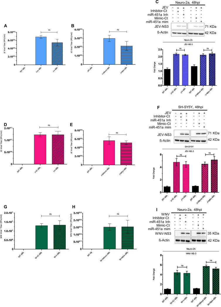
FIG 3.
miR-451a upregulation modulates neuronal apoptosis without affecting JEV/WNV propagation. Supernatant obtained from Neuro-2A transfected with miR-451a inhibitor/inhibitor-Ct (A) and miR-451a mimic/Mimic-Ct (B), and SH-SY5Y transfected with miR-451a-inhibitor/inhibitor-Ct (D) and miR-451a mimic/Mimic-Ct (E) for 24 h prior to infection by JEV (MOI 3) for another 48 h was subjected to plaque assay for determination of viral titer. Similarly, cell-culture media collected from Neuro-2A transfected with miR-451a inhibitor/inhibitor-Ct (G) and miR-451a mimic/Mimic-Ct (H) for 24 h followed by 48 h of WNV (MOI 3) infection were investigated for viral titer estimation utilizing plaque assay. Protein isolated from JEV- (C) and WNV-infected (I) Neuro-2A-cells and JEV-infected SH-SY5Y-cells (F) transfected with miR-451a inhibitor/Inhibitor-Ct and miR-451a mimic/Mimic-Ct, as indicated in the figure, was subjected to immunoblot analysis to determine abundance of JEV- and WNV-NS3 protein expression. The blots shown in the figure represent data from 3 independent experiments with similar results. ß-actin served as a loading-control. Stated beside each immunoblot is the molecular weight in kDa of the respective probed molecule. Quantitative analysis of immunoblots demonstrate the effect of miR-451-a inhibitor/inhibitor-control/miR-451a mimic/mimic-control transfection in JEV-infected Neuro-2A cells (C, bottom: ratio of JEV-NS3 to ß-actin), JEV-infected SH-SY5Y cells (F, bottom: ratio of JEV-NS3 to ß-actin), and WNV-infected Neuro-2A cells (I, bottom: ratio of WNV-NS3 to ß-actin) upon viral NS-3 protein abundance (normalized to ß-actin). Data in bar graphs are represented as mean ± SD from three experimental replicates with similar outcomes. Statistical significance of differences between data belonging to 2 different experimental conditions was determined using Student's t test. ns= non-significant, using two-tailed Student's t test.

