ABSTRACT
The Omicron variant of severe acute respiratory syndrome coronavirus 2 (SARS-CoV-2) has spread worldwide. As of March 2022, Omicron variant BA.2 is rapidly replacing variant BA.1. As variant BA.2 may cause more severe disease than variant BA.1, variant BA.2 requires continuous monitoring. The current study aimed to develop a novel high-resolution melting (HRM) assay for variants BA.1 and BA.2 and to determine the sensitivity and specificity of our method using clinical samples. Here, we focused on the mutational spectra at three regions in the spike receptor-binding domain (RBD; R408, G446/L452, and S477/T478) for the variant-selective HRM analysis. Each variant was identified based on the mutational spectra as follows: no mutations (Alpha variant); L452R and T478K (Delta variant); G446S and S477N/T478K (Omicron variant BA.1); and R408S and S477N/T478K (Omicron variant BA.2). Upon analysis of mutation-coding RNA fragments, the melting curves of the wild-type fragments were distinct from those of the mutant fragments. The sensitivity and specificity of this method were determined as 100% and more than 97.5%, respectively, based on 128 clinical samples (40 Alpha, 40 Delta, 40 Omicron variant BA.1/BA.1.1, and 8 Omicron variant BA.2). These results suggest that this HRM-based assay is a promising screening method for monitoring the transmission of Omicron variants BA.1 and BA.2.
IMPORTANCE This study seeks to apply a novel high-resolution melting (HRM) assay to identify and discriminate BA.1 and BA.2 sublineages of the SARS-CoV-2 Omicron variant. Variant BA.2 may cause more severe disease than variant BA.1, meaning that identifying this variant is an important step toward improving the care of patients suffering from COVID-19. However, screening for these variants remains difficult, as current methods mostly rely on next-generation sequencing, which is significantly costlier and more time-consuming than other methods. We believe that our study makes a significant contribution to the literature because we show that this method was 100% sensitive and over 97.5% specific in our confirmation of 128 clinical samples.
KEYWORDS: SARS-CoV-2, receptor-binding domain, Omicron variant, BA.1, BA.2, high-resolution melting
INTRODUCTION
The severe acute respiratory syndrome coronavirus 2 (SARS-CoV-2) has spread worldwide, and novel coronavirus disease 2019 (COVID-19) cases continue to rise rapidly. Multiple SARS-CoV-2 variants have been detected, and several new variants continue to emerge from various regions (1, 2). Some variants possess amino acid substitutions in the spike protein, which plays a key role in the SARS-CoV-2 infection in humans (3). Since the spike protein binds to human receptor angiotensin-converting enzyme 2 through its receptor-binding domain (RBD), the RBD mutations, such as N501Y and L452R, can increase viral infectivity and cause immune escape. The Alpha (B.1.1.7) and Delta (B.1.617.2) variants, harboring N501Y and L452R, respectively, greatly contributed to the COVID-19 pandemic. In late September 2021, the novel Omicron variant (B.1.1.529) emerged from South Africa and spread rapidly worldwide (4, 5). This variant has many spike mutations (6), and the vaccine breakthrough infection rate of the Omicron variant is reportedly higher than that of other variants (7, 8). Thus, we should strive to prevent the spread of Omicron infection, even though the vaccination program is proceeding.
Recent studies have reported that Omicron sublineages, such as BA.1, BA.2, and BA.3, have emerged in several countries (9, 10). Since late 2021, the BA.1 variant (including BA.1.1, which harbors R346K) has become the main Omicron sublineage worldwide. In early 2022, the BA.2 variant showed an increasing prevalence in some countries, such as Denmark and the United Kingdom (11, 12), while the BA.3 variant is unlikely to spread. Because the 6-nucleotide deletion at spike amino acids 69 to 70 (13) was recognized as a signature mutation of the Omicron variant, it was used to detect the Omicron variant. However, the BA.2 variant lacks this deletion, meaning that it goes undetected (14). The BA.2 variant was then called “Stealth Omicron.” A previous report demonstrated that the reproduction number of BA.2 was 1.4-fold higher than that of BA.1, and BA.2 was more pathogenic in hamsters than BA.1 (15). Therefore, the BA.2 variant may outcompete BA.1 in the near future, and BA.2 should be monitored with caution.
Next-generation sequencing (NGS) has been used to diagnose SARS-CoV-2 variants (16); however, this process requires several days to determine the genome sequence. Rapid screening tests can help diagnose any SARS-CoV-2 variant within a day. High-resolution melting (HRM) analysis is one of the PCR-based screenings for detecting single-nucleotide polymorphisms in DNA sequences (17, 18). Since the melting temperature of double-stranded DNA varies by its nucleotide sequences associated with its GC content and base distribution, HRM can discriminate among different PCR amplicons in a single-nucleotide resolution. Previously, we have reported that HRM assay could detect some SARS-CoV-2 mutations (19–21) First, we established an assay platform to detect the SARS-CoV-2 mutations using HRM analysis (19). Second, we successfully developed an HRM-based assay to identify Delta mutants (L452R and T478K) using nested PCR with an outer primer set prior to HRM analysis (first PCR), improving the detection limit (20). Third, we succeeded in discriminating the Omicron B.1.1.529 and Delta variants using two mutational regions (G446S/L452 and S477N/T478K for Omicron variants, G446/L452R and S477/T478K for Delta variants) (Table 1) (21). Although the S477N/T478K RBD substitutions are common mutations among Omicron sublineages, BA.2 possesses some unique RBD mutations, including R408S without the G446S substitution.
TABLE 1.
Receptor-binding domain (RBD) amino acid substitutions in Alpha, Delta, and Omicron variants
| WHO label | Pangolin | RBD amino acid substitutionsa |
|---|---|---|
| Alpha | B.1.1.7 | N501Y |
| Delta | B.1.617.2 | L452R, T478K |
| Omicron | B.1.1.529/BA.1 | G339D, S371L, S373P, S375F, K417N, N440K, G446S, S477N, T478K, E484A, Q493R, G496S, Q498R, N501Y, Y505H |
| Omicron | BA.2 | G339D, S371F, S373P, S375F, T376A, D405N, R408S, K417N, N440K, S477N, T478K, E484A, Q493R, Q498R, N501Y, Y505H |
| Omicron | BA.3 | G339D, S371F, S373P, S375F, D405N, K417N, N440K, G446S, S477N, T478K, E484A, Q493R, Q498R, N501Y, Y505H |
aSubstitutions detected by this high-resolution melting assay are marked as bold.
In this study, we developed an HRM-based assay to distinguish BA.1 (including BA.1.1) and BA.2 at another RBD site, R408. Moreover, we validated this HRM-based assay at three RBD regions, R408, G446/L452, and S477/T478, using 128 clinical samples (n = 40, Alpha variant; n = 40, Delta variant; n = 40, Omicron variant BA.1/BA.1.1; n = 8, Omicron variant BA.2).
RESULTS
HRM analysis of standard RNA fragments.
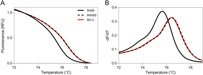
Although the Omicron sublineages BA.1 and BA.2 were found to be unique RBD substitutions, there was no BA.3-specific substitution in the RBD (Table 1). At present, there are far fewer new cases of the BA.3 variant than of either BA.1 or BA.2. Hence, the main aim of this assay is to discriminate between BA.1 and BA.2. In this study, we developed an HRM-based assay for the R408 site, where BA.2 has an R408S substitution but not BA.1. The normalized melting curves and melting peaks for R408, R408S, and BA.2 RBD are shown in Fig. 1. The R408S RBD plot was different from the R408 RBD plot. In addition, the BA.2 RBD plot was in good agreement with the R408S plot. Then, BA.2 RBD was analyzed with the HRM assay at G446/L452R and S477/T478, which we developed previously (21). The HRM assay correctly classified BA.2 as a G446/L452 and S477N/T478K variant (Fig. S1 and S2). These results suggest that our HRM assay can identify BA.2 and discriminate between BA.1 and BA.2 in the three RBD regions.
FIG 1.
Normalized melting curves and melting peaks of positive control RNAs for the R408 site. Normalized melting curve plots (A) and melting peak plots (B) for the R408 site were acquired using standard fragments of the R408 receptor-binding domain (RBD; solid black line), R408S RBD (dashed black line), and BA.2 RBD (solid red line).
HRM analysis of clinical samples.
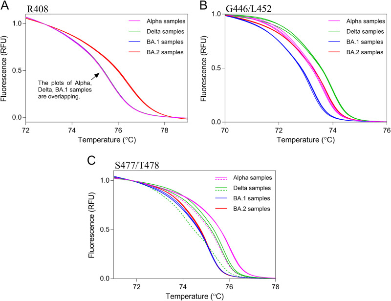
Finally, we confirmed the applicability of this HRM analysis to clinical samples. The Alpha variant was found to have no RBD substitutions at the R408/T478 sites (Table 1). Thus, this HRM-based assay identified a clinical sample containing the Alpha variant as the wild type. After whole-genome sequencing by NGS, 128 clinical samples (n = 40, Alpha; n = 40, Delta; n = 40, BA.1/BA.1.1; n = 8, BA.2 variants) were randomly selected (Table 2). Clinical samples were analyzed using HRM analysis together with positive control RNAs. After HRM analysis, Gene Scanning Software automatically classified samples into wild-type or mutant strains based on HRM curves. The results of the HRM analysis of three regions, R408, G446/L452, and S477/T478, are shown in Table 3, and the melting curve plots of representative samples are shown in Fig. 2. At the R408 site, all eight BA.2 samples were correctly classified as the R408S mutant, and all others were classified as wild type. At the G446/L452 site, 40 BA.1 variant samples were classified as the G446S mutant, and 40 Alpha samples, 40 Delta samples, and 8 BA.2 samples were correctly classified as the wild type. At the S477/T478 site, all BA.1 and BA.2 samples were correctly classified as S477N/T478K mutants. One Alpha sample and one Delta sample plot disagreed with each positive control plot (Fig. 2 and Fig. S3). Therefore, we calculated the sensitivity and specificity of the current HRM-based assay based on these results. The sensitivity and specificity of the assay for the three regions were 100% and over 97.5%, respectively (Table 4).
TABLE 2.
Pangolin of 128 clinical samples by whole-genome sequencing
| WHO label | Pangolin | No. of samples | |
|---|---|---|---|
| Alpha | B.1.1.7 | 40 | |
| Delta | AY.29 | 37 | |
| AY.29.1 | 1 | ||
| AY.46 | 1 | ||
| AY.92 | 1 | ||
| Omicron | BA.1 | 6 | |
| BA.1.1 | 34 | ||
| BA.2 | 8 |
TABLE 3.
Results of high-resolution melting analysis at R408, G446/L452, and S477/T478 sites
| HRM results | No. of Alpha samples | No. of Delta samples | No. of BA.1 samples | No. of BA.2 samples | |||
|---|---|---|---|---|---|---|---|
| R408 site | |||||||
| R408 | 40 | 40 | 40 | 0 | |||
| R408S | 0 | 0 | 0 | 8 | |||
| Unknown | 0 | 0 | 0 | 0 | |||
| G446/L452 site | |||||||
| G446/L452 | 40 | 0 | 0 | 8 | |||
| G446S/L452 | 0 | 0 | 40 | 0 | |||
| G446/L452R | 0 | 40 | 0 | 0 | |||
| Unknown | 0 | 0 | 0 | 0 | |||
| S477/T478 site | |||||||
| S477/T478 | 39 | 0 | 0 | 0 | |||
| S477/T478K | 1 | 39 | 0 | 0 | |||
| S477N/T478K | 0 | 0 | 40 | 8 | |||
| Unknown | 0 | 1 | 0 | 0 |
FIG 2.
Melting curves of clinical samples for three receptor-binding domain regions. Melting curve plots for the R408 (A), G446/L452 (B), and S477/T478 (C) sites were acquired using clinical samples with three Alpha variants (pink line), three Delta variants (green line), three BA.1/BA.1.1 variants (blue line), and three BA.2 variants (red line). Solid lines indicate true positive and true negative samples. Dashed lines indicate false negative samples.
TABLE 4.
Sensitivity and specificity of the high-resolution melting assay in comparison with next-generation sequencing
| Metrics | R408S detection | G446S detection | S477N/T478K detection |
|---|---|---|---|
| Sensitivitya | 100% (8/8) | 100% (40/40) | 100% (48/48) |
| Specificityb | 100% (120/120) | 100% (88/88) | 97.5% (78/80) |
Sensitivity = (true positives/true positives + false positives) × 100.
Specificity = (false negatives/false negatives + true negatives) × 100.
DISCUSSION
This study is the first report developing the screening test for discriminating Omicron variants BA.1 and BA.2 using HRM assay. This HRM-based assay had high sensitivity and specificity for BA.2 diagnosis. A previous study showed that the effective reproduction number of BA.2 is 1.4-fold higher than that of BA.1 (15). Additionally, the Delta variant was identifiable using this assay. New cases of infection with the Delta variant, such as the AY.29 sublineage (24), remain detectable in some countries, including Japan. The Delta variant increases the risk of hospitalization more than the Omicron variant (25). Therefore, rigorous global monitoring of both Omicron and Delta variants is warranted. Although the medical treatments are identical in people infected with these variants, variant tracking is essential to prevent their spread and reduce the occurrence of new variants. Additionally, this HRM-based assay is a valuable tool for rapid identification of these variants in a large number of samples since low-cost and high-throughput screening tests can be built using this assay.
All SARS-CoV-2 variants of concern have characteristic RBD substitutions in the spike protein, affecting the viral infectivity and immune evasion ability. In contrast, many mutations, excluding the RBD region, are not common among similar lineages, as the genomic sequence of SARS-CoV-2 continues to mutate over time. Direct detection of characteristic RBD mutations contributes to avoiding misdiagnosis of each variant. Therefore, we have developed HRM assays to detect SARS-CoV-2 RBD mutations (20, 21). Moreover, HRM analysis can detect unknown mutations unlike TaqMan probe assays. If a post-Omicron variant emerges, we can quickly develop an HRM-based assay to detect the RBD mutation in the new variant.
The TaqMan probe assay is typically used for single-nucleotide polymorphism detection. Multiple TaqMan probe kits for the detection of SARS-CoV-2 mutations are commercially available worldwide. As the Omicron RBD is highly mutated, the reactivity of the available specific probes may be affected, which is likely to increase the risk of false negatives. This HRM-based assay without a specific probe can be constructed more easily than a TaqMan probe assay. Thus, HRM analysis is useful in identifying highly mutated variants. However, the HRM assay may not be able to identify samples that contain low virus copies. Our previous study showed that when used with samples containing >107 copies/mL, the HRM assay could detect SARS-CoV-2 mutations (19). In this study, the two-step nested PCR improved the detection limit of the HRM assay, and low-copy virus samples could be identified, such as those having Ct values of 35. Moradzad et al. (26) reported that a one-step HRM assay can detect SARS-CoV-2 mutations with relatively high sensitivity and specificity, similar to the TaqMan probe assay. However, they used high-copy number viral RNAs (10 ng RNA in a tube) for HRM analysis. Additional work is required to improve the detection limit of our HRM-based assay using a one-step single-tube PCR assay.
This study needs to be interpreted in the context of its limitations. First, this study was performed on clinical samples from a limited area of Japan. Further studies are needed to confirm the utility of this HRM-based assay using a larger size of samples from various regions since the rates of false positive and false negative COVID-19 tests in clinical practice are essential information. Second, the low-copy virus samples with a Ct value less than 35 may more frequently result in a false positive or false negative. The limit of detection should be determined using low-copy virus samples. Third, our assay cannot discriminate between Omicron variants BA.1 and BA.3 because these sublineages possess the same mutational spectra at R408, G446S/L452, and S477N/T478K (Table 1). Even if the variant BA.3 replaces BA.1 and BA.2, we can identify BA.3 by determining the D405N mutation in combination with others.
In conclusion, we developed a novel assay to identify the main Omicron sublineages, BA.1 and BA.2, using HRM analysis. As this HRM-based genotyping assay does not require sequence-specific probes, unlike the TaqMan probe assay, it is easy to perform and is cost-effective. Our results suggest that the current HRM-based assay is a powerful high-throughput tool for determining the SARS-CoV-2 Omicron sublineages. Since this assay was verified using limited clinical samples, further studies using diverse samples are needed to validate this HRM-based assay among the various institutions.
MATERIALS AND METHODS
Ethics statement.
This project was approved by the Research Ethics Committee of Meijo University (Approval number: 2020-17-2) and Aichi Prefectural Institute of Public Health (Approval number: 20E-4) and was carried out according to the Infectious Diseases Control Law of Japan.
Preparation of standard RNA fragments: in vitro T7 transcription.
The SARS-CoV-2 sequence was obtained from NCBI (GenBank ID: MN908947), the GISAID database (www.gisaid.org), and the Pango nomenclature system (https://cov-lineages.org/lineages.html). Five RBD DNA fragments (wild type, R408S mutant, BA.1 variant mutant, BA.2 variant mutant, and Delta variant mutant; 600–1000 bp in length) with a 5′ T7 upstream promoter sequence were obtained from Eurofins Genomics K.K. (Tokyo, Japan). In vitro T7 transcription was performed as described previously (20). The synthesized single-stranded RNA fragments were used as reverse transcriptase (RT)-PCR amplification templates.
RT-PCR amplification: first PCR.
RT-PCR was performed in a single closed tube using a one-step RT-PCR kit (One Step PrimeScript III RT-qPCR Mix, with UNG; TaKaRa Bio Inc., Kusatsu, Japan) in accordance with the manufacturer’s instructions. The primer pairs used were as follows: R408 outer forward 5′-TGCTTGGAACAGGAAGAGAA-3′ and R408 outer reverse 5′-AACGCAGCCTGTAAAATCATC-3′; G466-T478 outer forward 5′-TTACAGGCTGCGTTATAG-3′ and G466-T478 outer reverse 5′-ACAAACAGTTGCTGGTGCAT-3′. Each DNA fragment was observed as a single, correctly sized band as follows: R408, 247 bp; G466-T478, 290 bp. RT-PCR amplification was performed as previously described (20). After amplification, the reaction mixture was diluted 10,000-fold with water and used as a template for the second PCR and HRM analyses.
HRM analysis: second PCR.
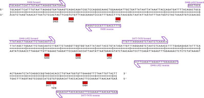
HRM was performed using an HRM reagent (MeltDoctor HRM Master Mix; Thermo Fisher Scientific, Waltham, MA, USA) according to the manufacturer’s instructions. The second primer pair was as follows: R408 forward 5′-TGCAGATTCATTTGTAATTAGAGGTGATGAAG-3′ and R408 reverse 5′-CTTTCCAGTTTGCCCTGGAG-3′; G446-L452 forward 5′-GGCTGCGTTATAGCTTGGAATTCTAACAATCTT-3′ and G446-L452 reverse 5′-TCAAAAGGTTTGAGATTAGACTTCC-3′; S477-T478 forward 5′- TTGTTTAGGAAGTCTAATCTCAAACC-3′ and S477-T478 reverse 5′-AAGTAACAATTAAAACCTTCAACACCATTACAAGG-3′. Each DNA fragment was observed as a single, correctly sized band as follows: R408, 64 bp; G466-L452, 104 bp; S477-T478, 107 bp. As shown in Fig. 3, the R408 forward primer design was based on the D405 coding sequence, the R408 reverse primer design was based on the K417 coding sequence, the G446-L452 forward primer design was based on the N440 coding sequence, and the S477–T478 reverse primer design was based on the E484 coding sequence to avoid the potential influences of the D405, K417, N440, and E484 mutations, respectively. Briefly, each reaction mixture (20 μL) contained 2 μL of the diluted RT-PCR mixture, 400 nmol/L of each primer, and 1× master mix. All reactions were performed in duplicate using a real-time PCR system (LightCycler 96 System; F. Hoff Mann-La Roche Ltd., Basel, Switzerland). PCR amplification was performed as previously described (20). HRM curves were analyzed using Gene Scanning Software, version 1.1.0.1320 (F. Hoffmann-La Roche Ltd.) under default settings. The normalized melting curve and melting peaks (−dF/dT) were acquired by setting the pre-melt and post-melt fluorescence at 100% and 0%, respectively.
FIG 3.
Schematic map of primer annealing sites for high-resolution melting analyses.
Clinical samples.
From July 2021 to January 2022, 128 nasopharyngeal swabs or saliva samples were collected from patients suspected of having COVID-19 and those detected with COVID-19 by Aichi Prefectural Institute of Public Health. The Ct values in the clinical quantitative PCR test for these samples ranged from 15 to 35. Viral RNA was extracted from clinical samples using spin columns (QIAamp Viral RNA Mini QIAcube Kit; Qiagen GmbH, Hilden, Germany) and an automated nucleic acid purification system (QIAcube Connect; Qiagen). Each eluent was reverse-transcribed, PCR-amplified, and subjected to library preparation using the QIAseq FX library kit (Qiagen), according to the protocol established by the National Institute of Infectious Diseases, Tokyo, Japan (22, 23). Whole-genome sequencing was performed using a next-generation sequencer (MiSeq system; Illumina Inc., San Diego, CA, USA). The obtained NGS reads were mapped to the SARS-CoV-2 Wuhan-Hu-1 reference genome sequence (29.9 kb single-strand RNA; GenBank ID: MN908947). For lineage assignment, Pangolin version 3.1.20, with pangoLEARN version 2022-02-28 was used.
ACKNOWLEDGMENTS
We thank the staff of the Laboratory of Virology, Department of Microbiology and Medical Zoology, Aichi Prefectural Institute of Public Health, who performed COVID-19 PCR testing, RNA purification from clinical samples, and NGS analysis.
All authors participated in the interpretation of the results. A.A. designed and conducted HRM analysis and wrote the original draft of the manuscript. H.A. and M.I. conducted NGS analysis and edited the manuscript. Y.M. performed sequence analysis. Y.O. planned the study and edited the manuscript. K.S., K.O., T.S., and H.J. planned the study and supervised the project. All the authors critically reviewed and approved the final version of the manuscript.
This work was supported in part by the Daiko Foundation and Meijo University Research Project for Countermeasures Against COVID-19.
We declare no conflict of interest.
Footnotes
Supplemental material is available online only.
Contributor Information
Hideto Jinno, Email: jinno@meijo-u.ac.jp.
Jian Li, Hubei University of Medicine.
REFERENCES
- 1.Rahimi A, Mirzazadeh A, Tavakolpour S. 2021. Genetics and genomics of SARS-CoV-2: A review of the literature with the special focus on genetic diversity and SARS-CoV-2 genome detection. Genomics 113:1221–1232. doi: 10.1016/j.ygeno.2020.09.059. [DOI] [PMC free article] [PubMed] [Google Scholar]
- 2.Tian D, Sun Y, Zhou J, Ye Q. 2022. The global epidemic of SARS-CoV-2 variants and their mutational immune escape. J Med Virol 94:847–857. doi: 10.1002/jmv.27376. [DOI] [PMC free article] [PubMed] [Google Scholar]
- 3.Ni W, Yang X, Yang D, Bao J, Li R, Xiao Y, Hou C, Wang H, Liu J, Yang D, Xu Y, Cao Z, Gao Z. 2020. Role of angiotensin-converting enzyme 2 (ACE2) in COVID-19. Crit Care 24:422. doi: 10.1186/s13054-020-03120-0. [DOI] [PMC free article] [PubMed] [Google Scholar]
- 4.Petersen E, Ntoumi F, Hui DS, Abubakar A, Kramer LD, Obiero C, Tambyah PA, Blumberg L, Yapi R, Al-Abri S, Pinto TCA, Yeboah-Manu D, Haider N, Asogun D, Velavan TP, Kapata N, Bates M, Ansumana R, Montaldo C, Mucheleng'anga L, Tembo J, Mwaba P, Himwaze CM, Hamid MMA, Mfinanga S, Mboera L, Raj T, Aklillu E, Veas F, Edwards S, Kaleebu P, McHugh TD, Chakaya J, Nyirenda T, Bockarie M, Nyasulu PS, Wejse C, Muyembe-Tamfum J-J, Azhar EI, Maeurer M, Nachega JB, Kock R, Ippolito G, Zumla A. 2022. Emergence of new SARS-CoV-2 variant of concern Omicron (B.1.1.529) - highlights Africa's research capabilities, but exposes major knowledge gaps, inequities of vaccine distribution, inadequacies in global COVID-19 response and control efforts. Int J Infect Dis 114:268–272. doi: 10.1016/j.ijid.2021.11.040. [DOI] [PMC free article] [PubMed] [Google Scholar]
- 5.Karim SSA, Karim QA. 2021. Omicron SARS-CoV-2 variant: a new chapter in the COVID-19 pandemic. Lancet 398:2126–2128. doi: 10.1016/S0140-6736(21)02758-6. [DOI] [PMC free article] [PubMed] [Google Scholar]
- 6.Hong Q, Han W, Li J, Xu S, Wang Y, Xu C, Li Z, Wang Y, Zhang C, Huang Z, Cong Y. 2022. Molecular basis of receptor binding and antibody neutralization of Omicron. Nature 604:546–552. doi: 10.1038/s41586-022-04581-9. [DOI] [PubMed] [Google Scholar]
- 7.Ren SY, Wang WB, Gao RD, Zhou AM. 2022. Omicron variant (B.1.1.529) of SARS-CoV-2: mutation, infectivity, transmission, and vaccine resistance. World J Clin Cases 10:1–11. doi: 10.12998/wjcc.v10.i1.1. [DOI] [PMC free article] [PubMed] [Google Scholar]
- 8.Christensen PA, Olsen RJ, Long SW, Snehal R, Davis JJ, Saavedra MO, Reppond K, Shyer MN, Cambric J, Gadd R, Thakur RM, Batajoo A, Mangham R, Pena S, Trinh T, Kinskey JC, Williams G, Olson R, Gollihar J, Musser JM. 2022. Signals of significantly increased vaccine breakthrough, decreased hospitalization rates, and less severe disease in patients with coronavirus disease 2019 caused by the Omicron variant of severe acute respiratory syndrome coronavirus 2 in Houston, Texas. Am J Pathol 192:642–652. doi: 10.1016/j.ajpath.2022.01.007. [DOI] [PMC free article] [PubMed] [Google Scholar]
- 9.Kumar S, Karuppanan K, Subramaniam G. 2022. Omicron (BA.1) and sub-variants (BA.1, BA.2 and BA.3) of SARS-CoV-2 spike infectivity and pathogenicity: a comparative sequence and structural-based computational assessment. bioRxiv. doi: 10.1101/2022.02.11.480029. [DOI] [PMC free article] [PubMed]
- 10.Desingu PA, Nagarajan K, Dhama K. 2022. Emergence of Omicron third lineage BA.3 and its importance. J Med Virol 94:1808–1810. doi: 10.1002/jmv.27601. [DOI] [PMC free article] [PubMed] [Google Scholar]
- 11.Dhawan M, Choudhary OP. 2022. Emergence of Omicron sub-variant BA.2: Is it a matter of concern amid the COVID-19 pandemic? Int J Surg 99:106581. doi: 10.1016/j.ijsu.2022.106581. [DOI] [PMC free article] [PubMed] [Google Scholar]
- 12.Desingu PA, Nagarajan K. 2022. Omicron BA.2 lineage spreads in clusters and is concentrated in Denmark. J Med Virol 94:2360–2364. doi: 10.1002/jmv.27659. [DOI] [PMC free article] [PubMed] [Google Scholar]
- 13.Vogels CBF, Breban MI, Ott IM, Alpert T, Petrone ME, Watkins AE, Kalinich CC, Earnest R, Rothman JE, Goes de Jesus J, Morales Claro I, Magalhaes Ferreira G, Crispim MAE, Singh L, Tegally H, Anyaneji UJ, Network for Genomic Surveillance in South A, Hodcroft EB, Mason CE, Khullar G, Metti J, Dudley JT, MacKay MJ, Nash M, Wang J, Liu C, Hui P, Murphy S, Neal C, Laszlo E, Landry ML, Muyombwe A, Downing R, Razeq J, de Oliveira T, Faria NR, Sabino EC, Neher RA, Fauver JR, Grubaugh ND, Brazil UKCGN . 2021. Multiplex qPCR discriminates variants of concern to enhance global surveillance of SARS-CoV-2. PLoS Biol 19:e3001236. doi: 10.1371/journal.pbio.3001236. [DOI] [PMC free article] [PubMed] [Google Scholar]
- 14.Rahimi F, Talebi Bezmin Abadi A. 2022. The Omicron subvariant BA.2: Birth of a new challenge during the COVID-19 pandemic. Int J Surg 99:106261. doi: 10.1016/j.ijsu.2022.106261. [DOI] [PMC free article] [PubMed] [Google Scholar]
- 15.Yamasoba D, Kimura I, Nasser H, Morioka Y, Nao N, Ito J, Uriu K, Tsuda M, Zahradnik J, Shirakawa K, Suzuki R, Kishimoto M, Kosugi Y, Kobiyama K, Hara T, Toyoda M, Tanaka YL, Butlertanaka EP, Shimizu R, Ito H, Wang L, Oda Y, Orba Y, Sasaki M, Nagata K, Yoshimatsu K, Asakura H, Nagashima M, Sadamasu K, Yoshimura K, Kuramochi J, Seki M, Fujiki R, Kaneda A, Shimada T, Nakada T-a, Sakao S, Suzuki T, Ueno T, Takaori-Kondo A, Ishii KJ, Schreiber G, Sawa H, Saito A, Irie T, Tanaka S, Matsuno K, Fukuhara T, Ikeda T, Sato K. 2022. Virological characteristics of the SARS-CoV-2 Omicron BA.2 spike. Cell 185:2103–2115.e19. doi: 10.1016/j.cell.2022.04.035. [DOI] [PMC free article] [PubMed] [Google Scholar]
- 16.Wang J, Hawken SE, Jones CD, Hagan RS, Bushman F, Everett J, Miller MB, Rodino KG. 2022. Collaboration between clinical and academic laboratories for sequencing SARS-CoV-2 genomes. J Clin Microbiol 60:e0128821. doi: 10.1128/JCM.01288-21. [DOI] [PMC free article] [PubMed] [Google Scholar]
- 17.Vossen RH, Aten E, Roos A, den Dunnen JT. 2009. High-resolution melting analysis (HRMA): More than just sequence variant screening. Hum Mutat 30:860–866. doi: 10.1002/humu.21019. [DOI] [PubMed] [Google Scholar]
- 18.Zhou L, Palais RA, Paxton CN, Geiersbach KB, Wittwer CT. 2015. Copy number assessment by competitive PCR with limiting deoxynucleotide triphosphates and high-resolution melting. Clin Chem 61:724–733. doi: 10.1373/clinchem.2014.236208. [DOI] [PubMed] [Google Scholar]
- 19.Aoki A, Mori Y, Okamoto Y, Jinno H. 2021. Development of a genotyping platform for SARS-CoV-2 variants using high-resolution melting analysis. J Infect Chemother 27:1336–1341. doi: 10.1016/j.jiac.2021.06.007. [DOI] [PMC free article] [PubMed] [Google Scholar]
- 20.Aoki A, Adachi H, Mori Y, Ito M, Sato K, Okuda K, Sakakibara T, Okamoto Y, Jinno H. 2021. A rapid screening assay for L452R and T478K spike mutations in SARS-CoV-2 Delta variant using high-resolution melting analysis. J Toxicol Sci 46:471–476. doi: 10.2131/jts.46.471. [DOI] [PubMed] [Google Scholar]
- 21.Aoki A, Mori Y, Okamoto Y, Jinno H. 2022. Simultaneous screening of SARS-CoV-2 Omicron and Delta variants using high-resolution melting analysis. Biol Pharm Bull 45:394–396. doi: 10.1248/bpb.b21-01081. [DOI] [PubMed] [Google Scholar]
- 22.Itokawa K, Sekizuka T, Hashino M, Tanaka R, Kuroda M. 2020. A proposal of alternative primers for the ARTIC network’s multiplex PCR to improve coverage of SARS-CoV-2 genome sequencing. bioRxiv. doi: 10.1101/2020.03.10.985150. [DOI]
- 23.Itokawa K, Sekizuka T, Hashino M, Tanaka R, Eto S, Someno R, Kuroda M. 2021. nCoV-2019 sequencing protocol for illumina V.5 protocols.io. https://www.protocols.io/view/ncov-2019-sequencing-protocol-for-illumina-eq2ly398mgx9/v5. [Google Scholar]
- 24.Shimura T, Kosaki K. 2022. Global spread of a Japan-originated Delta lineage of SARS-CoV-2 after the Tokyo Olympics is most likely unrelated to the Olympics. J Travel Med 29:taac017. doi: 10.1093/jtm/taac017. [DOI] [PMC free article] [PubMed] [Google Scholar]
- 25.Veneti L, Boas H, Brathen Kristoffersen A, Stalcrantz J, Bragstad K, Hungnes O, Storm ML, Aasand N, Ro G, Starrfelt J, Seppala E, Kvale R, Vold L, Nygard K, Buanes EA, Whittaker R. 2022. Reduced risk of hospitalisation among reported COVID-19 cases infected with the SARS-CoV-2 Omicron BA.1 variant compared with the Delta variant, Norway, December 2021 to January 2022. Euro Surveill 27:2200077. [DOI] [PMC free article] [PubMed] [Google Scholar]
- 26.Moradzad M, Soltani H, Salehi H, Rahmani K, Khateri D, Rahimi MZ, Az D, Fakhari S. 2022. A screening strategy for identifying the dominant variant of SARS-COV-2 in the fifth peak of Kurdistan- Iran population using HRM and probe-based RT-PCR assay. J Virol Methods 304:114514. doi: 10.1016/j.jviromet.2022.114514. [DOI] [PMC free article] [PubMed] [Google Scholar]
Associated Data
This section collects any data citations, data availability statements, or supplementary materials included in this article.
Supplementary Materials
Supplemental material. Download spectrum.01367-22-s0001.pdf, PDF file, 1.0 MB (981.5KB, pdf)


