Abstract
Basaloid squamous cell carcinoma of the lung is now considered a subtype of squamous cell carcinoma as per the 2015 WHO classification and remains a relatively unknown type of lung cancer due to its rarity. Here we report two cases of basaloid squamous cell carcinoma of the lung and their CT findings to clarify some of the radiologic features of this type of cancer. Two patients aged 85 and 68 years with lung basaloid squamous cell carcinoma visited our institution and underwent surgical resection. On CT, the lesions were 3.1 and 2.8 cm in size, respectively, well-defined, round in shape with lobulated margins and prominent intratumoral necrosis. The latter case was followed after operation for 20 months, and there was no recurrence of the disease on CT. Although very rare, basaloid squamous cell carcinoma should be considered a subtype of lung cancer in tumors sharing these CT findings.
Keywords: Squamous Cell Carcinoma; Lung; Computed Tomography, X-Ray
Abstract
폐의 기저세포양 편평세포암은 2015년 WHO 분류체계에서 편평세포암의 한 아형으로 분류되었고 폐암에서는 드물기에 잘 알려지지 않은 유형이다. 저자들은 두 기저세포양 편평세포암 증례를 보고하고 이들의 전산화단층촬영 영상소견을 명시하고자 한다. 85세 그리고 68세의 기저세포양 편평세포암을 가진 환자가 내원하여 수술적 절제를 시행 받았다. 전산화단층촬영에서 두 병변은 각각 3.1과 2.8 cm 크기로 경계가 좋으며, 둥글면서 소엽상 경계였고 내부는 괴사를 시사하는 저음영이 풍부하였다. 후자는 수술 후 20개월 동안 추적 관찰하였고 전산화단층촬영에서 재발은 없었다. 비록 드문 아형의 폐암이지만 이러한 전산화단층촬영 소견을 공유한다면 기저세포양 편평세포암을 고려해 볼 수 있겠다.
INTRODUCTION
Basaloid squamous cell carcinoma of the lung is now considered a subtype of squamous cell carcinoma as per the 2015 WHO classification of tumors of the lung, pleura, thymus, and heart (1). The cancer is known for its characteristic rapid growth rate and clinical progression as well as poor prognosis compared to non-basaloid squamous cell carcinoma (2,3). Several case studies have reported on the prognosis and clinicopathological findings of basaloid squamous cell carcinoma of the lung (4,5,6). However, because of its rarity, the radiologic appearance of this cancer remains to be investigated.
We hereby present two cases of surgically confirmed basaloid squamous cell carcinoma of the lung and discuss their radiological characteristics to add to the CT imaging database of the cancer. This is to prevent diagnostic delays in the early stages and ensure timely administration of proper treatment.
CASE REPORTS
CASE 1
An 85-year-old man visited our institution due to incidental finding of a solid pulmonary nodule in a chest CT scan taken during a routine health check-up. He was an ex-smoker who quit the habit before 35 years. Laboratory findings including peripheral blood and urine analysis were within normal limits. The results of pulmonary function tests were within normal range.
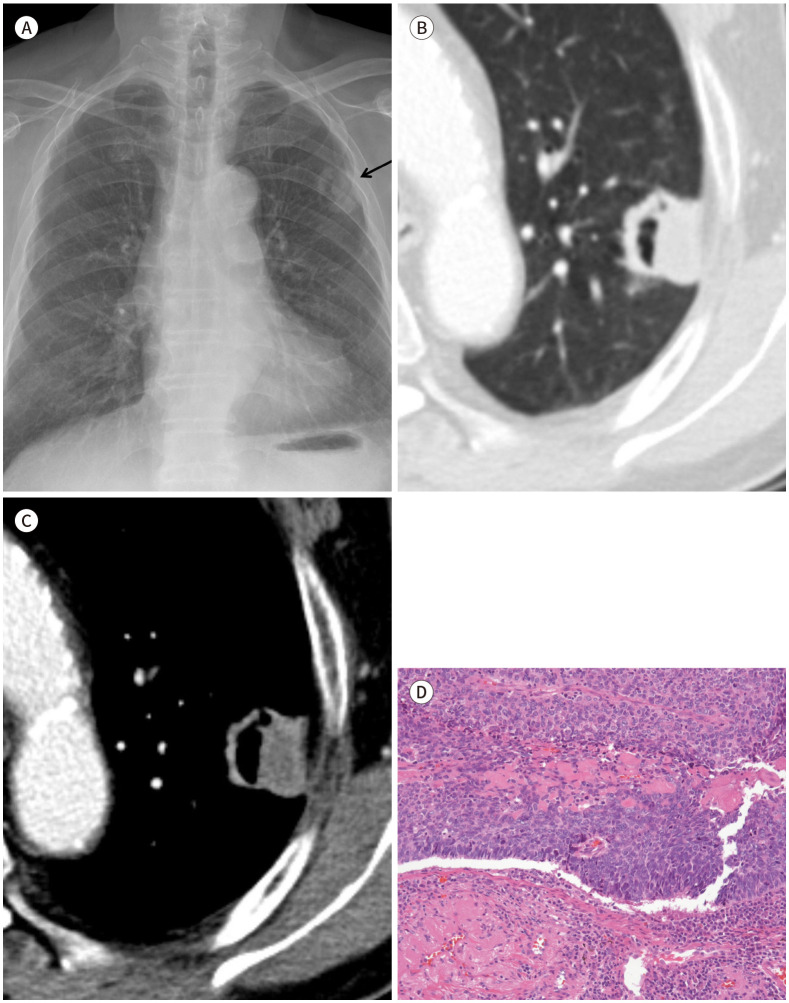
The posteroanterior and lateral radiographs of the chest revealed a well-defined cavitary nodule in the left upper lobe (Fig. 1A). The post-contrast chest CT scan demonstrated a 3.1 cm-sized, well-defined mass with cavitation at the periphery of the upper division of the left upper lobe (Fig. 1B, C). Compared with the pre-contrast CT scan, net Hounsfield units (HUs) for the lesion were 15 to 20 (pre-contrast: 35 to 40 HUs, post-contrast: 50 to 60 HUs). The mass showed lobulated margins and profuse internal necrosis (Fig. 1C). Because the mass was tightly approximated to and retracted the adjacent pleura, pleural invasion was suspected on CT scan. PET-CT showed a marked increase in fluorine-18 fluoro-deoxy-glucose uptake [maximum standardized uptake value (SUVmax) = 16.8] in the cavitary nodule with possible involvement of the adjacent pleura. Percutaneous needle biopsy was conducted twice for pathologic confirmation; however, both examinations showed non-neoplastic lung parenchyma cells.
Fig. 1. An 85-year-old man with basaloid squamous cell carcinoma of the lung.
A–D. The posteroanterior (A) chest radiographs showed a cavitary nodular opacity in the left upper lobe (arrow). Lung (B) and mediastinal window (C) images of chest CT scan demonstrate a 3.1 cm-sized, peripherally located, well-defined nodule with lobulated margin and cavitation in the left upper lobe. Mediastinal window image (C) shows significant intratumoral necrosis and peripheral contrast enhancement. Corresponding microscopic features (haematoxylin and eosin stain, × 200) (D) show tumor cells with squamous differentiation, high mitotic rate, and palisading pattern.
The patient underwent left upper lobectomy with mediastinal lymph node dissection. The resected specimen measured 3.5 × 3.0 × 2.1 cm in size and was pathologically confirmed as basaloid squamous cell carcinoma with poor differentiation. The diagnosis of basaloid carcinoma was based on the criteria defined by Brambilla et al. (7). Microscopic features of the lesion showed tumor cells with squamous differentiation, high mitotic rate, and a palisading pattern. Intratumoral necrosis was found on pathological examination. The lesion did extend to the visceral pleura without lymph node metastasis or lymphovascular and perineural invasion, and was staged as T2aN0M0 (Fig. 1D). Unfortunately, the patient suffered acute lung injury with ventilator-associated pneumonia during post-operative care, and died due to multi-organ failure one month after the operation.
CASE 2
A 68-year-old man visited our institution for a routine health check-up and follow up for chronic obstructive pulmonary disease. Two to three months before his visit, he started experiencing exacerbation in his symptom of cough. He was an active smoker who smoked two packs of cigarettes per day for 50 years.
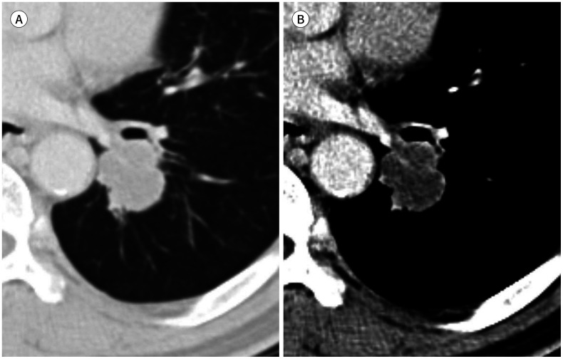
The posteroanterior and lateral radiographs of the chest revealed a nodular opacity at the central portion of the left lower lobe, and chest CT scan showed a well-defined 2.8 cm-sized nodule with lobulated margins and prominent intratumoral necrosis in the central portion of the left lower lobe (Fig. 2A, B). Compared with the pre-contrast CT scan, little enhancement was seen on post-contrast CT (pre- and post-contrast: ~30 HUs). PET-CT demonstrated a hypermetabolic mass in the left lower lobe (SUVmax = 9.5). Bronchoscopic biopsy was conducted on the left lower lobe and revealed moderately differentiated squamous cell carcinoma.
Fig. 2. A 68-year-old man with basaloid squamous cell carcinoma of the lung.
A, B. Lung (A) and mediastinal window (B) images of chest CT scan reveal a well-defined, 2.8 cm-sized nodule with lobulated margin and prominent intratumoral necrosis in the left lower lobe.
Consequently, the patient underwent left lower lobectomy with mediastinal lymph node dissection. The resected specimen measured 3.8 × 3.6 × 3.2 cm in size and was pathologically confirmed as basaloid squamous cell carcinoma showing squamous differentiation, high mitotic rate, and a palisading pattern. Intratumoral necrosis was also found on pathological examination. Visceral pleural, lymphovascular, and perineural invasion were not identified, and all regional lymph nodes examined were negative for cancer cells. Thus, the final TNM staging was pT2aN0M0. After the operation, the patient was discharged and has been receiving adjuvant chemo-radiotherapy ever since, maintaining complete remission status over 20 months.
DISCUSSION
Prior to release of the 2015 WHO classification, basaloid carcinoma was defined as a variant of either squamous-cell or large-cell carcinoma (7). However, after basaloid carcinomas were recognized to express squamous markers, they were excluded from the subtype of large cell carcinoma and were re-categorized as a subtype of squamous cell carcinoma. Therefore, subtypes of squamous cell carcinoma are now thought to consist of keratinizing, non-keratinizing, and basaloid variants (1).
Basaloid squamous cell carcinoma of the lung is a very rare subtype of squamous cell carcinoma. It is commonly found in the proximal bronchi as a single solitary lesion (7); however, occasionally it may be found admixed with other types of non-small cell lung carcinomas (NSCLCs). This cancer is often observed in male patients older than 60 years with a heavy smoking history (7). Histologically, diagnosis of basaloid carcinoma is based on four criteria: 1) Invasive finger-like growth of a solid lobular or anastomotic trabecular pattern from the bronchial and/or glandular duct lining; 2) small cuboidal to fusiform cells with a mean diameter of 12–15 µm, moderately hyperchromatic nuclei, and no prominent nucleoli (there may be scant nucleoli with visible cytoplasm and no nuclear molding); 3) peripheral palisading pattern with radially arranged cells at lobule peripheries; and 4) a high rate of mitosis between 15–44 per 10 high-power fields (7). Basaloid squamous cell carcinoma of the lung is a highly malignant and aggressive tumor with high rates of metastasis and death (2,3). Thus, this subtype of lung squamous cell carcinoma is distinguished from the majority of non-basaloid squamous cell carcinomas based on prognosis and should be diagnosed at an early stage for active and timely therapeutic intervention.
Although radiologic findings of squamous cell carcinomas are well-reported (8), specific details and differences in imaging features of their subtypes remain unclear. Moreover, there are limited data on imaging findings of basaloid squamous cell carcinoma of the lung and only clinical aspects have been reported in previous studies (2,3,9,10). In this study, we observed that basaloid squamous cell carcinoma tumors were well-defined, round in shape, and had lobulated margins on CT scan. On post-contrast CT, the lesions showed prominent intratumoral necrosis despite their small sizes (~3 cm was the largest diameter). Lobulated margins and prominent intratumoral necrosis on CT were common radiologic findings that may reflect tumor subtype or aggressive behavior. However, we cannot robustly conclude this as these findings may be subtype-specific because cavitary or necrotic lung nodules are one of the most typical presentations of squamous lung cancer on CT, irrespective of subtype (8). Nevertheless, we believe that the results of the two cases described herein are meaningful in terms of being the first to present radiologic findings for basaloid squamous cell carcinoma of the lung, which contributes to baseline research data for subsequent relevant studies.
According to previous reports, basaloid carcinoma has poor prognosis compared to other NSCLCs, with a median survival of 20–29 months even in stage I and II resected tumors (2,3,10). In contrast, one of our cases was followed for over 20 months after operation, and there was no recurrence of the disease on CT scan. Although this case needs further follow-up, it has shown favorable prognosis thus far, as in few previous case reports of lung basaloid carcinoma (4,5). At present, therapy for basaloid squamous cell carcinoma in various organs follows guidelines of conventional therapy for squamous cell carcinoma. In addition, there is no consensus on treatment for patients with basaloid squamous cell carcinoma of the lung. Therefore, further large-scale studies should be implemented to observe the clinical course of basaloid carcinoma and evaluate the presence of other beneficial treatment options. In addition, the clinicoradiologic and pathologic features to be used for prediction of basaloid squamous cell carcinoma of the lung should be documented.
Acknowledgments
This study was supported by a Veterans Health Service (VHS) Medical Center Research Grant, Republic of Korea (grant number: VHSMC 18040).
Footnotes
- Conceptualization, Y.H.J.
- Data curation, Y.H.J., K.J.H.
- Formal analysis, K.J.H., L.E., K.E.J.
- Funding acquisition, Y.H.J.
- Investigation, K.J.H., L.E., K.E.J.
- Methodology, K.J.H., Y.H.J.
- Project administration, Y.H.J.
- Resources, Y.H.J., K.E.J.
- Validation, Y.H.J.
- Visualization, K.J.H., K.E.J.
- Writing — original draft, K.J.H.
- Writing — review & editing, Y.H.J., L.E., K.J.H.
Conflicts of Interest: The authors have no potential conflicts of interest to disclose.
References
- 1.Travis WD, Brambilla E, Nicholson AG, Yatabe Y, Austin JHM, Beasley MB, et al. The 2015 World Health Organization classification of lung tumors: impact of genetic, clinical and radiologic advances since the 2004 classification. J Thorac Oncol. 2015;10:1243–1260. doi: 10.1097/JTO.0000000000000630. [DOI] [PubMed] [Google Scholar]
- 2.Wang LC, Wang L, Kwauk S, Woo JA, Wu LQ, Zhu H, et al. Analysis on the clinical features of 22 basaloid squamous cell carcinoma of the lung. J Cardiothorac Surg. 2011;6:10. doi: 10.1186/1749-8090-6-10. [DOI] [PMC free article] [PubMed] [Google Scholar]
- 3.Moro-Sibilot D, Lantuejoul S, Diab S, Moulai N, Aubert A, Timsit JF, et al. Lung carcinomas with a basaloid pattern: a study of 90 cases focusing on their poor prognosis. Eur Respir J. 2008;31:854–859. doi: 10.1183/09031936.00058507. [DOI] [PubMed] [Google Scholar]
- 4.Foroulis CN, Iliadis KH, Mauroudis PM, Kosmidis PA. Basaloid carcinoma, a rare primary lung neoplasm: report of a case and review of the literature. Lung Cancer. 2002;35:335–338. doi: 10.1016/s0169-5002(01)00427-5. [DOI] [PubMed] [Google Scholar]
- 5.Cakir E, Demirag F, Ucoluk GO, Kaya S, Memis L. Basaloid squamous cell carcinoma of the lung: a rare tumour with a rare clinical presentation. Lung Cancer. 2007;57:109–111. doi: 10.1016/j.lungcan.2007.01.021. [DOI] [PubMed] [Google Scholar]
- 6.Nagakawa H, Hiroshima K, Takiguchi Y, Yatomi M, Takahashi Y, Mikami M, et al. Basaloid squamous-cell carcinoma of the lung in a young woman. Int J Clin Oncol. 2006;11:66–68. doi: 10.1007/s10147-005-0533-6. [DOI] [PubMed] [Google Scholar]
- 7.Brambilla E, Moro D, Veale D, Brichon PY, Stoebner P, Paramelle B, et al. Basal cell (basaloid) carcinoma of the lung: a new morphologic and phenotypic entity with separate prognostic significance. Hum Pathol. 1992;23:993–1003. doi: 10.1016/0046-8177(92)90260-a. [DOI] [PubMed] [Google Scholar]
- 8.Hollings N, Shaw P. Diagnostic imaging of lung cancer. Eur Respir J. 2002;19:722–742. doi: 10.1183/09031936.02.00280002. [DOI] [PubMed] [Google Scholar]
- 9.Kim DJ, Kim KD, Shin DH, Ro JY, Chung KY. Basaloid carcinoma of the lung: a really dismal histologic variant? Ann Thorac Surg. 2003;76:1833–1837. doi: 10.1016/s0003-4975(03)01296-7. [DOI] [PubMed] [Google Scholar]
- 10.Moro D, Brichon PY, Brambilla E, Veale D, Labat F, Brambilla C. Basaloid bronchial carcinoma. A histologic group with a poor prognosis. Cancer. 1994;73:2734–2739. doi: 10.1002/1097-0142(19940601)73:11<2734::aid-cncr2820731114>3.0.co;2-4. [DOI] [PubMed] [Google Scholar]


