. 2022 Aug 12;11:e77825. doi: 10.7554/eLife.77825

Figure 2. PAM analysis for each Cas9 nuclease.

(A) Example of indel sequences measured by deep sequencing for Nsp2Cas9. The GFP coding sequences are shown in green; an 8 bp random sequence is shown in orange; black dashes indicate deleted bases; red bases indicate insertion mutations. (B) The PAM WebLogos for Nme1Cas9 orthologs containing an aspartate residue corresponding to the Nme1Cas9 H1024. PAM positions for each WebLogo are shown below. The PAM WebLogos for Nme2Cas9, Nsp2Cas9, PutCas9, SmuCas9, NarCas9, PstCas9 are generated from the first round of PAM screening and the PAM WebLogos for others are generated from the second round of PAM screening. PAM positions in the screening assay are shown on the bottom right. (C) The PAM WebLogos for Nme1Cas9 orthologs containing histidine, or asparagine residues corresponding to the Nme1Cas9 H1024. PAM positions for each WebLogo are shown below. The PAM WebLogo for Nan2Cas9 is generated from the first round of PAM screening and the PAM WebLogos for others are generated from the second round of PAM screening.

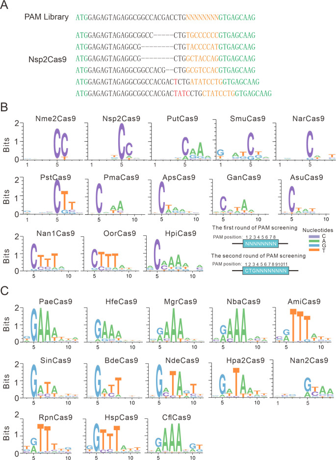
Figure 2—source data 1. The number of unique PAM sequences and the median coverage of every individual PAM variant for the Figure 2B and C.

Figure 2.

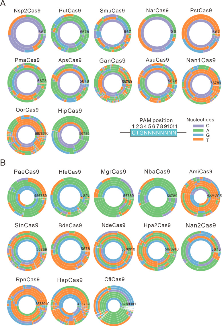
Figure 2—figure supplement 1. PAM wheels for Nme1Cas9 orthologs.

Figure 2—figure supplement 1.

(A) PAM wheels for Nme1Cas9 orthologs containing an aspartate residue corresponding to the Nme1Cas9 H1024. PAM positions in the screening assay are shown on the bottom right. (B) PAM wheels for Nme1Cas9 orthologs containing histidine, or asparagine residues corresponding to the Nme1Cas9 H1024. PAM wheels start in the middle of the wheel for the first 5’ base exhibiting sequence information.
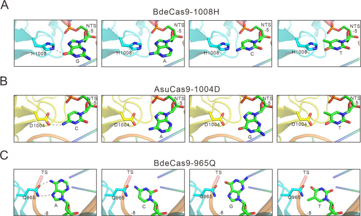
Figure 2—figure supplement 2. The specificity between amino acids and bases in calculated structural models.

Figure 2—figure supplement 2.

(A) Calculated structural model of Bdecas9. The amino acid near the 5 position of the NTS is histidine. Histidine’s side chain forms a potential hydrogen bond with the 6-hydroxyl group of guanine, if the guanine is other bases, this hydrogen bond would be not formed because of the too close distance (cytosine or thymine) or the lack of a hydroxyl group (adenine). (B) Calculated structural model of Asucas9. The amino acid near the 5 position of NTS is aspartic, and it forms a potential hydrogen bond with the 4-amine group of the cytosine. if the cytosine is replaced by other bases, this hydrogen bond would be abolished because of increased distance (adenine or guanine) or the lack of an amine group (thymine). (C) Calculated structural model of Bdecas9. The amino acid near the 8 position of TS is glutamine, and it forms a potential hydrogen bond with the 6-amine group of the adenine on the TS. If the cytosine is replaced by other bases, this hydrogen bond would be not formed because of too close distance (cytosine or thymine) or the lack of an amine group (guanine). TS: target strand, NTS: non-target strand.